Downloaded from https://journals.lww.com/plasreconsurg by BhDMf5ePHKav1zEoum1tQfN4a+kJLhEZgbsIHo4XMi0hCywCX1AWnYQp/IlQrHD3TDbD+Y6NAIFxHnYbs9WOGpNX/Mwl/WKMPZXO+SeqrJQ= on 12/23/2018 Downloadedfrom https://journals.lww.com/plasreconsurgby BhDMf5ePHKav1zEoum1tQfN4a+kJLhEZgbsIHo4XMi0hCywCX1AWnYQp/IlQrHD3TDbD+Y6NAIFxHnYbs9WOGpNX/Mwl/WKMPZXO+SeqrJQ=on 12/23/2018
The Effect of Epidural Anesthesia on Muscle
Flap Tolerance to Venous Ischemia
Cenk Cayci, M.D. Can Cinar, M.D. Osman A. Yucel, M.D. Turgay Tekinay, Ph.D. Jeffrey A. Ascherman, M.D. New York, N.Y.; and Istanbul and Ankara, Turkey
Background: Venous ischemia is a major cause of failure after free tissue transfers and replantations. The combination of general and epidural anesthesia leads to vasodilatation and improves tissue perfusion. Postoperative pain relief and sympa-thetic blockage are additional benefits of epidural anesthesia. The purpose of this study was to determine whether epidural anesthesia has benefits on microcircula-tion and neutrophil funcmicrocircula-tions in muscle flaps subjected to venous ischemia.
Method: Thirty Sprague-Dawley rats were divided into three groups: group I, gen-eral anesthesia; group II, spinal anesthesia; and group III, epidural anesthesia. Cremaster flaps were prepared, postcapillary venules were selected under intravital videomicroscopy, and flaps were subjected to venous ischemia. Images were re-corded from preselected postcapillary venules before venous ischemia (baseline) and following reperfusion. Neutrophil rolling and adhesion, functional capillary density, and diameters of postcapillary venules were evaluated.
Results: The increase in rolling neutrophils in group III was significantly lower than in groups I and II at 60 and 120 minutes. Change of adherent neutrophils in group III was significantly lower than in groups I and II at 15, 60 and 120 minutes. There was significantly more reduction in inner diameter of postcapillary venules in groups I and II compared with group III. Functional capillary density in groups I and II was significantly lower than in group III.
Conclusion: Epidural anesthesia regulated neutrophil functions, salvaged func-tional capillaries, and prevented vasoconstriction of postcapillary venules in cre-master muscle flaps subjected to venous ischemia. Spinal and general anesthesia, however, were found to be ineffective in improving microcirculation of muscle flaps subjected to venous ischemia. (Plast. Reconstr. Surg. 125: 89, 2010.)
M
icrosurgical methods are used for a variety of purposes. Although advancements in microsurgical techniques have improved the success of microsurgical tissue transfer, reper-fusion injury following ischemia is still a major cause of morbidity, prolonged hospitalization, and higher costs.Tissues are more tolerant to primary ischemia than venous ischemia.1Secondary ischemia
asso-ciated with venous dysfunction is observed more frequently and causes more serious damage than arterial or global ischemia.2Venous ischemia
usu-ally occurs due to errors in microvascular surgical methods. Venous obstruction causes venous pool-ing, cessation of arterial flow, extravasation of both erythrocytes and neutrophils, deposition of fibrin, and thrombus formation. The tissue dam-age is progressive and tissue necrosis may develop even after restoration of the venous flow (no-re-flow phenomenon).3–5 Although the mechanism
of this tissue damage is not completely under-stood, it was shown in various studies that lipid
From the Department of Surgery, New York Presbyterian Hospital–Weill Cornell Medical College, the Division of Plas-tic Surgery, New York Presbyterian Hospital–Columbia Uni-versity Medical Center, the Department of Plastic and Re-constructive Surgery, I.U. Cerrahpasa Medical School, and the UNAM, Institute of Materials Science and Nanotech-nology, Bilkent University.
Received for publication February 1, 2009; accepted July 15, 2009.
Presented at the 24th National Congress of the Turkish Society of Plastic, Reconstructive and Aesthetic Surgery, in Ankara, Turkey, October 25 through 29, 2002, and awarded the prize for the second best experimental research paper in the residents category.
Copyright ©2009 by the American Society of Plastic Surgeons
DOI: 10.1097/PRS.0b013e3181c49544
Disclosures: This study was supported by
depart-mental funds. None of the authors has a financial interest in any of the products, devices, or drugs mentioned in this article.
peroxidation,6,7prostanoid mechanisms,8and free
radicals have important roles in its pathophysiol-ogy. The course of the necrosis is different from the all-or-none phenomenon observed in global or arterial ischemia.
There is a debate on selection of the best an-esthesia technique and anesthetic drugs in micro-vascular surgery.9 –11When used concurrently with
general anesthesia, epidural anesthesia may lead to vasodilatation that increases perfusion and reg-ulates microcirculation, and provides effective pain control in the postoperative period. How-ever, with regard to regional anesthesia being an ideal method for anesthesia in microsurgery, there is ongoing debate, and controversial data exist in the literature.9,10,12–16The objective of this
study was to determine whether the positive effects of epidural anesthesia on microcirculation would improve circulation in flaps with venous ischemia.
MATERIALS AND METHODS
Thirty male Sprague-Dawley rats, weighing 175 ⫾ 36.3 g, were divided into three groups: general anesthesia in group I (n ⫽ 10), spinal anesthesia in group II (n ⫽ 10), and epidural anesthesia in group III (n⫽ 10). All experiments were performed according to protocols approved by the Institutional Animal Care and Use Com-mittee and complied with the Guide for the Care and
Use of Laboratory Animals.
Placement of epidural and spinal catheters was done as described by Bahar et al.1750l and 25 l
of lidocaine 2% (Jetokain; Adeka Ltd., Samsun, Turkey) were administered via a catheter for epi-dural (group III) and spinal anesthesia (group II), respectively. Lidocaine injection at a rate of 10 l/minute was continued until the rat was not carrying its own weight and lied down with its belly and chest on the ground. General anesthesia was achieved with intramuscular ketamine 6 mg/kg (Ketalar; Pfizer, New York, N.Y.) and xylazine 0.4 mg/kg (Rompun; Bayer, Leverkusen, Germany). The rat cremaster muscle island flap was pre-pared as described by Grant et al.18,19The vascular
pedicle was completely skeletonized during the dissection, rendering the flap microvasculature completely denervated. In group I, all flaps were elevated under general anesthesia. In groups II and III, spinal and epidural catheters were first placed under general anesthesia. In these groups, the flap elevation procedure was delayed for 24 hours to exclude the rats that developed neuro-logic deficits and to be sure that the rats totally recovered from general anesthesia. Cremaster flaps were then harvested under spinal and
epi-dural anesthesia.18,20 Rats were placed on a fixed
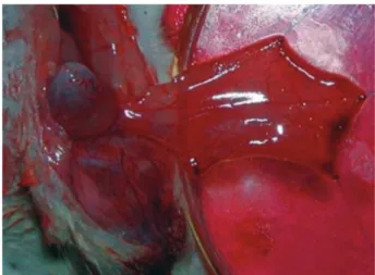
stage under the microscope. The muscle flap was secured on an upside down Petri dish by 5-0 nylon sutures (Fig. 1). The cremaster muscle flap was covered with a stretch film and kept in distilled water for 24 hours to avoid drying. The muscle was kept moist with lactated Ringer’s solution.
To evaluate tissue response to secondary ve-nous ischemia, microcirculation of cremaster muscles was assessed by using intravital videomi-croscopy. The optic apparatus of the intravital videomicroscopy consisted of an Olympus light microscope, CX41 (4⫻, 10⫻, 40⫻, 100⫻), equipped with a custom-made adaptor to ensure the connection with the ocular region of the dig-ital video camera (Sony TRV 120E; Sony, Tokyo, Japan). The magnification was 1800⫻ for the 40⫻ objective of the light microscope.
After flap elevation and a 30-minute waiting period, a vascular map of each flap was prepared under 10⫻ magnification. The postcapillary venules were determined for each flap for the measurement of experimental parameters, and were marked on the vascular map to relocate the same postcapillary venules for the subsequent reperfusion recordings. The baseline images were recorded from those veins for 45 seconds under 40⫻ magnification. Afterward, 30 minutes of ve-nous ischemia was created with a microvascular approximator clamp (vessel approximator clamp 0.4 g/mm2; S&T Marketing Ltd., Neuhausen,
Swit-zerland) applied to the pudic-epigastric vein. After the release of the vascular clamp, recordings were made from the preselected postcapillary venules at 15, 60, and 120 minutes of reperfusion. The images were recorded on the videotapes for future
Fig. 1. The cremaster muscle flap was secured on an upside
down Petri dish by 5-0 nylon sutures. The muscle was kept moist with lactated Ringer’s solution.
slow-motion evaluation by a blinded investigator for the following analyses:
Neutrophil rolling: Neutrophils making the
charac-teristic rolling movement in the postcapillary venules were counted for 30 seconds.
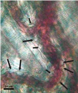
Neutrophil adhesion: Neutrophils which already
ad-hered, or were becoming adad-hered, to the en-dothelium within a certain area (100 m) of the postcapillary venules were counted for 30 seconds (Fig. 2).
Functional capillary density: Under 1800⫻
magnifi-cation, nine areas from the distal, nine areas from the middle, and nine areas from the prox-imal part of each flap were evaluated for 15 sec-onds each. Twenty-seven areas from every flap were thus examined, with each area measuring approximately 340⫻ 420m2. Functional
capil-laries with flow were recorded to estimate the functional capillary density for each flap.21
Postcapillary venule inner diameter change: Static
im-ages captured from the video imim-ages were an-alyzed by an image processing software (Image Tool version 2.0) to calculate the inner and outer diameter of postcapillary venules.
Statistical Evaluation
For the comparisons between the groups, mea-surements taken at minute 15, first (minute 60), and second (minute 120) hours were standardized according to the baseline values (Delta-⌬ values) of the same groups in the same postcapillary
venules. No correction was made for multiple test-ing. Statistical analysis of the data were done using SPSS version 11.5 for Windows (SPSS, Inc., Chi-cago, Ill.). Comparisons within the groups were made by the t test. Comparisons between the groups were performed with the analysis of vari-ance test. The cut-off for significvari-ance was p⬍ 0.05.
RESULTS
Neutrophil Behavior Neutrophil Rolling
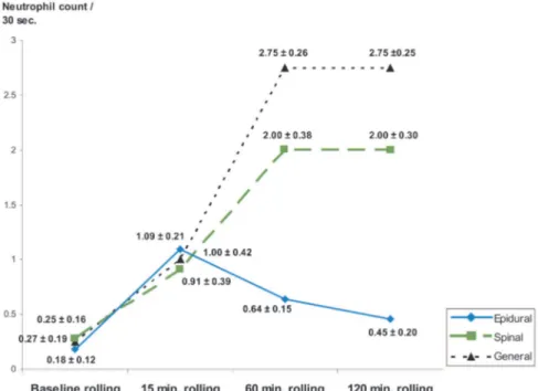
Rolling neutrophil counts before venous isch-emia and 15, 60, and 120 minutes after reperfu-sion are shown in Figure 3. In the epidural anes-thesia group, there was no statistically significant increase in neutrophil rolling during reperfusion except at 15 minutes. Significant statistical differ-ences were detected at 60 minutes and 120 min-utes for the spinal and 60 and 120 minmin-utes for the general anesthesia groups.
Intergroup evaluation revealed no significant differences between any of the groups at 15 min-utes. No significant differences between the gen-eral and spinal anesthesia groups were detected at 60 minutes and at 120 minutes. Statistically signif-icant differences between epidural anesthesia and general and spinal anesthesia groups at 60 and 120 minutes were detected (Fig. 4).
Neutrophil Adhesion
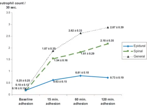
The number and course of the adherent neu-trophils from the general, spinal, and epidural an-esthesia groups recorded before venous ischemia (baseline) and at 15, 60, and 120 minutes of reper-fusion are shown in Figure 5. In the epidural anes-thesia group, no statistically significant differences were detected. In the general anesthesia group, the numbers of adherent neutrophils at 15, 60, and 120 minutes were significantly increased compared with the baseline levels. In the spinal anesthesia group, the numbers of neutrophils adherent to the endothelium significantly increased at 15, 60, and 120 minutes compared with the baseline levels.
Intergroup evaluation disclosed that there was no difference between the general and spinal an-esthesia groups (p⬎ 0.05). However, differences between epidural and general anesthesia were sig-nificant at 15, 60, and 120 minutes. The differ-ences between epidural and spinal anesthesia groups were statistically significant at 15, 60, and 120 minutes (Fig. 6).
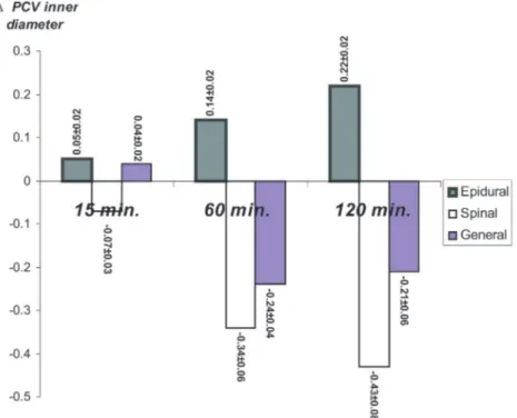
Inner Diameter of Postcapillary Venules
The changes in the postcapillary venule inner diameters compared with baseline diameters at Fig. 2. Intravital videomicroscopic image showing rolling and
adherent neutrophils (black arrows) to the postcapillary venules. Bar represents 20m.
Fig. 4. Changes of rolling movements of the neutrophils compared with baseline
(⌬-rolling)inthreegroupswereanalyzed.Nosignificantdifferencewasdetectedat 15 minutes, but the epidural anesthesia group had significant differences com-pared with both general and spinal anesthesia at 60 and 120 minutes. Units for the numbers are number of neutrophils per 30 seconds;⫾ values represent SEM (anal-ysis of variance multiple comparison test; *p⬍ 0.05 significant).
Fig. 3. Rolling neutrophil counts before venous ischemia and after reperfusion. Analysis
within the groups revealed that the epidural anesthesia group had no statistically signif-icant difference compared with baseline except at 15 minutes (p⬍0.01).Bothgeneraland spinal anesthesia groups had significant differences compared with baseline at 60 and 120 minutes. Units for the numbers are number of neutrophils per 30 seconds;⫾ values represent the standard error of mean (t test; *p⬍ 0.05 significant).
Fig. 5. Analysis of neutrophil adhesion within groups. There were no statistically
signif-icant differences in the epidural anesthesia group. Both general and spinal anesthesia groups had significant differences compared with baseline at 15, 60 and 120 minutes. Units for the numbers are number of neutrophils per 30 seconds;⫾ values represent SEM (t test; *p⬍ 0.05 significant).
Fig. 6. Changes of adhesion of neutrophils compared with baseline (⌬-adhesion) in the
three groups were analyzed. No statistically significant difference was detected between general and spinal anesthesia groups. Both of these groups had a statistically significant difference from the epidural anesthesia group at 15, 60, and 120 minutes. Units for the num-bers are number of neutrophils per 30 seconds;⫾ values represent SEM (analysis of variance multiple comparison test; *p⬍ 0.05 significant).
15, 60, and 120 minutes of reperfusion are illus-trated in Figure 7.
A statistically significant diameter change in the postcapillary venules compared with baseline diameter was detected at 15 minutes between the epidural and spinal anesthesia groups, and be-tween the spinal and general anesthesia groups, but not between the epidural and general anes-thesia groups.
Statistically significant change in postcapillary venules was detected when the epidural anesthesia group was compared with both general and spinal anesthesia groups at 60 minutes and at 120 min-utes. Postcapillary venule inner diameter change was not statistically significant between the general and spinal anesthesia groups either at 60 minutes or at 120 minutes (Fig. 7).
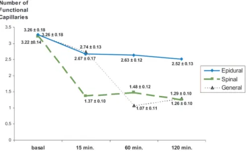
Functional Capillary Density
In the epidural anesthesia group, statistically significant reductions in functional capillary den-sity at 15 and 60 minutes and a more significant reduction at 120 minutes were detected (Fig. 8). In the spinal anesthesia group, statistically sig-nificant reductions of functional capillary density
with respect to the baseline value were detected at 15, 60, and 120 minutes of reperfusion. In the general anesthesia group, statistically significant reductions in functional capillary density at 15, 60, and 120 minutes were detected (Fig. 8).
The intergroup comparisons change in tional capillary density, calculated by the func-tional capillary counts at 15, 60, and 120 minutes of venous ischemia subtracted from baseline value for all groups are shown in Figure 9. At 15 minutes, spinal anesthesia had a statistically significant dif-ference from the epidural and general anesthesia groups. Change in functional capillary density of the epidural anesthesia groups were statistically significant compared with other groups at 60 and 120 minutes.
DISCUSSION
Venous ischemia usually results from an error in microsurgical technique. Secondary ischemia associated with venous dysfunction appears more frequently and causes more damage than arterial or total ischemia. In one study, irreversible dam-age occurred very rapidly in muscle following ve-nous ischemia, and necrosis developed in 75
per-Fig. 7. The change of the postcapillary venule inner diameters compared with
base-line diameters (⌬PCV) at 15, 60, and 120 minutes of reperfusion. Analysis of ⌬PCV between groups revealed a statistically significant difference when epidural anesthe-sia was compared with general and spinal anestheanesthe-sia groups at 60 and 120 minutes of reperfusion. Units for the numbers are micrometers;⫾ values represent SEM (analysis of variance multiple comparison test; *p⬍ 0.05 significant).
Fig. 8. Analysis of functional capillary density within groups. In all three groups a statistically
significant reduction in functional capillary density was detected at 15, 60, and 120 minutes compared with baseline. Units for the numbers are functional capillaries per unit area;⫾ values represent SEM (t test; *p⬍ 0.05 significant).
Fig. 9. The change of functional capillary density (FCD) compared with baseline
change in functional capillary density. Analysis of change in functional capillary density between groups revealed a statistically significant difference between the epidural anesthesia group and the spinal and general anesthesia groups at 60 and 120 minutes. Units for the numbers are functional capillaries per unit area;⫾ values represent SEM (analysis of variance multiple comparison test; *p⬍ 0.05 significant).
cent of the muscle tissue studied after 60 minutes of venous ischemia, making the early detection of venous occlusion critical to the correction of this complication.22 The critical venous ischemia
du-ration for muscle flaps is shorter than that for skin flaps. High metabolic activity and rigid fascial structure makes muscle tissue more vulnerable to the pressure necrosis resulting from edema and hemorrhage following venous occlusion.22In
ve-nous occlusion, arterial blood flow continues and causes hemorrhaging from microvessels into the extravascular space.23,24This leads to an increase in
the external pressure on vessels and collapse in capillaries. The edema formed in the interstitial space after venous ischemia is not beneficial for flap survival because it prevents oxygen diffusion.25,26
This explains the transition of flaps to an anaerobic state with continued arterial blood flow following venous ischemia.
Venous ischemia almost always requires mi-crosurgical revision of the venous anastomosis. Poor outcomes after microsurgical revision of the failed venous anastomosis attracted researchers’ attention toward finding measures to decrease the progressive tissue damage following this microsur-gical catastrophe. Angel et al. used deferoxamine, a free oxygen radical scavenger, in venous isch-emia flaps and reported an intermediate benefit. This study reported an improvement in flap sur-vival in the deferoxamine-treated group, and sug-gested free radical involvement in the mechanism of ischemia-reperfusion injury in secondary ve-nous ischemia.8Yucel et al. used cyclosporine A in
a rat inferior epigastric artery skin flap subjected to secondary venous ischemia, aiming to reduce lipid peroxidation. This study reported decreased malondialdehyde content, lower myeloperoxi-dase activity and improved flap survival in the treatment group, suggesting improved flap sur-vival after secondary venous ischemia by reducing lipid peroxidation by cyclosporine A treatment.27
We investigated the contribution of different anesthesia techniques to the microcirculation of muscle flaps subjected to venous ischemia. As freely transferred tissues are completely dener-vated and already sympathectomized, it has been questioned whether a sympathetic block induced by epidural anesthesia may actually decrease free flap perfusion by diverting the blood flow to the healthy vessels with lower vascular resistance.14
Other authors have suggested reduction of free flap blood flow because of a decrease in mean arterial blood pressure and cardiac output.12,28
The use of general anesthesia combined with epidural anesthesia is controversial, although
there is data recommending the use of this com-bined anesthesia.16,29 –31Scott et al. compared the
efficacy of epidural and general anesthesia to gen-eral anesthesia alone in patients undergoing free tissue transfers to the lower extremity. There was a significantly lower number of major microvas-cular complications and a better flap survival in the combined epidural and general anesthesia group.16Neurogenic vasoconstriction, which may
result in marked reduction of blood flow through injured or revascularized tissues, is mediated through sympathetic pathways which may be blocked by long-acting local anesthetics resulting in optimal intravascular pressures and elevated flow. Phelps and colleagues showed that distal sym-pathetic blockade using bupivacaine protects the digital circulation against vasospasm elicited by a strong stimulus for a minimum of 7 to 8 hours.29
Because free tissue transfers are lengthy inter-ventions, general anesthesia is preferred.9,10,12–16,32
Use of a combined epidural anesthesia is recom-mended in addition to this in suitable cases.13,14,16,33
Although the benefits of an epidural anesthesia are clinically known due to pain control and prevention of vasoconstriction (sympatholytic activity), there is controversy about the beneficial effect of epidural anesthesia on flap microcirculation.12,14,28 Epidural
anesthesia has beneficial effects on lower extremity venous hemodynamics.34Delis et al. quantified the
venous hemodynamic changes in the lower limb during and immediately after abdominal surgery performed under epidural anesthesia and general anesthesia combined, versus under general anes-thesia alone. Researchers investigated mean ve-locity, peak veve-locity, and volume flow measured at the level of the popliteal vein using duplex ultra-sonography. Epidural anesthesia administered as part of general anesthesia was associated with a significant enhancement of mean volume and vol-ume flow, improving lower extremity venous hemodynamics.34
In our study, microcirculation of the venous isch-emic flaps was improved by epidural anesthesia. The beneficial effect was observed as decreased numbers of rolling and adherent leukocytes, decreased vaso-constriction in the postcapillary venules, and in-creased numbers of perfused capillaries in the epi-dural anesthesia group.
The activation of the neutrophils and eventual endothelial cell interaction consists of three steps: rolling, adhesion, and migration.35 The
interac-tion between neutrophils and endothelial cells is mediated by a well-orchestrated sequence of in-teractions between adhesion molecules on both endothelium and neutrophils. The neutrophils
are recruited to the reperfused muscle by chemo-tactic factors, and begin to interact with the en-dothelium through a process called “rolling.”36,37
This initial loose adherence is an obligatory step that is necessary for later firm adherence leading to transendothelial migration into muscle paren-chyma and the physiologic sequelae, including the no-reflow phenomenon and tissue necrosis.38 – 41
Pronounced statistically significant differences in neutrophil functions and functional capillary den-sity were detected, particularly at minute 120 of reperfusion, between epidural and other groups in the late reperfusion period, rendering the re-sults more meaningful.
We found that epidural anesthesia played a protective role in the functional capillary count following venous ischemia, whereas there was a distinct reduction in the functional capillary count in the spinal and general anesthesia groups. This effect became more pronounced in the late pe-riod of reperfusion. Our study revealed decreased vasoconstriction in postcapillary venules to a sta-tistically significant extent with epidural anesthe-sia following venous ischemia, compared with spi-nal and general anesthesia.
We have found no studies in the literature investigating the effects of spinal anesthesia on the circulation of free flaps, and we could not find any benefits of spinal anesthesia on muscle flap mi-crocirculation. Our current experiment was de-signed to assess the outcomes but not the mech-anisms of different anesthesia types on flap microcirculation. The reason for the detrimental effect of spinal anesthesia cannot be explained with certainty. Possible mechanisms include he-modynamic changes and the pronounced sympa-thectomy effect of spinal anesthesia, causing the vascular steal phenomenon which may result in a reduction of free flap blood flow. To further study these possible mechanisms, additional experi-ments are required.
CONCLUSIONS
In conclusion, we found that epidural anes-thesia improves microcirculation and flap perfu-sion in the cremaster muscle island flap subjected to venous ischemia by enabling dilation of the postcapillary venules and regulating neutrophil functions. Epidural anesthesia, when adminis-tered as part of the anesthesia regimen in micro-surgery, may therefore generate a marked venous flow enhancement, thus aiding in the prevention of peri-operative and postoperative venous stasis, and in cases of venous insufficiency, it may protect against venous ischemic tissue damage.
Jeffrey A. Ascherman, M.D. Columbia University Medical Center 161 Fort Washington Avenue, Suite 607 New York, N.Y. 10032 [email protected]
REFERENCES
1. Kerrigan CL, Daniel RK. Critical ischemia time and the fail-ing skin flap. Plast Reconstr Surg. 1982;69:986–989.
2. Kerrigan CL, Zelt RG, Daniel RK. Secondary critical ischemia time of experimental skin flaps. Plast Reconstr Surg. 1984;74: 522–526.
3. Heden P, Sollevi A. Circulatory and metabolic events in pig island skin flaps after arterial or venous occlusion. Plast Re-constr Surg. 1989;84:475–481; discussion 482–483.
4. Knight KR, Angel MF, Lepore DA, et al. Secondary ischaemia in rabbit skin flaps: The roles played by thromboxane and free radicals. Clin Sci (Lond.) 1991;80:235–240.
5. May JW Jr, Chait LA, O’Brien BM, Hurley JV. The no-reflow phenomenon in experimental free flaps. Plast Reconstr Surg. 1978;61:256–267.
6. Angel MF, Narayanan K, Swartz WM, et al The etiologic role of free radicals in hematoma-induced flap necrosis. Plast Reconstr Surg. 1986;77:795–803.
7. Mellow CG, Knight KR, Angel MF, O’Brien BM. The effect of thromboxane synthetase inhibition on tolerance of skin flaps to secondary ischemia caused by venous obstruction. Plast Reconstr Surg. 1990;86:329–334.
8. Angel MF, Mellow CG, Knight KR, O’Brien BM. The effect of deferoxamine on tolerance to secondary ischaemia caused by venous obstruction. Br J Plast Surg. 1989;42:422–424. 9. Jakubowski M, Lamont A, Murray WB, de Wit SL. Anaesthesia
for microsurgery. S Afr Med J. 1985;67:581–584.
10. Macdonald DJ. Anaesthesia for microvascular surgery: A physiological approach. Br J Anaesth. 1985;57:904–912. 11. Faura A, Izquierdo E, Pelegri MD. Epidural vs. intradural
anesthesia in ambulatory surgery (in Spanish). Rev Esp Anes-tesiol Reanim. 1999;46:256–263.
12. Banic A, Krejci V, Erni D, Petersen-Felix S, Sigurdsson G. Effects of extradural anesthesia on microcirculatory blood flow in free latissimus dorsi musculocutaneous flaps in pigs. Plast Reconstr Surg. 1997;100:945–955; discussion 956. 13. Berger A, Tizian C, Zenz M. Continuous plexus blockade for
improved circulation in microvascular surgery. Ann Plast Surg. 1985;14:16–19.
14. Erni D, Banic A, Signer C, Sigurdsson GH. Effects of epidural anaesthesia on microcirculatory blood flow in free flaps in patients under general anaesthesia. Eur J Anaesthesiol. 1999; 16:692–698.
15. Robins DW. The anaesthetic management of patients un-dergoing free flap transfer. Br J Plast Surg. 1983;36:231–234. 16. Scott GR, Rothkopf DM, Walton RL. Efficacy of epidural anesthesia in free flaps to the lower extremity. Plast Reconstr Surg. 1993;91:673–677.
17. Bahar M, Rosen M, Vickers MD. Chronic cannulation of the intradural or extradural space in the rat. Br J Anaesth. 1984; 56:405–410.
18. Anderson GL, Acland RD, Siemionow M, McCabe SJ. Vas-cular isolation of the rat cremaster muscle. Microvasc Res. 1988;36:56–63.
19. Grant RT. The effects of denervation on skeletal muscle blood vessels (rat cremaster). J Anat. 1966;100:305–316. 20. Acland RD, Anderson G, Siemionow M, McCabe S. Direct in
distal to a small-vessel anastomosis. Plast Reconstr Surg. 1989; 84:280–288; discussion 289.
21. Ayhan S, Tugay C, Norton S, Araneo B, Siemionow M. Dehy-droepiandrosterone protects the microcirculation of muscle flaps from ischemia-reperfusion injury by reducing the expression of adhesion molecules. Plast Reconstr Surg. 2003;111:2286–2294. 22. Gabriel A, Chaney N, Stephenson LL, Zamboni WA. Effect
of total venous occlusion on capillary flow and necrosis in skeletal muscle. Plast Reconstr Surg. 2001;108:430–433. 23. Hjortdal VE, Hansen ES, Hauge E. Myocutaneous flap
isch-emia: Flow dynamics following venous and arterial obstruc-tion. Plast Reconstr Surg. 1992;89:1083–1091.
24. Marzella L, Jesudass RR, Manson PN, Myers RA, Bulkley GB. Functional and structural evaluation of the vasculature of skin flaps after ischemia and reperfusion. Plast Reconstr Surg. 1988;81:742–750.
25. Angel MF, Mellow CG, Knight KR, O’Brien BM. Secondary ischemia time in rodents: Contrasting complete pedicle in-terruption with venous obstruction. Plast Reconstr Surg. 1990; 85:789–793; discussion 794–795.
26. Vollmar B, Westermann S, Menger MD. Microvascular re-sponse to compartment syndrome-like external pressure el-evation: An in vivo fluorescence microscopic study in the hamster striated muscle. J Trauma 1999;46:91–96.
27. Yucel A, Senyuva C, Andican G, et al. Secondary venous ischemic injury associated with neutrophil infiltration and lipid peroxidation: Amelioration of injury by cyclosporin A in a rat inguinal island flap. Ann Plast Surg. 2000;45:54–60. 28. Van Twisk R, Gielen MJ, Pavlov PW, Robinson PH. Is additional epidural sympathetic block in microvascular surgery contrain-dicated? A preliminary report. Br J Plast Surg. 1988;41:37–40. 29. Phelps DB, Rutherford RB, Boswick JA Jr. Control of
vaso-spasm following trauma and microvascular surgery. J Hand Surg (Am.) 1979;4:109–117.
30. Sigurdsson GH, Thomson D. Anaesthesia and microvascular surgery: Clinical practice and research. Eur J Anaesthesiol. 1995;12:101–122.
31. Taras JS, Behrman MJ. Continuous peripheral nerve block in replantation and revascularization. J Reconstr Microsurg. 1998; 14:17–21.
32. Sigurdsson GH, Banic A, Wheatley AM, Mettler D. Effects of halothane and isoflurane anaesthesia on microcirculatory blood flow in musculocutaneous flaps. Br J Anaesth. 1994;73: 826–832.
33. Inberg P, Tarkkila PJ, Neuvonen PJ, Vilkki S. Regional an-esthesia for microvascular surgery: A combination of brachial plexus, spinal, and epidural blocks. Reg Anesth. 1993;18:98– 102.
34. Delis KT, Knaggs AL, Mason P, Macleod KG. Effects of epidural-and-general anesthesia combined versus general anesthesia alone on the venous hemodynamics of the lower limb: A randomized study. Thromb Haemost. 2004; 92:1003–1011.
35. Angel MF, Ramasastry SS, Swartz WM, Basford RE, Futrell JW. Free radicals: Basic concepts concerning their chemistry, pathophysiology, and relevance to plastic surgery. Plast Re-constr Surg. 1987;79:990–997.
36. Elgebaly SA, Hashmi FH, Houser SL, Allam ME, Doyle K. Cardiac-derived neutrophil chemotactic factors: Detection in coronary sinus effluents of patients undergoing myocar-dial revascularization. J Thorac Cardiovasc Surg. 1992;103:952– 959.
37. Dreyer WJ, Smith CW, Michael LH. Canine neutrophil ac-tivation by cardiac lymph obtained during reperfusion of ischemic myocardium. Circ Res. 1989;65:1751–1762. 38. Kubes P, Jutila M, Payne D. Therapeutic potential of
inhib-iting leukocyte rolling in ischemia/reperfusion. J Clin Invest. 1995;95:2510–2519.
39. Davenpeck KL, Gauthier TW, Albertine KH, Lefer AM. Role of P-selectin in microvascular leukocyte-endothelial interac-tion in splanchnic ischemia-reperfusion. Am J Physiol. 1994; 267:H622–H630.
40. Geng JG, Bevilacqua MP, Moore KL, et al. Rapid neutrophil adhesion to activated endothelium mediated by GMP-140. Nature 1990;343:757–760.
41. Jerome SN, Dore M, Paulson JC, Smith CW, Korthuis RJ. P-selectin and ICAM-1-dependent adherence reactions: Role in the genesis of postischemic no-reflow. Am J Physiol. 1994; 266:H1316–H1321.