Conscientious Design of Zn-S/Ti
‑N Layer by Transformation of
ZnTiO
3
on Electrospun ZnTiO
3
@TiO
2
Nano
fibers: Stability and
Reusable Photocatalytic Performance under Visible Irradiation
Kugalur Shanmugam Ranjith
*
and Tamer Uyar
*
Institute of Materials Science & Nanotechnology and UNAM-National Nanotechnology Research Center, Bilkent University, Ankara 06800, Turkey
*
S Supporting InformationABSTRACT: Herein, we report the rational design of Zn-S/Ti-N on TiO2 as a hierarchical nanoarchitecture from the
ZnTiO3@TiO2 nanofibers (NFs) through electrospinning followed by a hydrothermal process using L-cysteine as an S/N
source. Controlling the hydrothermal temperature, the hierarchical form of NFs exhibited highly efficient visible catalytic behavior for organic dye (i.e., Rhodamine B) degradation since S and N based surface function on the oxide surface resulted in unique interlayer induced strain coupled surface defects. The surface functionalization of the ZnTiO3surface with S and N was
solidly confirmed by X-ray photo-electrospectroscopy (XPS) and energy-dispersive X-ray (EDX) with elemental mapping results. Inducing the S/N functionality at higher hydrothermal temperature reverses the structural arrangement of ZnTiO3
favoring the interaction of S preferably with Zn and Ti with N for the formation of ZnS/TiN@TiO2NFs. The tunable band function through the Zn-S/Ti-N cofunctionalization exhibited effective long-term catalytic performance under UV and visible irradiation with a degradation rate of 0.0362 and 0.0313 min−1, which is nearly 3.1 and 1.3 times higher than that of the ZnTiO3@TiO2and ZnTiO3−S/N@TiO2NFs, respectively. The catalysts are highly photoactive after multiple photocatalytic
cycles with stable surface and structural features under visible irradiation. The study could provide new opportunities for designing hierarchical structures in ternary form of nanoscale architectures for effective visible photocatalytic activity.
KEYWORDS: ZnTiO3nanofibers, Electrospinning, Hierarchical heterostructures, ZnS shell layers, S/N codoping,
Visible photocatalysis, Solar irradiation
■
INTRODUCTIONOver the concern of extreme textile and organic industrial wastewater pollution, environment remediation is taken up to enable the world to step forward with various technologies for the development of wastewater treatment.1,2 Photoresponsive advanced oxidization processes through a heterostructural catalyst are intended toward the degradation or removal of organic pollutants from wastewater. In this context, TiO2and
ZnO are the most promising semiconductor photocatalysts with their wide band gap function facilitating their exploration for wastewater treatment. However, both these semiconductors
have their individual advantages and disadvantages when it comes to their catalytic efficiency under UV irradiation owing to the favorable wide band gap (>3.2 eV).3,4 Alloying these wide band gap materials in the form of ternary oxides such as ZnTiO3 favors a similar band function, overcoming their
structural disadvantages.5,6It is a known fact that UV photon energy accounts only for nearly 5% of total solar energy, which
Received: May 28, 2018
Revised: July 26, 2018
Published: August 26, 2018
pubs.acs.org/journal/ascecg
Cite This:ACS Sustainable Chem. Eng. 2018, 6, 12980−12992
Downloaded via BILKENT UNIV on February 26, 2019 at 13:50:27 (UTC).
is very low when compared to 45% of visible photon energy.7 In order to commercialize advanced oxidation process based wastewater treatments, solar energy has to be utilized, and semiconductor nanostructures have to be developed in such a way that they effectively drive visible catalytic behavior.8 Doping and decoration of metallic or nonmetallic elements with wide band gap semiconductors improved visible catalytic behavior by narrowing the band gap or its band function.9−12 In that, doping of nonmetallic elements such as N, C, S, and F plays an effective role as anionic impurities in the host lattice, thereby narrowing the band gap toward visible catalytic behavior.13 Among the nonmetallic ions, S and N have garnered notable attention over the wide band gap materials with extended thermal stability and improvised visible response.14 Doping with the cofunctionality of S and N has become attractive because of its tunable electronic structure which permits visible absorption in wide band gap semi-conductors.15 The favorable morphology with the 1D structural feature directs electron flow with high surface area and promotes the light responsive nature. Without disturbing the morphological features, inducing the S/N codopants in the single set synthesis process is highly complicated and only leads to a low content of S impurities on the catalyst surface. Postsynthesis of S/N codoping functionality through a hydrothermal process shows promise in nonmetal doped catalyst platforms, which can be created through the diffusion of S and N ions. Further processing the postsynthesis process to modify the surface features of the nanostructures to S and N based compounds with a heterostructural finish helps in improving the visible response and promotes charge separation efficiency. In the case of codoping functionality,L-cysteine is
one of the effective sources of S and N ions to obtain improvised structural features on the catalytic surface. Previously, there were few investigations which found that,L
-cysteine plays a role in the construction of S/N cofunction-alized surfaces for tunable functional properties.16,17 Con-trolled release and interaction of S2−and N ions through the hydrothermal process favors the surface functionality with tunable band function over the semiconductor surface.18From
the observations attained from our previous investigations, Zn ions were more favorable for the sulfidation reaction, and Ti ions interact poorly with S ions, thereby leading to promising interactions with Zn sites.19,20 Further, there is an interesting possibility of an interaction of Ti−N sites on the surface by a nitration process at higher temperatures, which will lead to the hierarchical heterostructural finish.21,22 Combining the sul fi-dation and nitration process on the ZnTiO3 surface would create a new structural feature with the role of S and N based interstitial structures with tunable band functionalities of Zn−S and Ti−N interactions. The exploration of the dual functionality of S and N ions over the ZnTiO3nanostructures facilitates extended visible catalytic behavior.
This work signifies the synthesis of the dual functionalization of S and N cocatalyzed ZnTiO3@TiO2 nanofibers (NFs)
through electrospinning followed by a hydrothermal process as shown in Scheme 1. The temperature influences the hydro-thermal process to modify the S and N impurities to form as a ZnS-TiN surface functionality on TiO2 NFs through the
selective interaction with Zn ions by sulfidation and Ti by the nitration process. Electrospun NFs of titanium butoxide, zinc acetate, and PVP composites were calcined to form TiO2− ZnTiO3NFs depending on the ionic concentration of Zn ions
on the fiber surface under high annealing temperature. Also, the process of hydrothermal treatment withL-cysteine serves as
the S and N source for obtaining dual functionality. The temperature of the hydrothermal process plays a crucial role in the S ionic state and structural morphology of thefibers with the Zn-S/Ti-N layer by transforming ZnTiO3over TiO2. After
adhering to the presence of the S/N functionality over the ZnTiO3@TiO2 NFs through the EDAX and XPS studies,
visible catalytic activity and long-term catalytic stability were investigated. After repeated photocatalytic cycles, the stable structural form of the S/N-ZnTiO3 surface finish exhibited efficient catalytic activity under visible irradiation.
■
EXPERIMENTAL SECTIONMaterials. Polyvinylpyrrolidone (PVP, Mw = 1,300,000,
Sigma-Aldrich, CAS No.: 9003-39-8), zinc acetate (Zn(OAc)2,
Sigma-Scheme 1. Synthesis Process for the Hierarchical Formation of S/N Modified ZnTiO3@TiO2NFs
Aldrich, CAS No.: 5970-45-6), titanium(IV) butoxide (Ti(OBu)4,
Sigma-Aldrich, CAS No.: 5593-70-4), ethanol (C2H5OH,
Sigma-Aldrich, CAS No.: 64-17-5), L-cysteine (97%, HSCH2CH(NH2
)-CO2H, Sigma-Aldrich, CAS No.: 52-90-4), acetic acid (CH3COOH,
Aldrich, CAS No.: 64-19-7), rhodamine B (RhB, Sigma-Aldrich, CAS No.: 81-88-9), methylene blue (MB, Sigma-Sigma-Aldrich, CAS No.: 122965-43-9), 4-chorophenol (99%, 4-CP, Alfa Aesar, CAS No.: 106-48-9), p-benzoquinone (98%, BQ, Alfa Aesar, CAS No.: 106-51-4), triethanolamine, (99%, TEOA, Sigma-Aldrich, CAS No.: 102-71-6), and isopropyl alcohol (99.5% IPA, Sigma-Aldrich, CAS No.: 67-63-0) were procured and were used as received without any further purification.
Electrospinning of ZnTiO3@TiO2NFs. TiO2−ZnTiO3NFs were
synthesized using an electrospinning process followed by the high temperature calcination process. PVP (1 g) was dissolved in 5 mL of ethanol by constant stirring as an electrospinning solution. As the Ti and Zn precursors, 1 mL of Ti(OBu)4and 4% (w/v) of Zn(OAc)2
were mixed in 2 mL of ethanol and 0.6 mL of acetic acid solution and stirred. After attaining a homogeneous clear solution, it was added into the polymeric solution and stirred overnight before the electrospinning process. The attained homogeneous solution was loaded in a 3 mL plastic syringe with a needle diameter of 0.4 mm and placed horizontally on a syringe pump (KD Scientific, KDS101). Under the applied high electricfield (15.0 kV) from a high voltage power supply (Spellman, SL series, USA) with a solutionflow rate of 0.5 mL h−1, the electrospun NFs were collected on the aluminum foil cover metal collector placed at a distance of 15 cm from the needle tip. This whole process was carried out at 22 °C and 19% relative humidity in a Plexiglas box. The obtained electrospun Zn(OAc)2/
Ti(OBu)4/PVP composite NFs were further calcined at 500°C for 3
h in air to remove the organic moiety to form ZnO-TiO2composite
NFs, followed by annealing at 800 °C for 3 h, which favors the formation of TiO2−ZnTiO3 (ZT-SN 00) NFs. Annealing at high
temperature causes Zn ions to move toward the surface and be substituted with Ti ions to form the ZnTiO3surfacefinish with a wall
thickness of 100 nm. The wall thickness depends on the ionic concentration of the Zn in the compositefibers, and herein, we use 4 wt % of Zn precursor concentration.
Synthesis of S/N-Doped-ZnTiO3@TiO2 and ZnS/TiN@TiO2
NFs. To synthesize the hierarchical functionality of the S/N modified ZnTiO3surface, pristine ZT-SN 00 NFs (0.05 g) andL-cysteine (0.02
g) were added to 40 mL of deionized water under stirring. After continuous stirring for 30 min at room temperature, the solution was transferred to a 50 mL Teflon autoclave and held at different temperatures (100, 150, and 200°C) for 6 h. As per the hydrothermal temperature, the samples were named as SN 00, SN 100, ZT-SN 150, and ZT-ZT-SN 200 for the pristine, 100, 150, and 200°C of hydrothermally grown samples. After the sample cooled down, precipitates were centrifuged and washed with water and ethanol thrice and dried at 80°C for 60 min. Dried samples were calcined at 200°C for 2 h in vacuum, which resulted in the ZT-SN NFs with different S and N functionalities.
Characterization. Scanning electron microscopy (SEM, FEI-Quanta 200 FEG) equipped with an energy dispersive X-ray spectrometer (EDS) was used to characterize the morphological and elemental composition of the electrospun nanostructures. The structural features of the ZT-SN samples were measured by a PANalytical X’Pert multipurpose X-ray diffractometer with Cu Kα radiation. Transmission electron microscopy (FEI-Tecnai G2 F30) with an accelerating voltage of 200 kV was used to investigate morphological features of the NFs. Elemental mapping was carried out using scanning transmission electron microscopy (STEM, Tecnai G2 F30, FEI). UV−vis diffuse reflectance spectra were obtained on an UV−vis spectrophotometer (Shimadzu UV-3600 spectrophotome-ter), and the band gap of the surface modified nanostructures were calculated through the transformed Kubelka−Munk relationship. X-ray photoelectron spectroscopy (XPS, Thermo K-alpha-monochro-mated) was employed to analyze the surface chemical composition. Photoluminescence (PL) measurements were performed by using a time-resolved fluorescence spectrophotometer (FL-1057 TCSPC)
with an excitation wavelength at 350 nm. The specific surface area of the samples were analyzed using the Brunauer−Emmet−Teller (BET) method carried out on a Quantachrome Instrument autosorb (iQ2)
analyzer after degassing the samples for 3 h at 200 °C. Dye degradation was analyzed by the UV−vis absorbance spectra of pollutants using an UV−vis spectrophotometer (Varian Cary 100).
Photocatalytic Degradation. Catalytic degradation was done under UV irradiation (Ultra-Vitalux Ultraviolet high pressure lamp (300 W, Osram) λ ∼ 365 nm, 272 lx of photon energy), visible irradiation (75 W, Osram), xenon lamp with UVfilter (λ > 400 nm, 560 lx of photon energy), and natural sunlight (λ > 400 nm, 860 × 100 lx of photon energy) (with UV cutoff filter (Lee type 226 (USA)) at room temperature using RhB as the probe molecule. In separate quartz cuvettes, 1 mg/mL of electrospun ZT-SN based NFs were immersed with RhB dye solution (10 ppm) and placed in the dark for 20 min to attain the adsorption/desorption equilibrium of dye on the NF surface before photoirradiation. The working distance between the lamp and the sample was set to be 15 cm, and a series of samples and controls (without catalyst) were simultaneously irradiated. The degradation of RhB dye molecules was monitored from the real time absorption intensity at 554 nm, and the relative concentrations of the pollutants (C/C0) evaluated the photocatalytic activities with respect
to time using a UV−vis spectrophotometer. The catalytic behavior was investigated in sunlight (>400 nm) under the shadow from 9:00 am to 3:00 pm during a sunny day. The average light intensity of sunlight was 860 × 100 lx. In order to investigate the long-term stability, the reusability of the catalysts over 10 consecutive cycles was tested under visible irradiation. The degradation efficiency of dye solution was calculated by ((C0 − C)/C0) × 100, where C and C0
denote the UV-absorption peak intensity before and after UV and visible irradiation. Further, photocatalytic degradation activity over another dye molecule (MB (15 ppm)) and colorless organic waste (4-CP (10 ppm)) were investigated.
Active Species Trapping Experiments. To quantify the production of active radicals such as hydroxyl radicals (°OH), superoxide radicals (O2°), and holes (h+) on the photocatalytic
reaction, 1.0 mM BQ (a quencher of O2°), IPA (a quencher of °OH),
and TEOA (a quencher of h+) were added to the photocatalytic
reaction with the catalyst.19 The method was similar to a former photocatalytic activity test.
■
RESULTS AND DISCUSSIONStructural and Morphological Studies. Scheme 1
represents the systematic procedure for preparing Zn-S/Ti-N and S/N-ZnTiO3 layers from the pristine ZnTiO3 layer on
TiO2NFs through the feasible construction of electrospinning followed by a hydrothermal process. The bead-free fibrous network having an averagefiber diameter of 625 ± 45 nm with the composition of Zn(OAc)2/Ti(OBu)4/PVP was revealed
from the SEM images inFigure S1. After calcination at 500°C for 3 h, it exhibited the formation of ZnO-TiO2composite NFs
with a fiber diameter of 425 ± 85 nm (Figure S2) by completely removing the organic part (PVP polymer matrix and acetate and butoxide group). Furthermore, on annealing the composite NFs at 800°C, the formation of ZnTiO3NFs
was favored. The obtained NFs were subjected to function-alization of S/N through a hydrothermal process, favoring the formation of S/N doped ZnTiO3NFs. InFigure 1, crystalline phases of S/N modified ZnTiO3 NFs were investigated
through an X-ray powder diffraction (XRD) pattern. Figure 1a exhibited the crystalline nature of the pristine ZnTiO3NFs
annealed at 800°C. On annealing the electrospun NFs at 800 °C, they change to the crystal phase of the hexagonal ZnTiO3,
which matches with the JCPDS card no. of 26-1500 with a cubic phase. No additional peaks representing TiO2or ZnO in
the pattern was found, which confirmed the purity of the ZnTiO3nanostructure (Figure 1a). After annealing at 800°C,
ZnTiO3 NFs were formed, while annealing below 800 °C exhibited a mixed phase consisting of ZnTiO3, ZnO, and TiO2.
However, the actual assembly of the NFs consists of the ZnTiO3@TiO2 based structural finish, which was later
confirmed by the TEM image mapping analysis. Because of the highly crystalline solid 100 nm wall thickness of the ZnTiO3layer, peaks corresponding to TiO2were absent in the pattern but were present as a central core on the annealed NF finish (Figure S3). On treating ZnTiO3NF withL-cysteine by
the hydrothermal process, no other additional diffraction peaks were observed until 150 °C (Figure 1b,c), but while treating ZnTiO3withL-cysteine by hydrothermal process at 200°C, a
change in diffraction pattern representing the crystal planes of anatase phase of TiO2 (JCPDS card no. 21-1272) and the
hexagonal phases of ZnS (JCPDS card no. 36-1450) with the suppressed diffraction peaks of ZnTiO3 was exhibited.
Additionally, a mild trace of new peaks at 37.1 and 43.2 was identified, which corresponds to the (111) and (200), respectively, of the crystal structure of the cubic TiN (JCPDS-38-1420). This observation suggest that the wall of ZnTiO3 was transformed to ZnS and TiN after the 200 °C hydrothermal treatment of L-cysteine. To observe the clear
origin of the spectral intensity of the ZnS and TiN crystal lattice, the pattern is presented in the log scale (Figure S4).
The SEM (Figure 2) and TEM (Figure 3) investigations reveal the structural features of NF morphology and exhibit the surface features respective to the thermal treatment and sulfur functionalization. As shown inFigure S2, after calcination at 500 °C to remove the organic content, the NFs exhibited grains with the average diameter of 425 ± 85 nm. Further annealing of the NFs at 800°C (diameter of 400 ± 50 nm) reveals the formation of densely packed grains with the size of 90 ± 10 nm, as shown in Figure 2a,b; the surface grains appeared to be bound to each other and exhibited a solidfiber network. On hydrothermally treating the ZnTiO3NF with L
-cysteine at 100°C, the surface of the NFs had a morphology
similar to that of ZT-SN 00 NFs (Figure 2c,d), but on treating thefibers under 150 °C, the surface of ZnTiO3NFs was quite
modified with nanograins decorated around with a size of 20− 30 nm. On further increasing the hydrothermal temperature, ZnTiO3 NFs clearly exhibited a change in morphology after the S/N modification at 200 °C (Figure 2g,h). A notable change in surface features after the hydrothermal process implies S/N modification over the ZnTiO3 NFs. The S/N
modification favors the considerable increase in surface features with smaller grains, without disturbing the morphol-ogy of the NFs.Figure 2h clarifies the smaller grain assembly after the hydrothermal process with the average size ca. 40 nm of surficial grains. EDAX spectra reveals the prominent exhibition of S ions after 200°C of hydrothermal treatment on the ZnTiO3layer (Figure S5) as compared with the pristine ZT-SN 00 NFs and low temperature hydrothermally treated samples. Formation of S/N modified ZnTiO3 NFs can be explained by the diffusion migrations of S ions over the metal Figure 1.XRD patterns of the ZT-SN based NFs (a) ZT-SN 00 NFs,
(b) ZT-SN 100, (c) ZT-SN 150, and (d) ZT-SN 200.
Figure 2.SEM and magnified SEM images of the S modified ZT-SN NFs. (a,b) Pristine ZT-SN 00 NFs, (c,d) ZT-SN 100, (e,f) ZT-SN 150, and (g,h) ZT-SN 200.
oxide surface through the Kirkendall effect under thermally activated conditions. It clearly exhibits that after the decomposition temperature ofL-cysteine (160 °C),
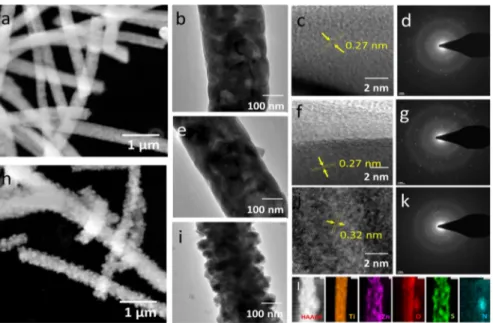
function-ally broken S and N ions favored the surface interaction with the ZnTiO3NF surface and modified its crystal structure.23,24 TEM images of pristine ZT-SN 00 NF reveal (Figure 3a,b) the solid core feature of thefiber morphology with a closely packed grain assembly. Each grain has an average size around 80−100 nm. The elemental mapping results evidence the intended core of Ti−O ionic distribution as compared with the surface level, which evidenced the formation of the ZnTiO3@
TiO2 NF feature. This may be due to the annealing effect,
which induces the mobility of Zn ions to the surface forming ZnTiO3 with the TiO2. Because of the solid grain assembly with higher crystalline quality, this feature was not detected through XRD analysis. Under the hydrothermal treatment at 150 °C with L-cysteine, there was no notable surface
modification apart from the exhibition of small surface grains (Figure 3e). HRTEM images (Figure 3f) revealed that the exhibition of unchanged lattice spacing of 0.27 nm represented the ZnTiO3lattice along the (104) planes.6On functionalizing
the ZnTiO3@TiO2NF withL-cysteine under the hydrothermal
process at 200°C, the surface interacted with the S and N ions and exhibited surface modified nanograined (20−40 nm) functionalization (Figure 3h, (i). The HRTEM images of the pristine and L-cysteine treated ZnTiO3@TiO2 NF clearly
revealed a change in surface grain lattice space variation from 0.27 to 0.32 nm, which indicates that the surface of ZnTiO3
was modified at higher temperature hydrothermal processes. The change in the lattice spacing difference after the hydrothermal process revealed that the outer surface was transformed to ZnS nanograins with the lattice spacing of 0.32 nm. The marked interplanar d spacing signifies the (011) zone axis which represents the hexagonal ZnS.25 After the deformation of the ZnS from ZnTiO3, the surface grains
were pragmatically exhibited in the form of loosely bonded individual nanograins at the surface without any secondary particle arrangement.26 Figure 3d, g, and k represents the SAED pattern of the as-prepared ZT-SN 00 NFs, ZT-SN 150 NFs, and ZT-SN 200 NFs, respectively. The SAED pattern
indicates the polycrystalline nature of the fiber surface with change in d-spacing with respect to the interaction of S ions on the surface. At high temperature, the surface was completely modified as the ZnS nanograin assembly with a solid TiO2
core, which is clearly evidenced from the EDAX elemental mapping analysis over the 200°C hydrothermally treated NFs. The 150 °C hydrothermally treated NFs show that S and N were functionalized on the NF surface without disturbing the morphology, which is shown in Figure 3l, Figure S6, and Figure S7.
The EDAX mapping profile reveals that when L-cysteine
interacts with the ZnTiO3surface, S ions interact with the Zn
sites and convert it as Zn-S, which forms the ZnS on TiO2NF,
and additionally, there is a possibility of N interaction of the fiber surface which evidence the presence of the Ti−N interface on thefiber morphology. The additional interesting feature observed from XRD and EDAX mapping is that in the hydrothermal process, at 200 °C there was a promising exhibition of TiO2 and ZnS/TiN crystal grains with
hierarchical assembly but that the surface was not covered uniformly because of the deformation of ZnTiO3into ZnS and
TiN. The deformation of ZnTiO3into Zn-S and Ti-N leads to
the possibility of leaching of unreacted metal ions, which causes the uncovered surface layer on TiO2 NF. In the
hydrothermal process (200 °C), S ions interact with the Zn ions, which later form as ZnS, and the N ions reacted with the Ti ions to form Ti-N on the surface. The presence of Ti ions on the ZnTiO3 NF surface was nearly left unreactive with S
ions and were dissolved in the precursor solution or leached from the surface during the hydrothermal process. This conventional process reveals the exhibition of ZnS, TiN, and TiO2phases in XRD, and in the image mapping, TiO2forms the core with the ZnS functionality with a mild trace of N throughout the NF surface. After surface modification through the hydrothermal process, the specific surface area of the ZT-SN samples increased due to the high surface features as 16.0 m2/g and 40.3 m2/g, for the pristine ZT-SN 00 and ZT-SN
200, respectively. From the comparative results, ZT-SN 200 shows the higher specific surface area, which is more than twice higher than the pristine NFs (Figure S8).
Figure 3.STEM, TEM, and HRTEM images of the as-prepared ZT-SN 00 NF (a,b,c,d), ZT-SN 150 NF (e,f), and ZT-SN 200 NF (h,i,j). (d,g,k) SAED pattern image of pristine ZT-SN 00, ZT-SN 150, and ZT-SN 200 NF. (l) EDAX mapping results of ZT-SN 200 NF.
Optical Studies. Figure 4 shows the UV−vis diffuse reflectance absorption spectra of the S/N-ZnTiO3 NF as a
function of hydrothermal temperature. On comparing with the pristine, ZT-SN NFs exhibited a blue shift at maximum absorption, but the absorption edge red-shifted toward the visible region. The inset shows the K-M plot of the ZT-SN NF with respect to the hydrothermal temperature. The pristine ZT-SN 00 NF reveals a sharp slope at the absorption edge in the UV region corresponding to the band edge absorption with a band gap of 3.01 eV. At lower hydrothermal temperature, the band gap slightly reduced (2.6 eV) due to the nitrogen and sulfur doping functionality above the valence band (VB), but interestingly, ZT-SN 200 samples show dual band edge absorption around 3.00 eV and shallow level of extended band edge absorption toward the visible region up to 1.8 eV. The possibility of the band edge at the UV region may be due to the TiO2 NF and ZnS (and the extended visible
absorption may be due to the interaction of Zn-S and Ti-N based surface functionality on the wide band gap TiO2NF or through the doping of N ion on the surface). The extended possibility of visible absorption may be due to the N subband levels on the ZnS and TiO2 surface, created during the
hydrothermal process. As observed from the previous investigations, the effective role of ZnS as a layer on a wide Figure 4.UV−vis diffuse reflectance spectra of the pristine and S/N
functionalized ZT-SN NFs. The inset shows the K-M function of the respective spectra.
Figure 5.XPS spectra of ZT-SN NFs. (a) Survey, (b) Ti 2p, (c) Zn 1S, (d) O 1s, (e) S 2p, and (f) N 1S signals taken from ZT-SN NFs synthesized at different hydrothermal temperatures.
band gap semiconductor would minimize the band function through the influence of band bending effect.19The effect of S functionality on the ZnTiO3 NF extends the broadened
absorption onset to 1.9 eV, which could be functionalized in the form of a type-II interface. The photographical images of the pristine and S/N modified ZT-SN NFs show the distinct color change from white to light orange after the hydrothermal process (Figure S9). The extended tail over the visible region represents band gap narrowing, which was attributed to the functionalization of the S and N atoms in the lattice of ZnTiO3. These functional atoms were occupied as the subband
level between the valence band (VB) and conduction band (CB) states and reduced the electron transition energy and favored the narrow band function.
The surface chemical composition was explored by the XPS spectra (Figure 5) for the S/N functionalized TiO2−ZnTiO3
NF. The survey spectra reveal (Figure 5a) a clear exhibition of O 1s, Ti 2p, Zn 2p, S 2p, N 1s, and C 1s core levels of the S/N modified ZnTiO3 surface. The presence of the C 1s peak at
284.8 eV is attributed to the adventitious hydrocarbons generated by the XPS instrument and has been used as an internal reference. The high resolution Zn 2p XPS spectra (Figure 5b) shows the sharp characteristic edges at 1021.4 and 1044.5 eV, which represent the Zn 2p3/2 and Zn 2p1/2,
respectively.19The pronounced ratio of Zn/Ti (1.2) with the dominant exhibition of Zn at the surface has appeared in pristine ZT-SN NF. Functionalization with the S/N at different hydrothermal temperatures increases the Zn/Ti ratio. The absorbed mild shift from 1021.4 to 1022.1 eV may be due to the possibility of Ti−O−Zn-S or through the N−Zn−S based surface moieties. The split difference of 23.1 eV between the Zn 2p3/2and Zn 2p1/2core level components
reveal the Zn2+ state nature.19 At higher hydrothermal temperature, the binding energy shifts with a notable difference which is reported to be the closest position of bulk ZnS, thereby signifying the Zn−S bond formation.25 The high resolution scan of Ti 2p shows (Figure 5c) that for the pristine NFs two peaks were absorbed at 458.7 and 464.5 eV, which represent Ti 2p3/2 and Ti 2p1/2, respectively. Sulfidation
induced the shift in the binding energy from 458.7 to 459.2 eV, which is closer to the known TiO2(459.8 eV).20The strong interaction of Zn-S which results in the structural variation from Ti−O−Zn to ZnS leads to the binding energy shift, but the spectral space separation evidenced the existence of the Ti4+and Zn2+oxidation states. It further leads to the evidence that there is an observation of trivalent Ti3+formation during
the hydrothermal process, which results from the Ti 2p3/2 spectra. The intensity of the Ti was reduced for the ZT-SN 200 due to the removal or leaching of Ti at the surface, and the presence of Ti spectra may be due to the mild trace presence of Ti−N interaction and due to the exposure of the core surface by the uncovered surface layer. The oxygen vacancies (Ov)
induced to uphold the electrostatic balance which was confirmed by the existence of Ti3+with the substitution ratio of Ti4+/Ti3+ in the ZnTiO
3structure. 27
The semiquantitative analysis of the as-synthesized ZT-SN NFs is summarized in
Table S1.
Figure 5d shows the higher resolution scanning of O 1s spectra of S/N functionalized samplesfitted with the Gaussian function. The three split fitted peaks through the Gaussian function are represented as lattice oxygen (OL), OV, and
chemisorbed oxygen ions (OC), respectively.19,20 From the spectral observation, it clearly reveals that the intensity of OV
centered at 531 ± 0.4 eV was influenced by the sulfidation process. The drastic change in the ratio of OV on the ZT-SN
200 sample may be due to the formation of Zn-S nanograins with the existence of Ti3+ functionality and the relative
percentages of OL and OV with the function of sulfidation
temperature as tabulated inTable S1. The reduction rate of OL on the ZT-SN 200 with other oxygen related peaks represents a promising interaction of the S ion with the ZnTiO3surface layer. The intensive modification of Ovand Ocon ZT-SN 200
samples reveals a promising surface modification through the sulfur interaction at higher temperature. The extended peak at 533.8 eV represents the interaction of C−O bonding on the fiber surface perhaps due to the decomposition of L-cysteine
favoring the C interaction on the catalyst surface.
High resolution scanning of S 2p spectra of the S/N functionalized samples at different hydrothermal temperature is represented in Figure 5e. With the Gaussian function, the high resolution S 2p signal is split into three sets of peaks with different intensities at 162.2 eV, 164.2 eV, and 168.8 eV, which in turn represent the S lattice interaction, S interstitials, and SO4at the catalytic surface. Moreover, at lower hydrothermal
(100 °C) temperature, S ions were just tagged with the ZnTiO3 layer as SO4 and S interstitial functionalities. On
increasing the hydrothermal temperature, the relative in-tensities of these peaks completely change with strong interaction of S 2p on the NF surface with the possibility of a trace amount of S interstitials, possibly due to S doping on the ZnTiO3layer. At higher temperature (200°C), the intense
deconvoluted S 2p peak at 162.2 and 164.2 eV could represent S2−and S interstitials by the deformed chemical functionality of Ti-S and Zn-S.28,29 At lower temperatures, the trace exhibition of SO4 functionality (168.4 eV) reveals the
interaction of oxygen sites of catalyst with the S ions. With increasing hydrothermal temperature, the lattice sulfur interaction with the metal ions increases in addition to the formation of S interstitials. In nature, the presence of S6+ and
S4+ work as a surface trap for the photogenerated carriers which minimize the carrier recombination.20,30 Further, the exhibition of S modification on the ZnTiO3NF surface induces
the change in band function for the promotion of visible absorption behavior. The exhibited broad peak at 400 eV could represent the presence of N ions or N-doping in the NF surface (Figure 5f).31,32
The high resolution N 1s XPS spectra reveal the surface interaction of N species on the nanofibers by exhibiting the peaks at 396.4 and 399.6 eV. The previous investigations reveal that the interaction of N 1s around 396 eV evidence the substitution of nitrogen at the oxygen lattice such as Ti-N/Zn-N.33,34The peaks located around 400 eV represents different interstitial nitrogen species with strong oxygen adsorption on the fiber surface with the function of hydrothermal temper-ature.35 The hydrothermal process induces the doping functionality of N ions on the fiber surface through the decomposition of L-cysteine where the N ions substitute the
site of the oxygen and sulfur anions in the ZnTiO3, ZnS, and TiO2lattice, the metal nitride functionality, and N doped ZnS
functionalities on the NF surface.31 The relative atomic concentration of N ion in hydrothermally treated samples has been found to be 1.6, 2.4, and 3.6 at % for SN 100, ZT-SN 150, and ZT-ZT-SN 200 NF samples, respectively, based on the XPS data.
Figure 6shows the photoluminescence spectra of the ZT-SN NF under an excitation wavelength of 350 nm. The sharp
emission around 397 nm represents the band edge emission of the ZnTiO3layer. The weak and broad emissions around 456
and 548 nm denote the shallow and deep trap states due to the surface defect states.36As compared with the pristine NF, on functionalizing, the S/N impurities suppress the deep trap state defect emission, which is caused by the oxygen vacancy states. Shift in the band edge emission reveals a change in band function with respect to the hydrothermal temperature, which is a result of surface functionalization by S and N ions. As the wall of ZnTiO3was converted to ZnS, the band edge emission
was blue-shifted due to the wide band function of the ZnS. The shift in the shallow-trap/self-trap state emission to 445 nm as a
function of the hydrothermal process temperature reveals the structural modification in the band level. This emission may be due to the self-trapped excitons (STE) located at TiO6 oxtahedra that arise owing to the favorable interaction of the Ti 3d orbital with the O 2p orbital due to the structural modification on the ZnTiO3 layer. The intense emission
signifies the presence of localized surface states within the band gap. As observed from the XPS results, the hydrothermal reaction with L-cysteine induces sulfur and nitrogen based
passivated traps which narrow the band function. As a function of hydrothermal temperature, the surface state emission at 445 nm gets intense due to the electron−hole recombination from impurity levels in the ZnTiO3layer. Additionally, the intense
emission at 445 nm may be due to the S interstitials based defect states on the ZnTiO3surface. The strong interaction of S2−with the function of hydrothermal temperature leads to the intense emission behavior. From this observation, hydro-thermal treatment of L-cysteine on the ZnTiO3 wall surface
initiates the sulfur based defect sites which leads to the narrow band function.
Photocatalytic Properties. The effect of S/N cofunction-ality effectively induced the enhanced visible absorption in the ZT-SN NFs. To utilize this extended functionality, photo-catalytic degradation of an organic dye molecule (RhB) was investigated under the UV (λ = 365 nm) and visible irradiation (λ > 400 nm), and their time responsive absorption spectra under photoirradiation is presented in Figure S10. The RhB dye molecule has exhibited a self-degradation of 7% under visible degradation in 180 min. In the dark, catalytic NFs exhibit nearly 4−7% of dye adsorption, which is due to absorption of dye molecules on the functionalized surface. An observation of ZT-SN 200 NFs shows a high absorption of dye Figure 6. Photoluminescence spectra of the pristine and S/N
functionalized ZT-SN based NFs.
Figure 7.Photocatalytic properties of surface modified ZT-SN NFs on RhB dye solution. (a,b) Photocatalytic degradation efficiency, (c,d) kinetic fits of ZT-SN NF under UV irradiation and visible irradiation, and (e) comparison study on the photocatalytic degradation rate under UV irradiation and visible-light irradiation. (f) Catalytic efficiency of the ZT-SN NFs for different irradiation sources in 60 min.
molecules in the dark as compared to the rest of the samples (Figure S11). InFigure 7a, ZT-SN NF exhibits the degradation efficiency of 88.8, 94.1, 96.3, 98.1, and 68.9% for pristine ZT-SN 00 NF, ZT-ZT-SN 100, ZT-ZT-SN 150, ZT-ZT-SN 200, and P25 catalysts, respectively, under UV irradiation of 120 min. Under visible irradiation, the same set of catalysts exhibits RhB decomposition levels of 70.1, 88.7, 93.6, 97.6, and 18.9% for pristine ZT-SN 00 NF, ZT-SN 100, ZT-SN 150, and ZT-SN 200 catalysts, respectively, in 120 min. Table S2 shows the photoresponsive behavior of the ZT-SN NF with respect to the hydrothermal sulfidation temperature. The pristine ZT-SN 00 NFs show a degradation activity around 70.1% in 120 min of visible irradiation, which represents narrow band function (3.01 eV) when compared with bulk ZnTiO3 (3.67 eV).37
When compared with the pristine NF, ZT-SN based samples show faster catalytic behavior under UV and visible irradiation, representing the S and N induced defect levels, thereby favoring effective carrier separation efficiency and promoting visible absorption behavior. The degradation activity fits the pseudo-first-order kinetics, and from the kinetic plot (Figure 7c,d), the degradation rates were calculated and compared with different photoirradiation (Figure 7e). Under visible irradiation, ZT-SN 200 samples exhibited the highest degradation rate of 0.0314 min−1, which is about 3.11 times, 1.78 times, and 1.46 times higher than that of pristine ZT-SN 00, ZT-SN 100, and ZT-SN 150 samples, respectively. A similar trend is observed in UV irradiation as well as natural sunlight irradiation, but the photonic intensity under UV irradiation was low as compared to visible irradiation. With the low photonic energy, catalysts were highly active under UV irradiation as with the photonic energy from the visible sources. TheFigure 7f shows the photocatalytic efficiency of a ZT-SN based catalyst under different irradiation sources at 60 min of photoirradiation. From the observation, ZT-SN 200 exhibited superior catalytic efficiency under all respective sources and exhibited promising catalytic activity in natural sunlight irradiation. Other organic pollutants such as MB and CP also exhibited the same degradation trend with nearly similar degradation rate differences (Figure S12).
The results show thatL-cysteine effectively works as a sulfur
and nitrogen source for the effect of band gap narrowing and with visible light activity. The reason behind the extended visible catalytic behavior of the S/N modified ZT-SN NFs was attributed to the S elements introduced on the ZnTiO3lattice,
which narrow the band function with the sulfur based defect states below the CB. In general, we can denote the substation role of N and S interaction with the band structures of the ZnTiO3layer for the favorable visible catalytic activity by the effective separation of carriers through the sub-band levels, but interestingly, we observe that at higher sulfidation temperature, ZnTiO3@TiO2 transformed into the ZnS/Ti−N@TiO2
hierarchical structure. On accounting their individual counter parts, both have a wide band gap nature, but in the form of heterostructural nature, they exhibited a promising visible catalytic activity. Further, the exhibition of CO in the XPS results of O 1s spectra reveals the presence of C functionality or doping on thefiber surface due to the decomposition ofL
-cysteine. On comparing with the low temperature growth ZT-SN NF, ZnS/TiN@TiO2 (ZT-SN 200) NFs shows faster
catalytic activity under UV and visible irradiation. The formation mechanism of this structure is not clearly under-stood, but the ZnS/TiN@TiO2form of hierarchical structure exhibited a superior catalytic efficiency. Similar to the catalytic
performance, optical responses evidence the enhancement toward the visible region, which is confirmed from the UV and PL results. In the bulk form, the CB and VB position of TiO2is
more positive when compared with the ZnS/TiN leading to the type-I band structure, which inhibits the carrier reaching the catalytic surface. However, the transformation of ZnTiO3 into ZnS/TiN based layer enhances the carrier lifetime by changing the band edge positions to result in the effective carrier separation with narrow band function at the heterostructural interface.
The presence of S and N ion on the ZnTiO3surface induces
change in spectral response toward the visible region. Under 150°C of hydrothermal treatment, the acquired NFs get the S and N doping functionality of the ZnTiO3layer, but at 200°C,
it clearly picturized the heterostructure structure of ZnS@TiO2 with the TiN intergrains. The presence of TiN intergrains was confirmed by the XRD and image mapping results (Figure 3i), but below 200°C of hydrothermal treatment, the sulfur ions interact in the form of SO4with interstitial ionic states on the
NF surface. Sulfur interaction on the catalytic surface with N doping leads to accessible visible absorption with spatial transition of charge carriers. The CB and VB positions were calculated over the surface modified ZT-SN NFs to endorse the band function and to study the possibility of carrier transportation under photoirradiation. The band position with respect to the surface defect states were implicated by calculating the valence and CB position with respect to the surface functionalities of the ZT-SN NF.Figure 8 shows the XPS VB spectra of the S/N form of the surface functionalized ZnTiO3layer. The positions representing A and B inFigure 8
were identified as valence and d-orbital peaks, respectively. The pristine ZT-SN NF has the VB maximum edge at 3.72 eV with the strong tail in valence band function up to 2.86 eV due to the presence of surface oxygen defect states. Further, coupling the calculated band gap from UV DRS spectra (3.01 eV), the position of the CB minimum would occur at−0.15 eV. With the hydrothermal treatment at 150 °C and below, the VB maxima shifted mildly to 2.72 eV band function with a tail at 2.03 eV (Figure S13), which might be due to the N and S doping on the ZnTiO3 layer. Under 200°C of hydrothermal
treatment, the VB maxima shifted to 2.31 eV with a mild band function tail at 1.81 eV (Figure S13), which represent the change in VB positions due to C and N doping on the NF surface. The shift toward the lower binding energy values (to the right) implies that the VB position has risen with respect to the sulfidation temperature. Correlating with the calculated band gap from the UV-DRS spectra, the CB minimum suggests ∼0.55 and −0.40 eV for the ZT-SN 100 and ZT-SN 150 NF, respectively, but because of the two band edges absorbed (in DRS spectra) on the ZT-SN 200 NF, the discrete CB minima lie between−0.91 eV and −0.01 eV. The CB minima at −0.91 eV represent the solid core feature of the ZnS or TiO2, and the
−0.01 eV represents the extended band function due to the transformation of the ZnTiO3 surface to ZnS with TiN
interfaces. Figure 8b represents the schematic illustration of density of states (DOS) of the surface modified ZT-SN NFs with narrow band function. The N doping functionality leads to a shift in the VB position. Furthermore, the role of S on the ZnTiO3 layer induces the narrow band gap by inducing a
change in CB position.
The possibility of the band energy shift in the CB and CB position was demonstrated by the XPS and UV visible DRS results. The huge shift in the VB spectra represented the
spacing peaks, confirming the transformation of ZnTiO3into ZnS/TiN during the hydrothermal process. The dual role of N and S based defects is to induce narrow band functionality with a shift in the d-spacing peaks. N-doping may induce an upper lift of the VB position, which minimizes the band function of the ZnTiO3 layer.38 The presence of sulfur interstitials and
sulfur defect bands creates discrete band levels below the CB position through the localized surface states, which minimize the CB position with narrow band function. The presence of S and N defects is attributed to the tail function in the CB position favoring the band narrowing functionality in the ZnTiO3layer. While modifying the ZT-SN NF withL-cysteine
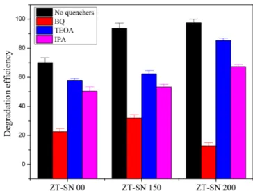
at different hydrothermal temperatures, we observed that the interactive nature of the S ions initiate the tailing of the CB through the surface defect states. The presence of S interstitials and the O−Zn−S based surface functionalities on the catalytic surface are promising defect functional levels which minimized the CB position with tailing in the band position. The experimental results motivated us to understand the possible visible catalytic response on the hybrid composite catalyst. The production rate of reactive oxygen species (ROS) was quantified to understand the feasible mechanism of the surface functionality of ZT-SN NF under visible irradiation (Figure S14). Under visible irradiation, the trapping results (Figure 9) with the production of photoinduced holes (h+), hydroxyl
radicals (°OH) and superoxide radicals (O2°−) were
investigated in a way similar to that in the photocatalytic experiment. For pristine ZT-SN NF, the catalytic activity over
RhB decreases effectively by the addition of BQ. On adding IPA and TEOA with RhB, the activity decayed under photoirradiation but not as noteworthy as with BQ addition. The observation proposed that the production rate of O2° is
promising in ZT-SN 00 NFs which is primarily responsible for the catalytic activity. For ZT-SN 150 samples, the photo-catalytic degradation activities for RhB decreases with the addition of IPA and BQ but not in the TEOA. It may be due to the effective separation of electron by S defect, which favors the production of superoxide, which later transforms to the hydroxyl radical. However, the photogenerated hole is not influenced greatly in the catalytic properties. However, for the ZT-SN 200 samples, TEOA does not respond much to the catalytic activity, but BQ gives good response and has mild activity under IPA, influencing a swell. The obtained results suggest that under visible light irradiation, ZnS/TiN@TiO2
(ZT-SN 200) NF samples exhibited effective production of superoxides through photogenerated electrons which play a major role compared to that of the photogenerated holes. As compared with the pristine ZT-SN 00 NF, the production of ROS was high in ZT-SN 150 NF, but in ZT-SN 200 NF samples, mostly superoxides were found to be active on the surface. This may be due to the contribution of the N-doping functionality with sulfur induced defect levels for effective carrier separation under photoirradiation.
Besides the tunable visible catalytic behavior, the catalytic stability was the notable factor which has disturbed the photocorrosion and self-poisoning effect. The reusable catalytic properties were investigated by consecutive degradation cycles under visible irradiation of RhB dye molecules for nearly 10 cycles (Figure 10). After each cycle, the degraded pollutants were removed and refilled with fresh dye solution for the next run. The catalyst exhibited more stable catalytic degradation efficiency for more than 3 cycles while using ZnS/TiN@TiO2
NFs as the visible active catalyst. After 10 cycles, visible active ZT-SN 150 and ZT-SN 200 NFs exhibited a degradation efficiency of 86.3% and 83.3% after 120 min, but pristine ZT-SN 00 NFs have stable catalytic efficiency even after 8 cycles under similar visible irradiation. After 10 cycles, the pristine ZT-SN 00 NFs exhibited a degradation efficiency of 68.3% after 120 min. These results solidify the surface stability and catalytic durability of the S/N modified ZT-SN NFs, which Figure 8.(a) Valence-band XPS spectra of the pristine ZT-SN 00 NF
and surface modified ZT-SN NF samples. (b) Schematic illustration of the DOS of surface modified ZT-SN NF, as compared to that of the pristine one.
Figure 9. Trapping experiment of the active species during the catalytic reaction with 120 min of Xe lamp irradiation with ZT-SN based catalysts.
could be a long-term stable catalyst under visible irradiation. The morphological and structural stability of the S/N functionalized ZT-SN NF was nearly stable with 1D assembly after the reusable process under visible irradiation (Figure S15). The XPS results (Figure S16) reveal that after reusable cycles, the atomic ratio of the S/N based functionalities on the catalytic surface started to leach through the photocorrosion process, which further led to minimizing its catalytic efficiency and surface stability. The ICP-MS results revealed 2−3% leaching of Zn ions from ZN-SN 200 indicating poor structural stability of the hierarchicalfinish, but the SN 150 and ZN-SN 00 NF catalysts do not exhibit any notable leaching effect during the catalytic cycle. When compared with the hierarchical structural feature, ZT-SN 150 NF catalytic surfaces are more stable because of closely packed grain assemblies,
which lead to effective long-term stability under visible irradiation.
Proposed Photocatalytic Mechanism. On the basis of the band function and ROS production rate quantification, the possible mechanism proposed under the visible irradiation of S/N functionalized ZT-SN NF is seen illustrated in the
Scheme 2. As ZT-SN 00 NF has a band gap energy of 3.01 eV, it will be active under visible irradiation (λ > 400 nm), and further, the presence of Ovleads to improved photogenerated
carrier separation, improving the visible activity. Functionaliz-ing the S/N into the ZnTiO3layer at low temperature (<200
°C) leads to SO4and S interstitials acting as trap levels below
the CB. Further, the functionality of N doped ions may have improvised the VB position for favorable band gap narrowing. The presence of the sulfur induced trap states leads to the delay in carrier recombination rate, and it narrows the band function to improvise the visible catalytic efficiency. At higher hydrothermal temperature (200°C), the catalyst surface was modified to ZnS/TiN@TiO2 hierarchical structure. The
constructed heterostructure leads to visible absorption through sulfur based defect levels in the band function and possible minimization of the VB position by the N- and C-doping nature on the catalyst surface. Furthermore, there are previous investigations on decorating the ZnS surface layer over the wide band gap semiconductors, which shows that it leads to narrow band function at the heterostructural interface due to strained conduction band minimum (CBM) energies.39 ZnS based surface functionality would influence the work function, and the band bending could be attributed to the electronic hybridization on the catalyst surface19,40,41with surface defect states. Further, the N-doping functionality of the ZnS layer leads to reduction in band function for it to be a visibly active one. Additionally, the presence of Ti−N impurities leads to promoted carrier separation and avoidance of the charge Figure 10. Long-term (10 cycles) consecutive photocatalytic
degradation of RhB dye solution by using ZT-SN catalysts (15 mg) under indoor sunlight (λ > 400 nm) irradiation.
Scheme 2. Schematic Representation of the Photocatalytic Mechanism on S/N Functionalized ZT-SN NFs with the Function of Sulfidation Temperature
recombination process. These will lead to improved visible catalytic activity through effective production of photo-generated carriers under visible irradiation.
Observation attained from the optical and XPS results (Figure 4 and Figure 5) reveal that the interaction of S/N functionality leads to narrow band function, which in turn improvised the visible catalytic behavior. Sulfur and nitrogen based defect states on the catalytic surface improvise the carrier separation rate, which also improvised the production rate of ROS on the catalyst surface. As the band potential of the pristine catalyst lies more negative than the reactive potential for the production of superoxide radicals, the photogenerated electrons effectively participate in the catalytic process. Owing to the higher carrier separation efficiency of photogenerated electron and holes on the catalytic surface through the S/N functionality, the heterostructures are a superior photocatalytic system when compared to the pristine one. The production rate of the ROS and effective separation of charge carriers determined the catalytic performance of the NF. On the surface, sulfur and nitrogen based defect level density favors the carrier separation rate by standing in as photogenerated hole trappers,42which leads to improvised carrier response for effective catalytic performance under UV and visible irradiation. Trends of the exhibited catalytic performance under UV and visible irradiation were similar. Further, the amounts of photonic density leads to improved catalytic performance as natural solar irradiation is used as the source.
■
CONCLUSIONVisible active S/N cofunctionalized Zn-S/TiN decorated TiO2
as a hierarchical assembly from ZnTiO3@TiO2 NFs was
synthesized through electrospinning followed by a hydro-thermal process withL-cysteine. Controlling the hydrothermal
temperature, the hierarchical surface features of the hetero-structural decorations were designed, and their visible responsive properties improved. The existence of N and S in the host lattice leads to narrowing the band function, and these defect levels lead to the improvised production rate of photogenerated charge carriers for effective visible catalytic performance. The hierarchical assembly of ZnS/TiN@TiO2
NFs displayed faster catalytic activity on RhB dye solution compared to that of the low temperature hydrothermal treated samples. The hydrothermal temperature influences the interaction of S and N with the catalytic surface, enhancing the catalytic rate. The contribution of S induced states with the N-doping functionality on the surface led to the narrow band function and minimized the valence band position, making the NFs visible active. The hierarchical functionality of Ti−N leads to higher carrier separation rate and reduces rapid carrier recombination rate. These features were absent for low temperature annealed hydrothermal treated NFs. Further, reusable properties of NF samples were nearly stable for multiple cycles for the catalytic degradation of RhB dye. To conclude, ZnTiO3@TiO2and ZnTiO3−S/N@TiO2NFs could
be used as high potential catalysts depending on their surface functionalities to enhance visible photocatalytic efficiency for the degradation of organic pollutants for wastewater treatment.
■
ASSOCIATED CONTENT*
S Supporting InformationThe Supporting Information is available free of charge on the
ACS Publications website at DOI: 10.1021/acssusche-meng.8b02455.
SEM images of the as electrospun Zn(OAc)2
/Ti-(OBu)4/PVP composite nanofibers and TiO2−ZnO
composite NFs; HAADF-STEM and STEM EDAX mapping images of ZT-SN 00 NFs; XRD spectrum of ZT-SN 200 NF; EDAX spectra of ZT-SN based NFs; TEM and STEM-EDAX mapping images of ZT-SN 150 NF and ZT-SN 200 NF; BET of ZT-SN 00 and ZT-SN 200 NF; optical images of the ZT-SN based NFs; table related to the XPS results of ZT-SN NFs; optical absorption spectra of RhB under UV and visible irradiation with the presence of catalyst; optical absorption spectra of RhB with catalyst in the dark and without catalyst in visible irradiation; table related to the photoresponsive behavior of ZT-SN based NFs; optical absorption of MB and CP with the presence of ZT-SN based NFs under visible irradiation; valence band spectra of ZT-SN based NFs; reactive oxygen species quantification; SEM images of the reused ZT-SN based NFs; and XPS spectra of the reused ZT-SN based NFs (PDF)
■
AUTHOR INFORMATIONCorresponding Authors
*(K.S.R.) Tel: +90-3122908987. E-mail:[email protected]. *(T.U.) E-mail:[email protected].
ORCID
Tamer Uyar:0000-0002-3989-4481 Notes
The authors declare no competingfinancial interest.
■
ACKNOWLEDGMENTSK.S.R. acknowledges The Scientific & Technological Research Council of Turkey (TUBITAK), BIDEB 2216-Fellowships for Research Fellowship Programme for Foreign Citizens for a postdoctoral fellowship.
■
REFERENCES(1) Khin, M. M.; Nair, A. S.; Babu, V. J.; Murugan, R.; Ramakrishna, S. A review on nanomaterials for environmental remediation. Energy Environ. Sci. 2012, 5, 8075−8109.
(2) Safdar, M.; Simmchen, J.; Janis, J. Light-driven micro- and nanomotors for environmental remediation. Environ. Sci.: Nano 2017, 4, 1602−1616.
(3) Ansari, S. A.; Khan, M. M.; Kalathil, S.; Nisar, A.; Lee, J.; Cho, M. H. Oxygen vacancy induced band gap narrowing of ZnO nanostructures by an electrochemically active biofilm. Nanoscale 2013, 5 (19), 9238−9246.
(4) Randorn, C.; Irvine, J. T. S. Synthesis and visible light photoactivity of a high temperature stable yellow TiO2photocatalyst.
J. Mater. Chem. 2010, 20 (39), 8700−8704.
(5) Wu, H.; Min, Y.; Zhang, Q.; Li, W.; Yuan, J.; Wu, Z.; Wang, S. Low-temperature synthesis of mesoporous ZnTiO3-graphene
compo-site for the removal of norfloxacin in aqueous solution. RSC Adv. 2016, 6 (105), 103822−103829.
(6) Pawar, R. C.; Kang, S.; Park, J. H.; Kim, J.; Ahn, S.; Lee, C. S. Evaluation of a multi-dimensional hybrid photocatalyst for enrich-ment of H2 evolution and elimination of dye/non-dye pollutants.
Catal. Sci. Technol. 2017, 7 (12), 2579−2590.
(7) Binas, V.; Venieri, D.; Kotzias, D.; Kiriakidis, G. Modified TiO2
based photocatalysts for improved air and health quality. J. Materiomics. 2017, 3, 3−16.
(8) Chen, D.; Wang, Z.; Ren, T.; Ding, H.; Yao, W.; Zong, R.; Zhu, Y. Influence of Defects on the Photocatalytic Activity of ZnO. J. Phys. Chem. C 2014, 118 (28), 15300−15307.
(9) Yan, H.; Wang, X.; Yao, M.; Yao, X. Band structure design of semiconductors for enhanced photocatalytic activity: The case of TiO2. Prog. Nat. Sci. 2013, 23 (4), 402−407.
(10) Zaleska, A. Doped-TiO2: A Review. Recent Pat. Eng. 2008, 2
(3), 157−164.
(11) Nada, A. A.; Nasr, M.; Viter, R.; Miele, P.; Roualdes, S.; Bechelany, M. Mesoporous ZnFe2O4@TiO2Nanofibers Prepared by
Electrospinning Coupled to PECVD as Highly Performing Photo-catalytic Materials. J. Phys. Chem. C 2017, 121 (44), 24669−24677.
(12) Nasr, M.; Balme, S.; Eid, C.; Habchi, R.; Miele, P.; Bechelany, M. Enhanced Visible-Light Photocatalytic Performance of Electro-spun rGO/TiO2Composite Nanofibers. J. Phys. Chem. C 2017, 121
(1), 261−269.
(13) Tan, Y. N.; Wong, C. L.; Mohamed, A. R. An Overview on the Photocatalytic Activity of Nano-Doped-TiO2 in the Degradation of
Organic Pollutants. ISRN Mater. Sci. 2011, 2011, 1.
(14) Xing, Z.; Ju, Z.; Zhao, Y.; Wan, J.; Zhu, Y.; Qiang, Y.; Qian, Y. One-pot hydrothermal synthesis of Nitrogen-doped graphene as high-performance anode materials for lithium ion batteries. Sci. Rep. 2016, 6, 26146.
(15) Shen, K.; Xue, X.; Wang, X.; Hu, X.; Tian, H.; Zheng, W. One-step synthesis of band-tunable N, S co-doped commercial TiO2/
graphene quantum dots composites with enhanced photocatalytic activity. RSC Adv. 2017, 7 (38), 23319−23327.
(16) Wang, P.; Lin, Z.; Su, X.; Tang, Z. Application of Au based nanomaterials in analytical science. Nano Today 2017, 12, 64−97.
(17) Niu, S.; Lv, W.; Zhou, G.; He, Y.; Li, B.; Yang, Q. H.; Kang, F. N and S co-doped porous carbon spheres prepared using L-Cysteine as a dual functional agent for high-performance lithium−sulfur batteries. Chem. Commun. 2015, 51 (100), 17720−17723.
(18) Miao, X.; Yan, X.; Qu, D.; Li, D.; Tao, F. F.; Sun, Z. Red Emissive Nitrogen Co-doped Carbon Dots and Their Application in Ion Detection and Theraonostics. ACS Appl. Mater. Interfaces 2017, 9 (22), 18549−18556.
(19) Ranjith, K. S.; Senthamizhan, A.; Balusamy, B.; Uyar, T. Nanograined surface shell wall controlled ZnO-ZnS core-shell nanofibers and their shell wall thickness dependent visible photo-catalytic properties. Catal. Sci. Technol. 2017, 7 (5), 1167−1180.
(20) Ranjith, K. S.; Uyar, T. Rational interjecting of Na and S co-catalyzed TiO2 based nanofibers: Presence of surface layered TiS3
shell grains and sulphur induced defects for efficient visible-light driven photocatalytic properties. J. Mater. Chem. A 2017, 5 (27), 14206−14219.
(21) Cao, G.; Li, Y.; Zhang, Q.; Wang, H. Fabrication of Hollow Tetrapod-Like TiN Nanostructures and Its Electrochemical Property. J. Am. Ceram. Soc. 2012, 95 (8), 2478−2480.
(22) Howell, I. R.; Giroire, B.; Garcia, A.; Li, S.; Aymonier, C.; Watkins, J. J. Fabrication of plasmonic TiN nanostructures by nitridation of nanoimprinted TiO2nanoparticles. J. Mater. Chem. C
2018, 6 (6), 1399−1406.
(23) Jiang, J.; Yu, R.; Yi, R.; Qin, W.; Qiu, G.; Liu, X. Biomolecule-assisted synthesis of flower-like NiS microcrystals via a hydrothermal process. J. Alloys Compd. 2010, 493 (1), 529−534.
(24) Chang, K.; Chen, W.L-Cysteine-Assisted Synthesis of Layered MoS2/Graphene Composites with Excellent Electrochemical
Per-formances for Lithium Ion Batteries. ACS Nano 2011, 5 (6), 4720− 4728.
(25) Shen, X.; Jiang, Z.; Gao, C.; Xu, Z.; Xie, Z.; Zheng, L. One-step construction of ZnS/C and CdS/C one-dimensional core−shell nanostructures. J. Mater. Chem. 2007, 17, 1326−1330.
(26) Brayek, A.; Chaguetmi, S.; Ghoul, M.; Ben Assaker, I.; Souissi, A.; Mouton, L.; Beaunier, P.; Nowak, S.; Mammeri, F.; Chtourou, R.; Ammar, S. Photoelectrochemical properties of nanocrystalline ZnS discrete versus continuous coating of ZnO nanorods prepared by electrodeposition. RSC Adv. 2016, 6 (37), 30919−30927.
(27) Bharti, B.; Kumar, S.; Lee, H. N.; Kumar, R. Formation of oxygen vacancies and Ti3+ state in TiO
2 thin film and enhanced
optical properties by air plasma treatment. Sci. Rep. 2016, 6, 32355.
(28) Ataman, E.; Isvoranu, C.; Knudsen, J.; Schulte, K.; Andersen, J. N.; Schnadt, J. Adsorption of L-Cysteine on rutile TiO2(110). Surf.
Sci. 2011, 605 (1−2), 179−186.
(29) Neal, A. L.; Techkarnjanaruk, S.; Dohnalkova, A.; McCready, D.; Peyton, B. M.; Geesey, G. G. Iron sulfides and sulfur species produced at hematite surfaces in the presence of sulfate-reducing bacteria. Geochim. Cosmochim. Acta 2001, 65 (2), 223−235.
(30) Veamatahau, A.; Jiang, B.; Seifert, T.; Makuta, S.; Latham, K.; Kanehara, M.; Teranishi, T.; Tachibana, Y. Origin of surface trap states in CdS quantum dots: relationship between size dependent photoluminescence and sulfur vacancy trap states. Phys. Chem. Chem. Phys. 2015, 17 (4), 2850−2858.
(31) Wang, M. Z.; Xie, W. J.; Hu, H.; Yu, Y. Q.; Wu, C. Y.; Wang, L.; Luo, L. B. p-type ZnS:N nanowires: Low-temperature solvothermal doping and optoelectronic properties. Appl. Phys. Lett. 2013, 103 (21), 213111.
(32) Chen, C.; Bai, H.; Chang, C. Effect of Plasma Processing Gas Composition on the Nitrogen-Doping Status and Visible Light Photocatalysis of TiO2. J. Phys. Chem. C 2007, 111 (42), 15228−
15235.
(33) Livraghi, S.; Chierotti, M. R.; Giamello, E.; Magnacca, G.; Paganini, M. C.; Cappelletti, G.; Bianchi, C. L. Nitrogen-Doped Titanium Dioxide Active in Photocatalytic Reactions with Visible Light: A Multi-Technique Characterization of Differently Prepared Materials. J. Phys. Chem. C 2008, 112 (44), 17244−17252.
(34) Zhang, Z.; Wang, X.; Long, J.; Gu, Q.; Ding, Z.; Fu, X. Nitrogen-doped titanium dioxide visible light photocatalyst: Spectro-scopic identification of photoactive centers. J. Catal. 2010, 276 (2), 201−214.
(35) Bian, J. M.; Li, X. M.; Gao, X. D.; Yu, W. D.; Chen, L. D. Deposition and electrical properties of N−In codoped p-type ZnO films by ultrasonic spray pyrolysis. Appl. Phys. Lett. 2004, 84 (4), 541. (36) Choudhury, B.; Dey, M.; Choudhury, A. Shallow and deep trap emission and luminescence quenching of TiO2 nanoparticles on Cu
doping. Appl. Nanosci. 2014, 4 (4), 499−506.
(37) Raveendra, R. S.; Prashanth, P. A.; Hari Krishna, R.; Bhagya, N. P.; Nagabhushana, B. M.; Raja Naika, H.; Lingaraju, K.; Nagabhushana, H.; Daruka Prasad, B. Synthesis, structural character-ization of nano ZnTiO3ceramic: An effective azo dye adsorbent and
antibacterial agent. Journal of Asian Ceramic Societies. 2014, 2 (4), 357−365.
(38) Wu, H. C.; Lin, Y. S.; Lin, S. W. Mechanisms of Visible Light Photocatalysis in N-Doped Anatase TiO2 with Oxygen Vacancies
from GGA+U Calculations. Int. J. Photoenergy 2013, 2013, 1−7. (39) Schrier, J.; Demchenko, D. O.; Wang, L. W.; Alivisatos, A. P. Optical properties of ZnO/ZnS and ZnO/ZnTe heterostructures for photovoltaic applications. Nano Lett. 2007, 7 (8), 2377−2382.
(40) Zhang, Z.; Yates, J. T. Band Bending in Semiconductors: Chemical and Physical Consequences at Surfaces and Interfaces. Chem. Rev. 2012, 112 (10), 5520−5551.
(41) Lin, Y. J.; Tsai, C. L. Changes in surface band bending, surface work function, and sheet resistance of undoped ZnO films due to (NH4)2Sx(NH4)2Sxtreatment. J. Appl. Phys. 2006, 100 (11), 113721.
(42) Khan, H.; Swati, I. K.; Younas, M.; Ullah, A. Chelated Nitrogen-Sulphur-Co doped TiO2: Synthesis, Characterization,
Mechanistic, and UV/Visible Photocatalytic Studies. Int. J. Photo-energy 2017, 2017, 1−17.