© 2019 Turkish Journal of Plastic Surgery | Published by Wolters Kluwer - Medknow 143
Abstract
Case Report
I
ntroductIonReduction mammoplasty is one of the most common surgeries done by plastic surgeons. Pyoderma gangrenosum is a rare inflammatory disease, characterized by ulcers with purplecolored borders and erythematous halo. Clinically, the patient has fever and severe local pain. Pyoderma gangrenosum can occur after any surgical procedure and the diagnosis is usually delayed. It is important for plastic surgeons and clinicians to be aware of this rare condition.
c
aser
ePortA previously well 38-year-old woman admitted to emergency room with fever, malaise, and severe local pain and rash on her breasts. She had undergone reduction mammoplasty by another surgeon 4 days ago. The patient had a history of rhinoplasty and minor esthetic procedures.
She had inverted T-scars due to breast reduction. There was minimal fat necrosis and no dehiscence was observed. Her examination revealed erythema and hyperalgesia on her breasts. The clinical presentation was prediagnosed as bilateral mastitis and the patient was consulted to the Department of Infection Diseases. Ultrasound findings were reported as bilateral mastitis and her inflammatory parameters were
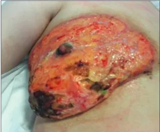
elevated (C-reactive protein: 460 mg/dL and leukocytes: 29730 uL). Intravenous and local antibiotherapy was started immediately. Twenty-four hours later, she had ulcers around the incisions and superficial epidermolysis developed. The borders were irregular and erythema was observed. Oddly, the nipple-areola complex was not affected with this condition [Figure 1]. The patient had no evidence of purulence or fluctuation. Her situation was progressive and she was admitted to operating room for irrigation, biopsy, and wound culture. All microbiological examinations were negative (aerobic, anaerobic, mycobacteria, and fungi) and pathology report identified a nonspecific inflammatory process with neutrophilic infiltration. Despite an aggressive antibiotherapy, her inflammatory parameters and fever raised and ulcers expanded. The patient had a fever of 38.9°C, C-reactive protein was counted 516 mg/dL, leukocytes were 39600 uL, and procalcitonin was 28,58 ng/ml. The patient was thought to have a severe infectious condition. However, she was not in a septic appearance and feeling well other than Pyoderma gangrenosum is a rare inflammatory disease, characterized by ulcers with purple-colored borders and erythematous halo. Clinically, the patient has fever and severe local pain. Pyoderma gangrenosum can occur after any surgical procedure and the diagnosis is usually delayed. It is important for surgeons and infectologists to be watchful about this rare condition because if misdiagnosed, it has serious results including severely painful ulcerations, prolonged therapy, repeated hospitalizations, psychological trauma, and extensive scarring. The authors report a postsurgical pyoderma gangrenosum case after reduction mammoplasty. This report emphasizes on the importance of awareness about this uncommon disease and its diagnosis.
Keywords: Breast reduction, posttraumatic, pyoderma gangrenosum
Address for correspondence: Dr. Nilufer Bahadirli,
Department of Plastic, Reconstructive and Aesthetic Surgery, Istanbul Medipol University, Istanbul, Turkey. E‑mail: [email protected]
Access this article online
Quick Response Code:
Website:
http://www.turkjplastsurg.org DOI:
10.4103/tjps.tjps_5_19
This is an open access journal, and articles are distributed under the terms of the Creative Commons Attribution‑NonCommercial‑ShareAlike 4.0 License, which allows others to remix, tweak, and build upon the work non‑commercially, as long as appropriate credit is given and the new creations are licensed under the identical terms.
For reprints contact: [email protected]
How to cite this article: Bahadirli N, Sutcu M, Akan M. A rare condition
to keep in mind: Pyoderma gangrenosum after breast reduction. Turk J Plast Surg 2019;27:143-6.
A Rare Condition to Keep in Mind: Pyoderma Gangrenosum
after Breast Reduction
Nilufer Bahadirli, Mustafa Sutcu, Mithat Akan
Department of Plastic, Reconstructive and Aesthetic Surgery, Istanbul Medipol University, Istanbul, Turkey
Bahadirli, et al.: Pyoderma gangrenosum after breast reduction
Turkish Journal of Plastic Surgery ¦ Volume 27 ¦ Issue 3 ¦ July-September 2019
144
the local pain. On the postoperative 3rd day, the diagnosis for pyoderma gangrenosum was suspected due to the appearance of the ulcers and her clinical situation. About 80 mg/day oral prednisone was held and the inflammatory parameters regressed dramatically. On the next day, her fever was 36°C, C-reactive protein was 117 mg/dL, leukocyte count was 27440 uL, and procalcitonin was6,51 ng/ml. Progression of the ulcers stopped and erythematous halo disappeared [Figures 2 and 3]. Cyclosporine (2 × 150 mg) was added to her treatment on the postoperative day 14. The pain control was provided nonsteroid anti-inflammatory drugs, tramadol, and narcotic analgesics. However, her dressings were changed once in 2–3 days with general anesthesia due to her severe pain. A practical solution for this excessive pain was needed because 6–8 months of healing process was expected. Grafting was the first choice of treatment. However, an autograft was not the best idea since a minimal trauma could aggravate the disease and we could have donor area ulcers. An autograft was thought to be very risky. Therefore, a xenograft was planned. After a month of local wound care with acide borique in powder form to aggravate
the granulation, her wounds were granulated and a synthetic porcine xenograft (EZ Derm®, © 2018 Mölnlycke Health Care AB, United Kingdom) was adapted to her granulated wounds. The first dressing was made with a noncohesive wound contact layer (Mepitel®, © 2018 Mölnlycke Health Care AB, United Kingdom). The pain decrease was dramatic. We were able to perform wound dressings without any analgesic drugs and she was discharged within a week. During ward rounds, it was observed that she had lost the whole graft within a month. However, the wounds remained pain free and the epithelization was in progress from the periphery. Six months later, 2-cm × 8-cm lesion was still present on her right breast and no granulation tissue was observed. After 2 weeks, a pomade with epidermal growth factor was applied and new granulation tissue was formed in a week. The healing kept on going. Epithelization was complete after a total of 8 months [Figure 4]. The scars were not matured and the anatomy was distorted. Despite all the deformation, the nipple-areola complexes were intact.
d
IscussIonPyoderma gangrenosum is a very rare disease. It is called as “pyoderma gangrenosum” as it was first thought to be
Figure 2: Ulcers on the 8th day after breast reduction and 3rd day after
irrigation, biopsy, and wound culture. Nipple‑areola complex is spared
Figure 1: Ulcers and erythematous halo around the incisions on the
5th day after breast reduction
Figure 4: Final scars after 8 months Figure 3: Ulcers on the 8th day after breast reduction and 3rd day after
irrigation, biopsy, and wound culture. Nipple‑areola complex is spared
Bahadirli, et al.: Pyoderma gangrenosum after breast reduction
145 145
Turkish Journal of Plastic Surgery ¦ Volume 27 ¦ Issue 3 ¦ July-September 2019 145 a streptococcal infection that results in gangrene. It is now
known as an autoimmune condition. In the majority of the cases, this disease is misdiagnosed as an infection because of its clinical similarity and leading to debridement which, in fact, exaggerates the problem.[1]
In a systematic review of postsurgical (breast, cardiothoracic, or abdominal surgeries) pyoderma gangrenosum, 220 cases were identified from all over the world and only 25% of them were after breast surgeries.[2]
This condition is defined as an inflammatory neutrophilic dermatosis that is noninfectious. The disease typically begins between after 4 days postoperatively to several weeks. It appears as a pustule that spreads dramatically under the healthy skin.[3] The very painful skin ulcers develop. Necrosis is in the middle and purplish borders with erythematous halo are at the periphery. Strangely, the ulcers rarely affect the nipple-areola complex. It spares the nipple and the ulcers spread around it. This parameter is a very interesting characteristic for this disease. The patient develops fever and the laboratory shows increased leukocytes and inflammatory markers including C-reactive protein and interleukin-8. In most of the cases, it is misdiagnosed as an infectious disease and multiple debridements are performed. This worsens the cutaneous ulcers and spreads the necrosis. Workup for pyoderma gangrenosum should include a detailed history and a physical examination that focuses on the risk factors and diseases associated with pyoderma gangrenosum. Wound culture is recommended. A full-thickness biopsy needs to be performed from the peripherally erythematous halo. Skin biopsies are nonspecific that show neutrophilic infiltrate, swelling of the endothelium, and necrosis. All microbiological examinations (aerobic, anaerobic, mycobacteria, and fungi) are negative if the situation is not complicated with secondary infection. Positive culture does not exclude the diagnosis of pyoderma gangrenosum.[4]
The treatment of pyoderma gangrenosum is mainly immune suppression, wound care, and pain relief. Corticosteroid treatment (40–120 mg/daily) is the first choice for avoiding rapid progression. Then, cyclosporine and other immune modulators, such as, sulfa drugs, dapson, and tumor necrosis factor alpha blockers are added to the treatment and corticosteroids are dozed off. The cyclosporine is maintained until all the wounds are healed and continued for 3 more months after the healing. The wound care mostly consists of moisturizing the area with local antibiotics and epithelizants. With local antibiotics, secondary infections are prevented. Epithelizants accelerates the healing process. Grafting is usually not the first option because early and late failures of the skin grafts have been reported. It should only be performed with patients who are under immunosuppression and show no sign of active disease. Long-term follow-ups (1 year or more) are needed since late loss of grafts is also reported. Some authors suggest to avoid the surgical intervention as much as possible. Assisting therapies such as vacuum-assisted closure and hyperbaric oxygen therapy are also recommended by some authors.[5]
The most frequent postsurgical pyoderma gangrenosum cases after breast surgery are after breast reductions, followed by breast reconstruction, lumpectomy or mastectomy without reconstruction, augmentation mammoplasty, and other breast procedures.[6] Postbreast reduction pyoderma gangrenosum is reported in only around 50 patients all around the world.[7] It is important to know and recognize this complication because it has severe outcomes if diagnosed late. Posttraumatic pyoderma gangrenosum has its own specific features which make it easier to recognize. First of all, after the surgery, the scar presents with small dehiscence followed by disproportionately painful ulcers. The wound progresses to larger areas and no granulation tissue is present. Second, it typically begins after 4–5 days after the surgery and it has no self-limitation. It grows larger even with antibiotics and local treatments. Third and strangely, the nipple-areola complex is spared in this disease. Moreover, finally, the response to immunosuppression is dramatic. The fever is dropped and inflammatory markers decrease within a day. One of the main problems with this disease is severe local pain. It prevents the medical team to perform wound care. Since skin autografting is not a wise option due to the pathergy phenomenon and although unheard of applying xenografts can be considered as a solution. Even though all the graft becomes lytic, it allows us to change the dressing pain free. With no pain, general anesthesia is not required; therefore, its complications are avoided. It increases the patient’s coherence with the treatment. Furthermore, it allows us to change the dressings more often and in an atraumatic way. Moreover, finally, a xenograft is a biological dressing, which is better than any kind of synthetic dressing.
We are alerting all plastic surgeons to be watchful about this very rare and extreme condition because it can occur after any kind of surgical procedure. We also recommend considering the usage of xenografts for reducing the unbearable pain and to be able to perform pain-free local wound care. It is especially very important since plastic surgery operations are mostly elective and esthetic expectations of the patient are very high. Declaration of patient consent
The authors certify that they have obtained all appropriate patient consent forms. In the form, the patient has given her consent for her images and other clinical information to be reported in the journal. The patient understands that name and initial will not be published and due efforts will be made to conceal identity, but anonymity cannot be guaranteed. Financial support and sponsorship
Nil.
Conflicts of interest
There are no conflicts of interest.
r
eferences1. Grillo MA, Cavalheiro TT, da Silva Mulazani M, Rocha JL, Semchechen D, da Cunha CA, et al. Postsurgical pyoderma gangrenosum complicating reduction mammaplasty. Aesthetic Plast Surg 2012;36:1347-52.
Bahadirli, et al.: Pyoderma gangrenosum after breast reduction
Turkish Journal of Plastic Surgery ¦ Volume 27 ¦ Issue 3 ¦ July-September 2019
146
2. Zuo KJ, Fung E, Tredget EE, Lin AN. A systematic review of post-surgical pyoderma gangrenosum: Identification of risk factors and proposed management strategy. J Plast Reconstr Aesthet Surg 2015;68:295-303.
3. Doren EL, Aya-ay ML. Pyoderma gangrenosum following breast reduction: Treatment with topical tacrolimus and steroids. Aesthet Surg J 2014;34:394-9.
4. Boughaza I, Al Haderi O, Mhamdi O, Lakhdar A, Baydada A, Zeraidi N,
et al. Pyoderma gangrenosum a diagnosis not to miss: Case report. Int
Ann Med 2017;1(5).
5. Niezgoda JA, Cabigas EB, Allen HK, Simanonok JP, Kindwall EP,
Krumenauer J, et al. Managing pyoderma gangrenosum: A synergistic approach combining surgical débridement, vacuum-assisted closure, and hyperbaric oxygen therapy. Plast Reconstr Surg 2006;117:24e-8e.
6. Abtahi-Naeini B, Bagheri F, Pourazizi M, Forozeshfard M, Saffaei A. Unusual cause of breast wound: Postoperative pyoderma gangrenosum. Int Wound J 2017;14:285-7.
7. Tuffaha SH, Sarhane KA, Mundinger GS, Broyles JM, Reddy SK, Azoury SC, et al. Pyoderma gangrenosum after breast surgery: Diagnostic pearls and treatment recommendations based on a systematic literature review. Ann Plast Surg 2016;77:e39-44.