Tumor Biology November 2018: 1–8 Ó The Author(s) 2018 Article reuse guidelines: sagepub.com/journals-permissions DOI: 10.1177/1010428318811766 journals.sagepub.com/home/tub
VDR gene FokI polymorphism as a poor
prognostic factor for papillary thyroid
cancer
Selvihan Beysel
1,2,3, Nilnur Eyerci
4, Ferda Alparslan Pinarli
4,
Mahmut Apaydin
1, Muhammed Kizilgul
1, Mustafa Caliskan
1,
Ozgur Ozcelik
1, Seyfullah Kan
1and Erman Cakal
1Abstract
This is the first study to investigate the effect of vitamin D receptor (VDR) gene single-nucleotide polymorphisms on the clinicopathologic features of papillary thyroid cancer in Turkey. A total of 165 patients with papillary thyroid cancer and 172 controls were included in this case–control study. VDR gene single-nucleotide polymorphisms FokI (rs2228570), BsmI (rs1544410), ApaI (rs7975232), and TaqI (rs731236) were evaluated using reverse-transcription polymerase chain reaction. VDR gene polymorphisms BsmI, ApaI, and TaqI did not differ between the papillary thyroid cancer group and control group (p . 0.05, each). BsmI, ApaI, and TaqI were not associated with papillary thyroid cancer risk. The VDR gene FokI CT/TT genotype was associated with an increased papillary thyroid cancer risk (CT vs CC: odds ratio = 1.71, 95% confidence interval = 1.15–2.76, p = 0.028; TT vs CC: odds ratio = 2.44, 95% confidence interval = 1.29–4.62, p = 0.005; CT/TT vs CC: odds ratio = 1.88, 95% confidence interval = 1.20–2.96, p = 0.006; CT/CC vs TT: odds ratio = 1.80, 95% confidence interval = 1.05–3.20, p = 0.041). VDR gene polymorphisms were not in linkage disequilibrium. The FokI TT genotype was associated with having T3 and T4, stage III/IV, extra-thyroidal invasion. The FokI CT/TT or TT genotype was associated with developing N1 status, multifocality, tumor size ø 10 mm, and treatment with radioiodine therapy. Persistence/recurrence did not differ between the FokI genotypes. Carriers of the FokI T allele were at an increased risk of more advanced tumor-node-metastasis stage, greater tumor size, multifocality, and extra-thyroidal invasion of papillary thyroid cancer compared with the CC genotype. VDR gene FokI T allele and TT genotype correlated with aggressiveness of papillary thyroid cancer; thus, FokI could be useful as a poor prognostic factor to assess the high risk of papillary thyroid cancer.
Keywords
VDR gene, papillary thyroid cancer, FokI polymorphism
Date received: 22 November 2017; accepted: 17 October 2018
Introduction
Thyroid cancer is a relatively rare neoplasm that affects an estimated 14.9 people per 100,000. It is the most common endocrine cancer and constituted predomi-nantly of its papillary subtype. Thyroid cancer accounts for \2% of all cancers in males and 1%–1.5% in females with a female-to-male ratio of 3:1 in almost all ethnic groups.1,2 Genetic variation contributes to the development of thyroid cancer; however, the pathogen-esis of thyroid cancer is not yet known exactly.2,3
1
Department of Endocrinology and Metabolism, Ankara Diskapi Yildirim Beyazit Teaching and Research Hospital, Ankara, Turkey
2
Department of Medical Biology, Basxkent University, Ankara, Turkey
3
Department of Endocrinology and Metabolism, Afyonkarahisar Saglik Bilimleri University, Afyonkarahisar, Turkey
4
Department of Genetic Research, Ankara Diskapi Yildirim Beyazit Teaching and Research Hospital, Ankara, Turkey
Corresponding author:
Selvihan Beysel, Department of Endocrinology and Metabolism, Afyonkarahisar Saglik Bilimleri University, Afyonkarahisar 03200, Turkey. Email: [email protected]
Creative Commons Non Commercial CC BY-NC: This article is distributed under the terms of the Creative Commons Attribution-NonCommercial 4.0 License (http://www.creativecommons.org/licenses/by-nc/4.0/) which permits non-commercial use, reproduction and distribution of the work without further permission provided the original work is attributed as specified on the SAGE and Open Access pages (https://us.sagepub.com/en-us/nam/open-access-at-sage).
Molecular biomarkers have not been identified for the diagnosis and prognosis of thyroid cancer.4
In addition to the effect on calcium and bone hemos-tasis, 1,25-diydroxyvitamin D3 (1,25(OH)2D3), the
biologically active form of vitamin D, has many extra-skeletal effects such as anti-proliferative, anti-apoptotic, and pro-differentiation properties.5–7Active vitamin D shows its biologic actions by tightly binding to the intracellular vitamin D receptor (VDR), a hormone-regulated transcription factor. VDR is a member of the nuclear hormone receptor family of transacting tran-scriptional regulatory factors, including the steroid hor-mone receptors, thyroid horhor-mone receptors, retinoic acid receptors, and retinoid-X receptors. The VDR gene is located on chromosome 12q13.1 and is composed of five promoters, eight coding exons, and six untranslated exons.5–8Its promoter region is able to generate multi-ple transcripts. During activation, VDR forms a hetero-dimer with the related retinoid-X receptor and regulates the expression of many target genes because it binds to the vitamin D response elements in the chromatin region. Active vitamin D regulates gene transcription by binding to the VDR. It shows anti-tumoral effect by stimulating differentiation and apoptosis.9 It reduces cellular proliferation, inflammation, angiogenesis, and invasion. VDR regulates other metabolic pathways such as immune response and cancer signaling.5–10 These are considered as VDR-mediated signaling path-ways. Studies have shown that VDR gene polymorph-isms are found in patients with cancer.11Bsml (A.G, rs1544410), ApaI (A.C, rs7975232), TaqI (T.C, rs731236), and FokI (C.T, rs2228570) are human VDR gene polymorphisms. VDR BsmI, ApaI, and FokI gene polymorphisms have shown to increase the risk of hav-ing breast cancer.12VDR FokI gene polymorphism increases the susceptibility for prostate cancer, BsmI polymorphism does so for malignant melanoma, and TaqI increases the risk for renal cell carcinoma.11 The FokI polymorphism was associated with an increased ovarian cancer risk.13FokI T/T genotype was associ-ated with higher progression rate in head and neck squamous cell carcinoma.14VDR BsmIpolymorphism was a risk factor for colorectal and skin cancer in a Caucasian population and the ApaI polymorphism was a risk factor for basal cell cancer in an Asian pop-ulation. The TaqI polymorphism was a risk factor for oral, breast, and basal cell cancer, and the FokI poly-morphism was a risk factor for ovarian and skin can-cer.15 However, to date, the association between the VDRgene polymorphism and thyroid cancer has only been investigated in two studies. The VDR ApaI CC and FokI TT polymorphisms were associated with decreased follicular thyroid cancer but haplotype Tabf was associated with an increased risk for follicu-lar thyroid cancer.10 The VDR gene polymorphism
was not associated with differentiated thyroid cancer.16
This study aimed to investigate the relation between the VDR gene polymorphism and the clinical features and prognostic significance of patients with papillary thyroid cancer (PTC) in a Turkish population. This is the first study to investigate the effects of VDR gene polymorphisms on the clinical features of PTC includ-ing cancer staginclud-ing, outcomes, pathologic findinclud-ings, and prognostic factors.
Patients and methods
Study population
A total of 165 patients with PTC (mean age: 46.89 6 13.22 years) and 172 control subjects (mean age: 45.25 6 4.89 years) were enrolled in this case-controlled study. The subjects with PTC were recruited among patients who were treated at the Department of Endocrinology and Metabolism at Diskapi Teaching and Research Hospital, Ankara, Turkey, between 2011 and 2015. Patients were followed up for a mean of 39.12 6 8.4 months. Subjects with anaplastic carci-noma, follicular carcicarci-noma, and nodular hyperplasia were excluded. PTC was confirmed by pathology. Controls with thyroid disease, cancer, autoimmune dis-ease, and severe disease or family history of thyroid carcinoma were excluded. The histopathologic features of thyroid carcinoma were assessed according to tumor size (T), tumor-node-metastasis (TNM) cancer staging, uni-/multi-focal tumor, extra-thyroidal invasion, lymph node metastasis (N), distance metastasis (M), and angiolymphatic invasion. The clinical features of sub-jects with thyroid carcinoma were assessed according to treatment with/without radioactive iodine treatment. TNM cancer staging and outcomes (disease-free and recurrent/persistent) were assessed according to the American Thyroid Association (ATA) guideline.17 Clinical follow-up was examined in all patients and outcomes were classified according to disease status. Disease-free survival was defined as negative radiologic examination with a serum thyroglobulin value \1 mg/L and undetectable thyroglobulin antibodies (\40 IU/L). Patients without these criteria were defined as having persistent disease. Tumor recurrence was defined as reappearance of tumor after complete cure. Each subject gave written informed consent. The study was approved by the local ethics committee of Diskapi Teaching and Research Hospital (2015/28).
Genotype analysis
Genotyping of VDR gene single-nucleotide polymorph-isms (SNPs) FokI (rs2228570), BsmI (rs1544410), ApaI (rs7975232), and TaqI (rs731236) were performed.
Genomic DNA was isolated from collected peripheral blood samples of the subjects using a DNA Isolation Kit (Roche Diagnostics, Indianapolis, IN, USA). VDR polymorphisms were evaluated using reverse transcription-polymerase chain reaction (RT-PCR). VDR gene SNPs were separately assessed using a fluorescence-based allele-specific PCR assay, KASPar (KBiosciences, Hoddesdon, UK), performed on a Rotor-Gene Q real-time cycler (Qiagen, Hilden, Germany). Allele discrimination was performed using Rotor-Gene Q software v.2.3.1 (Qiagen, Hilden, Germany). The genotype identification was performed blind without information on the phenotypes of the subjects.
Statistical analysis
Statistical analysis was performed using the SPSS 18.0 (SPSS, Inc.) software. Variables are presented as mean 6 standard deviation (SD) or median (min– max), percentages (%), odds ratios (OR), and 95% confidence intervals (CIs). Normality was tested using the Kolmogorov–Smirnov and Shapiro–Wilk W test. SNPs are expressed as allelic frequency (q) or preva-lence of genotypes (%). Categorical variables were analyzed using the chi-square test or Fisher’s exact test, where appropriate. Student’s t-test was used for normally distributed continuous variables or log-transformed variables between two groups. The Hardy–Weinberg equilibrium (HWE) at individual loci was assessed using the chi-square test. Multiple logistic regression analysis and Fisher’s exact test were tested using models: dominant (major allele homozy-gotes vs heterozyhomozy-gotes + minor allele homozyhomozy-gotes), recessive (major allele homozygotes + heterozygotes vs minor allele homozygotes), and codominant (major allele homozygotes vs heterozygote and minor allele homozygotes vs major allele homozygotes). Statistical significance was defined as p value \0.05. Pairwise linkage disequilibrium (LD) and correlation coeffi-cients (r2) were analyzed using the HAPLOVIEW pro-gram. We made a variable reflecting all possible combinations of BsmI-ApaI-TaqI genotypes for each SNP.
Results
Age (46.89 6 13.22 vs 45.25 6 4.89 years) and sex (women, 81.2% vs 86.8%) did not differ between the patients with PTC and controls (p . 0.05; Table 1). 25(OH)D3levels were lower in patients with PTC than
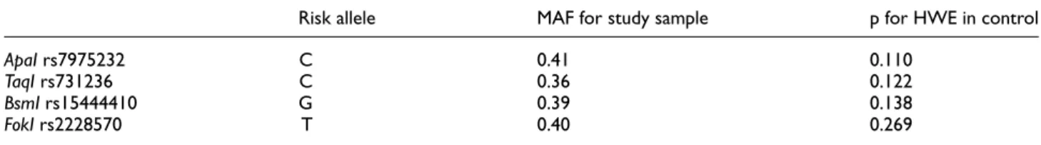
in controls (14.41 6 8.10 vs 17.47 6 10.32 ng/mL, p = 0.005). Four polymorphisms of the VDR gene were HWE in the control group. Minor allele frequency and HWE are shown in Table 2. Distributions of VDR gene polymorphisms are shown in Table 3. Frequency of
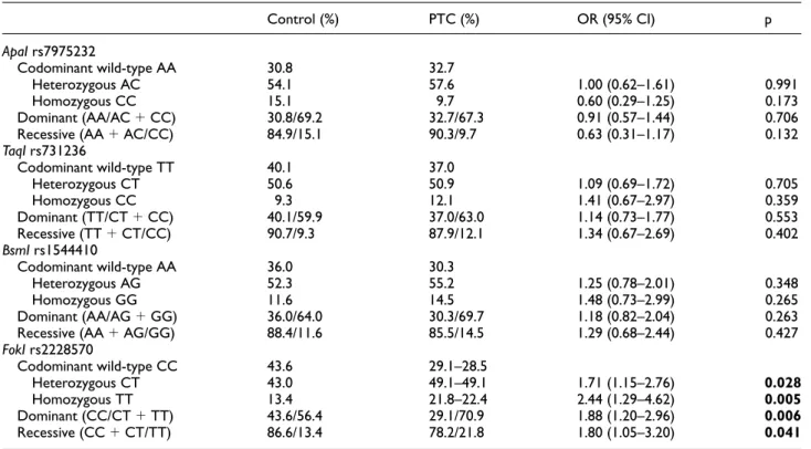
VDR gene ApaI rs7975232, TaqI rs731236, and BsmI rs1544410 polymorphisms did not differ between patients with PTC and controls in a codominant model and dominant model and recessive model (p . 0.05, each). VDR gene polymorphisms ApaI, TaqI, and BsmI were not associated with PTC. Frequency of VDR gene FokIrs2228570 increased in PTC patients than in con-trol (p \ 0.05). Haploview analysis shows that VDR gene polymorphisms are not in LD. Compared to con-trol, FokI TT genotype increased in patients with PTC in a recessive model (TT vs CT/CC, OR = 1.80, 95% CI = 1.05–3.20, p = 0.041) and FokI CT/TT genotype increased in patients with PTC in a dominant model (CT/TT vs CC, OR = 1.88, 95% CI = 1.20–2.96, p = 0.006). Compared to control, FokI CT and FokI TT genotypes increased in patients with PTC in a codo-minant model (CT vs CC, OR = 1.71, 95% CI = 1.15– 2.76, p = 0.028 and TT vs CC, OR = 2.44, 95% CI = 1.29–4.62, p = 0.005).
Association between VDR gene FokI rs2228570 and clinical features of PTC patients are shown in Table 4. Patients carrying FokI TT genotype increased risk for developing T3 and T4 PTC compared to patients carry-ing FokI CC genotype (TT vs CC: OR = 2.71, 95% CI = 1.13–6.53, p = 0.003) and C allele and CC geno-type (TT vs CT/CC: OR = 3.21, 95% CI = 1.76–6.09, p = 0.001, recessive model). The FokI T allele and TT genotype increased risk for developing N1 PTC in wild comparison, dominant and recessive model (TT vs CC: OR = 5.16, 95% CI: 1.31–10.42; p \ 0.001 and CT/TT vs CC: OR = 4.71, 95% CI: 1.25–12.51, p = 0.008; and TT vs CT/CC: OR = 5.14, 95% CI: 2.16–10.59; p \ 0.001, respectively). Patients carrying the FokI TT genotype had increased risk for developing stage III/IV PTC compared with patients carrying the CC genotype (TT vs CC: OR = 4.33, 95% CI: 1.07–10.74; p = 0.029) and C allele and CC genotype (TT vs CT/ CC: OR = 2.67, 95% CI: 1.04–6.83; p = 0.036, reces-sive model). FokI T allele and TT genotype increased risk of multifocal tumor in wild, heterozygote compari-son, dominant and recessive model (TT vs CC: OR = 5.20, 95% CI = 1.93–11.94; p = 0.001; CT vs CC: OR = 2.72, 95% CI = 1.25–5.93, p = 0.010; CT/ TT vs CC: OR = 2.26, 95% CI = 1.30–3.76, p = 0.002; TT vs CT/CC: OR = 2.55, 95% CI = 1.03–5.93, p = 0.026; respectively). The FokI T allele and TT gen-otype were more frequent in patients with PTC with tumors ø10 mm in size compared with tumors \10 mm in size (TT vs CC: OR = 4.09, 95% CI = 1.58–10.52, p = 0.003; CT vs CC: OR = 2.38, 95% CI = 1.10–5.15, p = 0.026; CT/TT vs CC: OR = 2.58, 95% CI = 1.25–5.33, dominant model). The FokI TT genotype was more common in patients with PTC with extra-thyroidal invasion compared with those without extra-thyroidal invasion (TT vs CC: OR = 15.26, 95% CI = 1.90–30.98; TT vs CT/CC:
OR = 2.4, 95% CI = 1.20–4.85, p \ 0.001, recessive model). The FokI TT genotype and T allele were more frequent in patients with PTC treated with radioiodine therapy compared with those without radioiodine ther-apy (TT vs CT: OR = 3.15, 95% CI = 1.20–8.27, p = 0.016; TT vs CC: OR = 7.2, 95% CI = 1.71–21.34, p = 0.002; CT/TT vs CC: OR = 5.10, 95% CI = 1.99– 8.28, dominant model; TT vs CT/CC: OR = 4.34, 95%
CI = 1.22–9.48, p = 0.007, recessive model). Patients with recurrent/persistent disease and disease-free status did not differ between the FokI genotypes (p . 0.05). The VDR gene polymorphisms ApaI, BsmI, and TaqI were not related to pathologic features (multifocality, capsule invasion, extra-thyroidal invasion, TNM-sta-ging, tumor size, lymph node metastasis), radioiodine therapy, and outcome.
Table 1. Characteristics of subjects.
PTC (n = 165) Control (n = 172) p
Age (years)a 46.89 6 13.22 45.25 6 4.89 0.308
Women (%) 81.2 86.6 0.175
Family history of thyroid cancer (%) 4.9 –
Preoperative 25(OH)D (ng/mL)a 14.41 6 8.10 17.47 6 10.32 0.005
Age at the diagnosis (years)a 44.70 6 13.55 –
Type (%)
Conventional papillary thyroid cancer 63.6
Follicular variant of papillary thyroid cancer 32.7
T classification (%) Tx 4.8 T1 and T2 78.2 T3 and T4 17.0 N classification (%) Nx 4.8 N0 78.8–78.2 N1 16.4–17.0 M classification (%) M1 1.3 M0 98.7 Clinical stage (%) I and II 85.4 III and IV 14.6 Multifocal (%) 56.8 Cancer size ø 1 cm (%) 54.1 Extra-thyroidal invasion (%) 9.6
Cervical lymph node metastasis (%) 17.2
Distance metastasis (%) 1.8
Capsular invasion (%) 14.7
Angiolymphatic invasion (%) 8.3
Perineural invasion (%) 1.9
I131 radiotherapy (%) 86.1
Time of follow-up (months)a 39.12 6 8.4
Disease free (%) 89.1
Recurrent/persistent (%) 7.9
PTC: papillary thyroid cancer; T: tumor size; N: lymph node metastasis; M: distance metastasis; 25(OH)D: 25-hydroxy-vitamin D. Bold value represents significant p-value.
a
Data are presented as mean 6 SD. Percentage was shown as (%).
Table 2. Minor allele frequency and Hardy–Weinberg equilibrium of VDR gene polymorphisms.
Risk allele MAF for study sample p for HWE in control
ApaI rs7975232 C 0.41 0.110
TaqI rs731236 C 0.36 0.122
BsmI rs15444410 G 0.39 0.138
FokI rs2228570 T 0.40 0.269
Table3. Genotype analysis of VDR gene polymorphisms. Control (%) PTC (%) OR (95% CI) p ApaI rs7975232 Codominant wild-type AA 30.8 32.7 Heterozygous AC 54.1 57.6 1.00 (0.62–1.61) 0.991 Homozygous CC 15.1 9.7 0.60 (0.29–1.25) 0.173 Dominant (AA/AC + CC) 30.8/69.2 32.7/67.3 0.91 (0.57–1.44) 0.706
Recessive (AA + AC/CC) 84.9/15.1 90.3/9.7 0.63 (0.31–1.17) 0.132
TaqI rs731236 Codominant wild-type TT 40.1 37.0 Heterozygous CT 50.6 50.9 1.09 (0.69–1.72) 0.705 Homozygous CC 9.3 12.1 1.41 (0.67–2.97) 0.359 Dominant (TT/CT + CC) 40.1/59.9 37.0/63.0 1.14 (0.73–1.77) 0.553 Recessive (TT + CT/CC) 90.7/9.3 87.9/12.1 1.34 (0.67–2.69) 0.402 BsmI rs1544410 Codominant wild-type AA 36.0 30.3 Heterozygous AG 52.3 55.2 1.25 (0.78–2.01) 0.348 Homozygous GG 11.6 14.5 1.48 (0.73–2.99) 0.265 Dominant (AA/AG + GG) 36.0/64.0 30.3/69.7 1.18 (0.82–2.04) 0.263
Recessive (AA + AG/GG) 88.4/11.6 85.5/14.5 1.29 (0.68–2.44) 0.427
FokI rs2228570 Codominant wild-type CC 43.6 29.1–28.5 Heterozygous CT 43.0 49.1–49.1 1.71 (1.15–2.76) 0.028 Homozygous TT 13.4 21.8–22.4 2.44 (1.29–4.62) 0.005 Dominant (CC/CT + TT) 43.6/56.4 29.1/70.9 1.88 (1.20–2.96) 0.006 Recessive (CC + CT/TT) 86.6/13.4 78.2/21.8 1.80 (1.05–3.20) 0.041
PTC: papillary thyroid cancer; OR: odds ratio; CI: confidence interval. Bold values represent significant p-values.
Table 4. Associations between VDR gene FokI polymorphism and clinical feature of papillary thyroid cancer.
Variables Dominant model Recessive model
Genotype (CT + TT/CC) (CC + CT/TT)
CC (wild) CT TT p p OR (95% CI) p OR (95% CI) p
n = 47 n = 81 n = 37 CT/CC TT/CC T classification (n) T1 and T2 39 68 22 T3 and T4 4 11 13 0.458 0.003 2.60 (0.84–7.99) 0.086 3.21 (1.76–6.09) 0.001 N classification (n) N0 40 67 22 N1 2 12 14 0.088 \0.001 4.71 (1.25–12.51) 0.008 5.14 (2.16–10.59) \0.001 Clinical stage (n) I and II 39 68 27 III and IV 3 11 9 0.250 0.029 2.28 (0.77–4.78) 0.095 2.67 (1.04–6.83) 0.036 Multifocal (n) 15 47 26 0.010 0.001 2.26 (1.30–3.76) 0.002 2.55 (1.03–5.93) 0.026 Cancer size ø 1 cm (n) 15 45 25 0.026 0.003 2.58 (1.25–5.33) 0.009 2.18 (0.98–4.84) 0.052 Extra-thyroidal invasion (n) 1 4 10 0.458 0.001 4.46 (0.66–20.1) 0.057 2.4 (1.20–4.85) \0.001 Angiolymphatic invasion (n) 1 5 7 0.322 0.010 3.81 (0.57–12.53) 0.094 1.73 (0.96–3.15) 0.005 Recurrent/persistent (n) 1 5 7 0.335 0.065 3.81 (0.57–12.56) 0.095 4.74 (1.48–11.15) 0.062 I131 radiotherapy (n) 30 71 35 0.016 0.002 5.10 (1.99–8.28) 0.001 4.34 (1.22–9.48) 0.007
OR: odds ratio; CI: confidence interval; T: tumor size; N: lymph node metastasis. The number of subjects is shown as (n).
Discussion
This is the first study to show the effects of VDR gene polymorphisms on the clinical features of PTC includ-ing TNM cancer staginclud-ing, outcomes, pathologic find-ings, and prognostic factors in a Turkish population. We found that the frequency of VDR gene ApaI, TaqI, and BsmI polymorphisms did not differ between PTC and controls with no association. The frequency of VDR gene FokI TT genotype and CT heterozygotes were increased in PTC compared with the controls. VDR gene FokI was associated with an increased risk of PTC. Haploview analysis showed that VDR gene polymorphisms were not in LD. Patients with PTC car-rying the FokI TT genotype were more likely to have adverse pathologic and prognostic factors including T3 and T4, stage III/IV, and extra-thyroidal invasion. Patients with PTC carrying the FokI CT/TT or TT gen-otype were more likely to develop N1, multifocal tumor, and tumors ø 10 mm. FokI CT/TT or TT geno-type carriers were more likely to be treated with radio-iodine therapy. VDR gene FokI was related to aggressiveness of PTC disease; however, it was not cor-related with persistence/recurrence. Thus, VDR gene FokI might be used as a biomarker for poor clinico-pathologic findings.
In humans, vitamin D shows a supportive effect on thyroid tissue. VDR gene BsmI and TaqI polymorph-isms were shown to be associated with decreased risk of autoimmune thyroid disease.18VDR gene ApaI, Bsml, and FokI polymorphisms were associated with Graves’ disease in an Asian population, whereas ApaI, Bsml, TaqI, and FokI were not associated with Graves’ disease in Caucasians.19TaqI CC and FokI TT were increased in Hashimoto’s thyroiditis in Turkey.20 Genetic and epigenetic determinants of thyroid cancer may have an effect on 1,25(OH)2D3 signaling.
Development and progression of thyroid cancer depends on impaired 1,25(OH)2D3signaling
21,22
which has been confirmed in clinical studies22–26and thyroid cancer cell lines.27 Local 1,25(OH)2D3-VDR signaling
is decreased in primary thyroid cancer with local lymph node metastasis, and complete loss of signaling is more significant in anaplastic thyroid cancer with distant metastasis.10,22–25Hence, thyroid cancer progression is characterized by the loss of proteins that play a role in vitamin D signaling or 1,25(OH)2D3 sensitivity.27 The
signaling effect of 1,25(OH)2D3-VDR in thyroid cancer
occurs via the local anti-tumoral effect of 1,25(OH)2D3.21,22This anti-tumoral effect occurs either
directly (via VDR binding) or indirectly (via the inter-action between other critical transcriptional regulators or cell signaling systems).21,28,29 Therapeutic use of vitamin D agonists in thyroid cancer requires the pres-ence of VDR. An experimental study showed that VDR FokI CC was associated with vitamin D analog
resistance in the treatment of thyroid cancer.30 Active vitamin D has many anti-cancer features including anti-proliferative, anti-apoptotic, pro-differentiating, and anti-inflammatory effects.5–10 These effects are mediated by VDR. VDR regulates also metabolic path-ways such as the immune response and cancer signal-ing.5–8VDR is stimulated by p53, a tumor-suppressing gene.16 An association was found between vitamin D deficiency and thyroid cancer.10,31,32
Epidemiologic studies reported that there was a rela-tionship between VDR gene and breast cancer, prostate cancer, lung cancer, colorectal cancer, and other gas-trointestinal cancers.4–8However, reliable data on thyr-oid cancer are available only in in vitro experimental studies.10,23,24,27–31VDRgene Apa, Bsml, Taq, and FokI genotypes were not associated with risk of PTC in a German population.10 In an Iranian population, fre-quency of VDR gene ApaI, FokI, Bsml, Taq, and Tru9 did not differ between differentiated thyroid cancer and controls, with no relationship.16However, these studies did not investigate the genotype-phenotype correlation such as TNM cancer staging, outcomes, pathologic findings, and prognostic factors. In this study, vitamin D deficiency was found in patients with PTC compared with controls. This study showed that frequency of VDRgene ApaI, TaqI, and BsmI did not differ between patients with PTC and controls. VDR gene ApaI, TaqI, and BsmI were not associated with a risk for PTC. VDR gene FokI rs2228570 showed significant differ-ences between patients with PTC and controls: VDR gene FokI (variant or heterozygotes) compared with wild type (CC) revealed a significant association. VDR gene FokI TT (TT vs CC: OR = 2.44, 95% CI = 1.29– 4.62, p = 0.005) and CT heterozygotes (CT vs CC: OR = 1.71, 95% CI = 1.15–2.76, p = 0.028) were asso-ciated with an increased PTC risk compared with the controls. Our study suggests that VDR gene FokI may be associated with the development of PTC. Allele fre-quency distribution showed a significant association of FokI variant allele (T) on susceptibility to PTC. These results revealed that VDR gene FokI might play a criti-cal role in the etiology of the PTC. VDR gene FokI TT genotype was associated with having T3 and T4, stage III/IV, and extra-thyroidal invasion of PTC. FokI CT/ TT or TT genotype was associated with developing N1, multifocality, and tumor size ø10 mm in PTC. Patients carrying FokI CT/TT or TT genotype were mostly treated with radioiodine therapy. The persis-tence/recurrence rate in PTC did not differ between FokI genotypes. Although VDR gene FokI was associ-ated with more advanced TNM stage, greater diameter of tumor, multifocality, and extra-thyroidal invasion, outcomes in follow-up did not differ. VDR gene FokI might be considered as a risk for poor clinicopathologic features and advanced stage of PTC. VDR gene FokI might be suggested as a poor prognostic factor of PTC.
However, the molecular mechanism needs to be identified.
Bsml, ApaI, and TaqI polymorphisms of VDR gene are found in the three primer untranslated region (3#-UTR) and have been shown to be in strong LD.33,34FokI polymorphism was reported as an inde-pendent marker of VDR gene because it has not been shown to be in LD with any other VDR polymorph-isms.34 Haploview analysis showed that VDR gene polymorphisms were not in LD. ApaI and BsmI poly-morphisms of the VDR gene, both in intron 8, are con-sidered as silent SNPs. These polymorphisms do not change the amino acid sequence of the encoded protein, but they might affect gene expression by modulating stability of messenger RNA (mRNA).11TaqI poly-morphism is located at codon 352 in exon 9 of the VDR gene. According to the Taq restriction site, prod-ucts are digested into two or three fragments. TT geno-type (absence of restriction site) is related to lower active vitamin D3.4–11 The only locus that has an impact on the structure of VDR protein is the FokI polymorphism, which is located on the 5# end region of VDRgene. VDR gene FokI polymorphism is functional because it is found in a coding sequence. FokI poly-morphism is located in the first ATG starting code of VDR protein. FokI is involved in thymine to cytosine (T/C) substitution at exon 2, the first translation initia-tion region is removed, and the transcripinitia-tional activity of VDR is changed.4–11,16It alters the ACG codon that is found 10 base pairs upstream from the translation starting codon and leads to generating an additional starting codon. Two different VDR isoforms occur with the transition of allele T to C in ATG. When the initiating translation starts from this alternative site in the thymine variant, it leads to the generation of a lon-ger VDR protein comprising 427 amino acids. The gene is transcribed in normal length if there is a restriction site. Thus, the C/C allele codes the 424-amino acid tein and the T/T allele codes the 427-amino acid pro-tein. The longer VDR protein has low activity in transcription; accordingly, activation is decreased in the target cell.4–11Arai et al.35reported that compared with FokI T/T genotype, FokI C/C showed 1.7-fold greater function in vitamin D-dependent transcrip-tional activation of a reporter through the regulation of a vitamin D response element. FokI rs2228570 poly-morphism is the only VDR gene polypoly-morphism involved in the generation of an altered protein expression.11
In this study, the control population was consistent with HWE. The small sample size, cross-sectional design, and seasonal change in 25(OH)D3 are
limita-tions of this study.
In conclusion, this study showed that VDR gene FokImight contribute to the susceptibility of PTC risk
in the Turkish population. VDR gene FokI correlated with aggressiveness of papillary thyroid cancer but not with outcome of disease. FokI could be useful as supple-mentary prognostic factor to assess high risk of papil-lary thyroid cancer and to identify patients who need more aggressive management and follow-up. Further studies in different populations are needed to confirm these results.
Availability of data and materials
All data are freely available for scientific purpose.
Acknowledgement
We special thanks to Professor Dr Erkan Yurtcu for genetic analysis of data.
Declaration of conflicting interests
The author(s) declared no potential conflicts of interest with respect to the research, authorship, and/or publication of this article.
Funding
The author(s) received no financial support for the research, authorship, and/or publication of this article.
ORCID iD
Selvihan Beysel https://orcid.org/0000-0001-6963-1503
References
1. Ukekwe FI, Olusina DB and Okere PCN. Patterns of thyroid cancers in Southeastern Nigeria: a 15 year histo-pathologic review (2000–2014). J Clin Diagn Res 2017; 11(8): EC16–EC19.
2. Figlioli G, Elisei R, Romei C, et al. A comprehensive meta-analysis of case-control association studies to eval-uate polymorphisms associated with the risk of differen-tiated thyroid carcinoma. Cancer Epidemiol Biomarkers Prev2016; 25: 700–713.
3. Kohler A, Chen B, Gemignani F, et al. Genome-wide association study on differentiated thyroid cancer. J Clin Endocrinol Metab2013; 98(10): E1674–E1681.
4. Bai Y-H, Lu H, Hong D, et al. Vitamin D receptor gene polymorphisms and colorectal cancer risk: a systematic meta-analysis. World J Gastroenterol 2012; 18(14): 1672–1679.
5. Gnagnarella P, Pasquali E, Serrano D, et al. Vitamin D receptor polymorphism FokI and cancer risk: a comprehen-sive meta-analysis. Carcinogenesis 2014; 35(9): 1913–1919. 6. Raimondi S, Pasquali E, Gnagnarella P, et al. BsmI
poly-morphism of vitamin D receptor gene and cancer risk: a comprehensive meta-analysis. Mutat Res 2014; 769: 17–34. 7. Serrano D, Gnagnarella P, Raimondi S, et al.
Meta-anal-ysis on vitamin D receptor and cancer risk: focus on the role of TaqI, ApaI, and Cdx2 polymorphisms. Eur J Cancer Prev2016; 25(1): 85–96.
8. Tuoresmaki P, Vaisanen S, Neme A, et al. Patterns of genome-wide VDR locations. PLoS ONE 2014; 9(4): e96105.
9. Tang C, Chen N, Wu M, et al. Fok1 polymorphism of vitamin D receptor gene contributes to breast cancer sus-ceptibility: a meta-analysis. Breast Cancer Res Treat 2009; 117(2): 391–399.
10. Penna-Martinez M, Ramos-Lopez E, Stern J, et al. Vita-min D receptor polymorphisms in differentiated thyroid carcinoma. Thyroid 2009; 19(6): 623–628.
11. Kostner K, Denzer N, Muller CS, et al. The relevance of vitamin D receptor (VDR) gene polymorphisms for can-cer: a review of the literature. Anticancer Res 2009; 29(9): 3511–3536.
12. Iqbal MUN and Khan TA. Association between Vitamin D receptor (Cdx2, Fok1, Bsm1, Apa1, Bgl1, Taq1, and Poly (A)) gene polymorphism and breast cancer: a systema-tic review and meta-analysis. Tumour Biol. Epub ahead of print 26 October 2017. DOI: 10.1177/101042 8317731280. 13. Liu Y, Li C, Chen P, et al. Polymorphisms in the vitamin
D Receptor (VDR) and the risk of ovarian cancer: a meta-analysis. PLoS ONE 2013; 8(6): e66716.
14. Hama T, Norizoe C, Suga H, et al. Prognostic signifi-cance of vitamin D receptor polymorphisms in head and neck squamous cell carcinoma. PLoS ONE 2011; 6(12): e29634.
15. Xu Y, He B, Pan Y, et al. Systematic review and meta-analysis on vitamin D receptor polymorphisms and can-cer risk. Tumour Biol 2014; 35(5): 4153–4169.
16. Haghpanah V, Ghaffari SH, Rahimpour P, et al. Vitamin D receptor gene polymorphisms in patients with thyroid cancer. Gene Ther Mol Biol B 2007; 11: 299–304.
17. Haugen BR, Alexander EK, Bible KC, et al. 2015 Ameri-can Thyroid Association Management guidelines for adult patients with thyroid nodules and differentiated thyroid cancer: the American Thyroid Association guide-lines task force on thyroid nodules and differentiated thyroid cancer. Thyroid 2016; 26(1): 1–133.
18. Feng M, Li H, Chen SF, et al. Polymorphisms in the vita-min D receptor gene and risk of autoimmune thyroid dis-eases: a meta-analysis. Endocrine 2013; 43(2): 318–326. 19. Zhou H, Xu C and Gu M. Vitamin D receptor (VDR)
gene polymorphisms and Graves’ disease: a meta-analy-sis. Clin Endocrinol 2009; 70(6): 938–945.
20. Yazici D, Yavuz D, Tarcin O, et al. Vitamin D receptor gene ApaI, TaqI, FokI and BsmI polymorphisms in a group of Turkish patients with Hashimoto’s thyroiditis. Minerva Endocrinol2013; 38(2): 195–201.
21. Clinckspoor I, Verlinden L, Overbergh L, et al. 1,25-dihy-droxyvitamin D3 and a superagonistic analog in combi-nation with paclitaxel or suberoylanilide hydroxamic acid have potent antiproliferative effects on anaplastic thyroid cancer. J Steroid Biochem Mol Biol 2011; 124(1–2): 1–9. 22. Clinckspoor I, Hauben E, Verlinden L, et al. Altered
expression of key players in vitamin D metabolism and signaling in malignant and benign thyroid tumors. J His-tochem CyHis-tochem2012; 60(7): 502–511.
23. Balla I, Tobias B, Kosa JP, et al. Vitamin D-neutralizing CYP24A1 expression, oncogenic mutation states and his-tological findings of human papillary thyroid cancer. J Endocrinol Invest2015; 38(3): 313–321.
24. Izkhakov E, Somjen D, Sharon O, et al. Vitamin D recep-tor expression is linked to potential markers of human thyroid papillary carcinoma. J Steroid Biochem Mol Biol 2016; 159: 26–30.
25. Khadzkou K, Buchwald P, Westin G, et al. 25-hydroxy-vitamin D3 1alpha-hydroxylase and 25-hydroxy-vitamin D receptor expression in papillary thyroid carcinoma. J Histochem Cytochem2006; 54(3): 355–361.
26. Schulten HJ, Al-Mansouri Z, Baghallab I, et al. Compar-ison of microarray expression profiles between follicular variant of papillary thyroid carcinomas and follicular adenomas of the thyroid. BMC Genomics 2015; 16(Suppl. 1): S7.
27. Somjen D, Grafi-Cohen M, Posner GH, et al. Vitamin D less-calcemic analog modulates the expression of estrogen receptors, vitamin D receptor and 1a-hydroxylase 25-hydroxy vitamin D in human thyroid cancer cell lines. J Steroid Biochem Mol Biol2013; 136: 80–82.
28. Liu W, Asa SL, Fantus IG, et al. Vitamin D arrests thyr-oid carcinoma cell growth and induces p27 dephosphory-lation and accumudephosphory-lation through PTEN/akt-dependent and -independent pathways. Am J Pathol 2002; 160(2): 511–519.
29. Verlinden L, Eelen G, Beullens I, et al. Characterization of the condensin component Cnap1 and protein kinase Melk as novel E2F target genes down-regulated by 1,25-dihydroxyvitamin D3. J Biol Chem 2005; 280(45): 37319–37330.
30. Sharma V, Fretwell D, Crees Z, et al. Thyroid cancer resistance to vitamin D receptor activation is associated with 24-hydroxylase levels but not the ff FokI poly-morphism. Thyroid 2010; 20: 1103–1111.
31. Penna-Martinez M, Ramos-Lopez E, Stern J, et al. Impaired vitamin D activation and association with CYP24A1 haplotypes in differentiated thyroid carci-noma. Thyroid 2012; 22(7): 709–716.
32. Sahin M, Ucan B, Ginis Z, et al. Vitamin D3 levels and insulin resistance in papillary thyroid cancer patients. Med Oncol2013; 30(2): 589.
33. Song GG and Lee YH. Vitamin D receptor FokI, BsmI, ApaI, and TaqI polymorphisms and susceptibility to ovarian cancer: a meta-analysis. Immunol Invest 2013; 42(7): 661–672.
34. Lurie G, Wilkens LR, Thompson PJ, et al. Vitamin D receptor rs2228570 polymorphism and invasive ovarian carcinoma risk: pooled analysis in five studies within the ovarian cancer association consortium. Int J Cancer 2011; 128(4): 936–943.
35. Arai H, Miyamoto K, Taketani Y, et al. A vitamin D receptor gene polymorphism in the translation initiation codon: effect on protein activity and relation to bone mineral density in Japanese women. J Bone Miner Res 1997; 12(6): 915–921.