The relationship between virulence factors and vancomycin resistance
among Enterococci collected from food and human samples in
Southern Turkey
Mevhibe TERKURAN1, Zerrin ERGİNKAYA1, Emel ÜNAL1, Mümtaz GÜRAN2 ,
Suna KIZILYILDIRIM2, Gökce UGUR2, Fatih KÖKSAL2
1 Çukurova University, Faculty of Agriculture, Department of Food Engineering; 2Çukurova University, Faculty of Medicine, Department of Clinical Microbiology, Adana, Turkey.
Summary: The aims of this research were to study the prevalence of potential virulence factors, vancomycin resistance and
also to evaluate a possible correlation that can exist between vancomycin resistance and potential virulence factors between 51
Enterococcus spp. isolated from food and 50 Enterococcus faecium strains from human in southern Turkey. Identification of the
isolates was determined by Vitek-II system. Antimicrobial susceptibility tests were performed by Vitek-II system and disc diffusion method. The presence of vanA and vanB as well as enterococcal virulence genes of cytolysin (cylA), the aggregation substance (asa1), gelatinase (gelE), enterococal surface protein (esp), hyaluronidase (hyl) were investigated by Polymerase Chain Reaction (PCR) method. Haemolysin production was also studied phenotypic method. Apart from one isolate, none of the food originated enterococci were resistant to vancomycin, and none carried vanA and vanB resistance genes. All clinical isolates were resistant to vancomycin and 84% of them carried vanA; 2%, vanB; and 14%, neither vanA nor vanB genes. Except for the cylA gene, all other virulence genes and vancomycin resistance were higher in human strains, and a positive correlation was observed between multivirulence genes and hemolytic activity. For all strains, a positive correlation existed between the esp gene positivity and vancomycin resistance, while for only E. faecium, esp, hyl gene positivity and vancomycin resistance a positive correlation could be seen. Furthermore, “silent cylA” genes were found in two food and one intestinal strains. Based on our findings, we can suggest that virulence increases in parallel to vancomycin resistance, and food may be a potential source for dissemination of gelE, asa1 and hyl virulence genes. Finally, esp and hyl genes presence should carefully be monitored in food originated enterococci.
Key words: Enterococci, relationship, vancomycin resistance, virulence factors.
Türkiye’nin güneyinde gıda ve insan örneklerinden toplanan Enterokok izolatlarının vankomisin direnci ve virülans faktörleri arasındaki ilişki
Özet: Bu çalışmanın amaçları, Türkiye’nin güneyinde gıda kaynaklı 51 adet Enterococcus spp. ile insan kaynaklı 50 adet
Enterococcus faecium izolatlarının potansiyel virülans faktörlerinin, vankomisin direnç özelliklerinin prevalansının belirlenmesi ve
vankomisin direnci ile virülans faktörler arasında muhtemel ilişkinin değerlendirilmesidir. İzolatlarının identifikasyonu Vitek-II sistemiyle, antimikrobiyel direnç testleri Vitek-II sistemi ve disk difüzyon metodlarıyla çalışıldı. vanA ve vanB ile sitolizin (cylA), agregasyon faktörü (asa1), jelatinaz (gelE), entrokok yüzey proteini (esp), hiyolüronidaz (hyl), genlerinden oluşan enterokokal virülans genler Polimeraz Zincir Reaksiyonu (PCR) metoduyla, hemolizin üretimi fenotipik yöntemle çalışıldı. Bir izolat dışında, gıda izolatlarının hiçbiri vankomisine dirençli değildi ve hiçbiri vanA ve vanB geni taşımamaktaydı. Klinik izolatların tümü vankomisine dirençliydi ve bu izolatların %84’ü vanA, %2’si vanB genlerini taşımakta; %14’ü vanA ve vanB genlerini taşımamaktaydı. cylA geni dışındaki test edilen diğer virülans genler ve vankomisin direnci, klinik izolatlarda daha yüksekti ve çoklu virülans genler ile vankomisin direnci arasında pozitif bir korelasyon gözlendi. Tüm izolatlar arasında esp gen pozitifliği ile vankomisin direnci, E. faecium türlerinde ise esp, hyl gen pozitiflikleri ile vankomisin direnci arasında pozitif bir korelasyon olduğu görüldü. Ayrıca, iki gıda ve bir intestinal enterokok suşunda ‘’sessiz cylA geni’’ bulundu. Bulgularımıza göre, virülans faktörler vankomisin direncine paralel olarak artabilir ve gıda kaynaklı enterokoklar gelE, asa1, hyl virülans genlerinin yayılmasında potansiyel kaynak olabilir. Sonuç olarak, gıda kaynaklı enterokoklarda esp ve hyl gen varlığı dikkatle izlenmelidir.
Anahtar sözcükler: Enterokok, ilişki, vankomisin direnci, virülans faktörler.
Introduction
Enterococci, widespread in nature, are part of the communal flora of gastrointestinal and genitourinary tracts of humans as well as digestive tract of animals.
enterococci can also present in foods, especially in those of animal origin such as meat, fermented sausages and cheeses. Several studies have indicated that strains of enterococci may have beneficial effects on the
production and ripening of cheeses as they may contribute to cheese flavor and taste with their metabolic and technological traits. However, enterococci may cause spoilage problems in certain cheeses or processed meat products (12, 20). Although fermented foods with enterococci contribution have a long history of safe use, the presence of enterococci in foods is still an important issue for the food industry and public health.
In some reports, enterococci are considered as emerging pathogens of humans and are often noted as important agents of hospital acquired infections with an increasing mortality rate of up to 61%. The reason behind this rise is their ability to develop resistance against a wide variety of antibiotics, especially, glycopeptides (vancomycin and teicoplanin) (9). In general, vancomycin resistance is found in enterococci and encoded by the
vanA gene cluster that carried on the mobile genetic
element Tn1546. This resistance can be transferred by conjugative plasmids (9, 12, 20). Although vancomycin has been used in the treatment of enterococci for many years, recently, vancomycin resistant enterococci (VRE) strains have emerged worldwide as important nosocomial agents. This has posed a serious problem in the treatment of enterococcal infections and other Gram positive bacteria such as Staphylococcus aureus as enterococci have the ability to transfer resistant determinants to other vancomycin-susceptible species horizontally (20).
Several studies have shown that enterococci may harbour putative virulence traits and virulence genes such as production of aggregation substances (agg), gelatinase (gelE), hemolysin, adhesine (ace), the adhesinlike E.
faecalis and E. faecium antigen A (efaAfs and efaAfm,
respectively) and enterococcal surface protein (esp) (12, 15, 21). Eventually, enterococci are not considered as “generally recognized as safe” (GRAS) microorganisms in the food industry due to reasons such as; spoilage problems, biogenic amine production and acquired resistance problems in combination with the virulence factors mentioned above.
In this study, we investigated the prevalence of phenotypic and genotypic (vanA and vanB genes) vancomycin resistance and five virulence genes of enterococal surface protein (esp), gelatinase (gelE), aggregation substances (asa1), cytolysin production (cylA), hyaluronidase (hyl) genes and hemolysis in human blood among enterococci derived from food products and human clinical samples to evaluate a possible correlation between vancomycin resistance and potential virulence factors.
Materials and Methods
Materials: A total of 101 enterococci isolates were
studied for the presence of virulence traits, vancomycin susceptibility and hemolytic activity. In this study, 51
Enterococcus spp., were isolated and identified from 80
food samples (n=28 cheese, n=26 fruit and vegetables
n=21 sucuk and n=5 chicken meat) purchasing in
Çukurova region from March to September 2010. Distribution of identified enterococci of food originated isolates; 24 from cheese samples, 10 from sucuk (a traditional Turkish meat product), 4 from raw chicken meat and 13 from fruit & vegetables foods. Clinical strains were collected over a period of 3 years (2010-2012) from clinical laboratories of the Balcalı Hospital Central Laboratory (Southern Turkey). A total of 50 clinical VRE strains, one isolate from each patient, were collected from infected hospitalized patients (n=25) and intestinally colonized hospitalized patients (n=25).
Isolation and identification of food and clinical enterococci: Isolation of food isolates was evaluated, as
described, by Abriouel et al.(1). Further species level identification of isolates with a typical enterococci morphology on kanamycin esculin azide agar (KEA) (Merck KGaA, Germany) media were examined by biochemical tests such as catalase production, Gram staining, gas production from glucose and growing in 6.5% NaCl. Clinical isolates were routinely grown on Columbia agar (Becton-Dickinson, Sparks, MD) supplemented with 5% defibrinated sheep blood. For identification, cultures were examined with VITEK-2 automated identification system in the University central laboratory.
All identified strains (food & clinical) were stored in Brain Hearth Broth (BHI) including 10% of human blood and 10% of glycerol at -20ºC until PCR analysis.
Screening of enterococci for glycopeptide resistance and virulence genes: Genomic DNA was extracted
mechanically by the “Mickle Sytem” (The Mickle Lab. Engeneering Co. Ltda, Gomshall, Surrey, UK) according to manufacturer’s instructions after overnight cultures in 5% defibrinated sheep-blood agar of enterococci. A spectrophotometer (UV-VIS Spectrophotometer CHEBIOS) was used for quantitation of DNA samples (100ng/µL DNA for each sample). Extracted DNAs of enterococci were stored at -20ºC until to use as a template for PCR amplifications.
Multiplex PCR was performed to screen specific virulence genes (esp, hyl, asa1, cylA and gelE) and vancomycin resistance genes (vanA, vanB) as described previously (25, 5, 14). The specific primers and PCR conditions were presented in Table 1. Amplicons were analyzed by electrophoresis on 2% agarose gels [PegGOLD Universal Agarose, 91052 Ertangen Deutschland, 2%(w/v)] containing 0.5% ethidium bromide in TBE buffer (40mM tris, 20mM boric acid, 1 mM EDTA, pH 8.3) for 30/60 minutes at 120/150 V in the presence of 50-100bp DNA ladder (Fermentas SMo.323-Lithuania). The gel was photographed on an UV transilluminator (Kodak Gellogic-1500 imaging system).
Assay of hemolytic activity: Production of hemolysis
was determined by streaking the Enterococcus spp. cultures on Mueller Hinton Agar plates supplemented with 5% human blood. Plates were aerobically incubated at 37ºC for 18-24 hours and were examined for hemolysis. Presence of zone of clearing around the colonies was interpreted as beta-hemolysis (19).
Vancomycin susceptibility test: Vancomycin
susceptibility of food originated enterococci was examined by disk diffusion method using the standard antibiotic discs with a concentration of 30 µg (Oxoid). Susceptibility or resistance was determined according to the recommendation of Clinical and Laboratory Standards Institute (CLSI) guidelines (7).
Vancomycin resistance patterns of clinical strains and confirmation for suspected results of food isolates were evaluated by using gram positive antibiotic susceptibility cards (Biomerieux Vitek-2-AST-P534-SA-France) in VITEK-2 automated identification system (Biomerieux, Durham, North Carolina, USA). The results were recorded after 18-24 h of incubation at 37ºC and evaluated by the manufacturer instructions using the breakpoints for enterococci proposed by the Clinical and Laboratory Standards Institute (7). Enterococcus faecalis 1047387 (vancomycin sensitive) and Enterococcus
faecium 1045803 (vancomycin resistant) were used as
reference strains in phenotypic antibiotic resistance tests.
Minimal inhibitory concentration (MIC) testing:
For all (resistant or suspected) isolates, MICs of vancomycin was determined by VITEK-2 AST compact panel (Biomerieux Vitek-2-AST-P534-SA-France) and confirmed by E-test (Biodisk, Solana, Sweden) according to the CLSI’s (7) guidelines.
Statistical analysis: Statistical analysis of data was
performed with S-PLUS 18 statistical program (S-PLUS
18.00 for Windows, Professional Edition). Number and percentage of categorical measures were summarized. Chi-square test was used to compare categorical measures between groups (Fisher's exact test where appropriate). A
P value of <0.05 was regarded as statistically significant.
Results
Among the food samples tested,a total of 51 isolates were identified as the following species; E.
faecium (20 isolates), E. faecalis (12 isolates), E. casseliflavus (7 isolates), E. gallinarum (4 isolates), E. durans (7 isolates) and E. raffinosus (1 isolates). A total
of 50 clinical enterococci (infection and intestinal isolates) were identified as E. faecium.
From a starting collection of 50 clinical isolates (corresponding to 25 patients with acute VRE infection and 25 patients without infection but with VRE colonization). A Total of 50 isolates were identified as E.
faecium. Most of isolates (9; 36%), (2; 8%), (2; 8%), (2;
8%) and (10; 40%) in nosocomial environment were from intensive care internal medicine, pediatric hematology, urology, brain surgery, and other clinics respectively, while most of the intestinal isolates (15; 60%), (4; 16%), (4; 16%) and (2; 16%) were from pediatric hematology and oncology, brain surgery, intensive care and burn unit clinic respectively.
In the present study, 39 (76.4%) of food enterococci were found to be sensitive to vancomycin, and 11 (21.5%) of these were found intermediate sensitive to vancomycin. Only one strain identified as E. casseliflavus and collected from a lettuce sample was found to be resistant to vancomycin. Its MIC value was found to be 64 µg/mL. E. faecium strains from all clinical samples were found to be resistant to vancomycin and teicoplanin, and their MIC values were above 128 µg/mL.
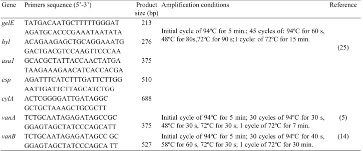
Table 1. List of primers and amplification conditions used in the present study. Tablo 1. Çalışmada kullanılan primerler ve amplifikasyon koşulları.
Gene Primers sequence (5’-3’) Product size (bp)
Amplification conditions Reference
gelE TATGACAATGCTTTTTGGGAT 213
Initial cycle of 94ºC for 5 min.; 45 cycles of: 94ºC for 60 s, 48ºC for 80s,72ºC for 90 s;1 cycle: of 72ºC for 15 min.
(25) AGATGCACCCGAAATAATATA hyl ACAGAAGAGCTGCAGGAAATG 276 GACTGACGTCCAAGTTCCCAA asa1 GCACGCTATTACCAACTATGA 375 TAAGAAAGAACATCACCACGA esp AGATTTCATCTTTGATTCTTGG 510 AATTGATTCTTAGCATCTGG cylA ACTCGGGGATTGATAGGC 688 GCTGCTAAAGCTGCGCTT vanA TCTGCAATAGAGATAGCCGC 375
Initial cycle of 94ºC for 5 min; 30 cycles of 94ºC for 30 s, 48ºC for 30 s, 72ºC for 30 s; 1 cycle of 72ºC for 7 min.
(5) GGAGTAGCTATCCCAGCATT
vanB TCTGCAATAGAGATAGCC GC
527
Initial cycle of 94ºC for 5 min; 30 cycles of 94ºC for 40 s, 58ºC for 60 s, 72ºC for 30 s; 1 cycle of 72ºC for 30 min.
(14) GGAGTAGCTATCCCAGCA TT
Table 2. Occurrance of virulence determinants among enterococci isolates. Tablo 2. Enterokok izolatları arasında virülans determinantların varlığı.
Na
Virülence Determinants
asa1 esp hyl gelE cylA β-hemolysis.
E.faecalis 5(41.7) 1(8.3) 7(58.3) 10(83.3) 1(8.3) 2(16.7)
E.faecium 22(31.4) 11(15.7) 22(31,4) 22(31.4) 6(8.6) 8(11.4)
E.gallinarum 1(25) 0 0 1(25) 0 0
E.casselliflavus 3(42.9) 0 1(14.3) 0 0 0
E.durans 5(71.4) 1(14.3) 1(14.3) 2(28.6) 1(14.3) 3(42.9)
a: Number of tested isolates; values in parenthesis indicate percentage
Table 3. The distribution of multi virulence genes of enterococci from different sources. Tablo 3. Farklı kaynaklı enterokoklarda çoklu virülans genlerin dağılımı.
Number of virulence genes Total P value
0 1 2 3 4 Food Na 23 10 13 3 2 51 P<0.001 % 45.10 19.60 25.50 5.90 3.90 100.00 Human Clinical Na 3 4 4 5 9 25 P<0.001 % 12.00 16.00 16.00 20.00 36.00 100.00 Human intestinal Na 18 7 0 0 0 25 P<0.001 % 72.00 28.00 0.00 0.00 0.00 100.00 Total Na 44 21 17 8 11 101 P<0.001 % 43.60 20.80 16.80 7.90 10.90 100.00 a: Number of isolates
Figure 1. The distribution of five virulence genes of enterococci from food, human clinical and intestinal isolates.
Şekil 1. Gıda, klinik enfeksiyon ve kolonizasyon kaynaklı enterokoklarda esp, hyl, gelE, asa1 ve cylA virülans genlerinin dağılımı.
Figure 2. Ethidium bromide-stained agarose gel electrophoresis of PCR-amplified products of enterococci. Lanes 11 and 28: 100 bp DNA markers; 17: cylA positive E. durans (parmesan cheese) isolate; 30: cylA positive E. faecalis (Urfa Cheese) isolate; 23: esp positive E. faecalis isolate (home made sucuk). The other strains are negative.
Şekil 2. Enterokok izolatlarının PCR ürünlerinin etidyum bromür ile boyanmış agaroz jel elektroforezi ile görüntüsü. Sıra 11 ve 28: 100 bp DNA cetveli; 17: cylA pozif E. durans (kaşar peyniri) izolatı; 30: cylA pozitif E. faecalis (Urfa Peyniri) izolatı; 23: esp positive E. faecalis (home made sucuk) izolatı. Diğer isolatlar negatiftir.
Table 4.The correlation of vancomycin resistance and virulence genes of E. faecium (n=70) isolates in present study.
Tablo 4. Çalışmada izole edilen E. faecium izolatlarında (n=70) vankomisin direnci ile virülans genler arasındaki korelasyon.
E. faecium
Ta esp asa1 hyl gelE cylA
S+Ib 20 0 5 2 5 1
Rc 50 11 17 20 17 5
Td 70 11 22 22 22 6
Pe 0.03f 0,57 0.02f 0.574 0.666 a. Total number of isolates
b. Isolates sensitive or intermediate sensitive to vancomycin c. Isolates resistant to vancomycin
d. Total number of E. faecium isolates
e. p values between number of isolates and virulence genes obtained with fisher`s exact test.
f. Statistically significant correlation between vancomycin resistance and virulence gene
Table 5. The correlation of vancomycin resistance and virulence genes of all enterococci isolates.
Tablo 5. Tüm enterokoklarda vankomisin direnci ile virülans genler arasındaki korelasyon.
Ta esp asa1 hyl gelE cylA
S+Ib 50 2 19 11 18 3
Rc 51 11 17 20 17 5
Td 101 13 36 31 35 8
Pe 0.02f 0.68 0.08 0.836 0.715
a. Total number of isolates
b. Isolates sensitive or intermediate sensitive to vancomycin c. Isolates resistant to vancomycin
d. Total Enterococcus spp. isolates
e. p values between number of isolates and virulence genes obtained with fisher`s exact test
f. Statistically significant correlation between vancomycin resistance and virulence gene
The prevalence of genes encoding vancomycin resistance in enterococci; none of food isolates carried
vanA and vanB resistance genes and 84% of clinical
enterococci carried vanA, 2% of them vanB and 14% them neither carried vanA nor vanB genes.
Hemolytic activities of food enterococci were found 32 (62.7%) non-hemolytic, 13 (25.4%) α-hemolytic, 6(11.7%), β –hemolytic, respectively. A total of 16(64%) human intestinal isolates were found α-hemolytic and 9(36%) were non-hemolytic; but 50% of clinical E.
faecium isolates were found to be α-hemolytic and 14%
were β-hemolytic.
In present study, E. faecium carried more virulence genes than E. faecalis and other Enterococcus spp. Occurrence of virulence determinants among enterococcal isolates were presented in Table 2. In addition, the presence of strains harbouring multiple virulence genes is shown in Table 3. A summary of the prevalence of the virulence genes among the Enterococcus spp. and their origin is shown in Figure1. The gel image of some virulence genes of different reservoirs were also shown in Figure 2. The gel image of vanA genes of clinical isolates were presented in Figure 3.
A summary of correlations between virulence factors and vancomycin resistance is presented in Tables 4 and 5.
Discussion and Conclusion
In previous studies, antimicrobial resistance and virulence were characterized in enterococci isolates from food, but some researchers have observed a correlation between the presence of antimicrobial resistance and of virulence determinants (10, 11, 13, 15, 17). Few studies have been conducted on resistance, virulence determinants and/or correlation of virulence and resistance in enterococci of food and clinical origins in Turkey (4, 18). In this study, vancomycin resistance and five virulence profiles of Enterococcus spp. isolated from food and clinical samples were characterized, and correlation between resistance to vancomycin was investigated.
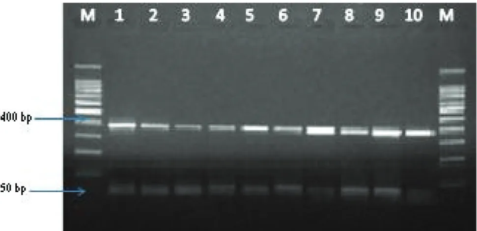
Figure 3. Agarose gel electrophoresis showing positive amplification of 375 base fragments specific for vanA of VRE. Faecium strains from clinical specimens. Lane M: Size marker (50-bp DNA ladder); lanes 1-10: VRE. Faecium strains
Şekil 3. Agaroz jel elektroforezi ile klinik kaynaklı VRE. faecium türlerinin 375 bp’de vanA gen fragmentleri görülmektedir. M: Moleküler belirteç (50-bp DNA ladder); 1-10: VRE. faecium türleri.
Literature suggests that E. faecalis and E. faecium have significant differences in terms of incidence of virulence factors as follows: (i) Franz et al., (11) reported that 10.4% of E. faecium strains were positive for one or more virulence determinants compared to 78.7% of E.
faecalis strains, (ii) E. faecalis strains tested by Eaton
and Gasson (10) possessed multiple determinants (between 6 and 11) and (iii) Vankerckhoven et al., (26) have concluded that E. faecium strains were generally free of virulence factors. By contrast, in our study, E.
faecium strains had significantly more virulence
determinants than E. faecalis strains and possessed multiple determinants (between 3 and 4 virulence genes), and were found with lower incidence (between 2, 3, and only one strain from Urfa cheese included 4 virulence genes) of multiple virulence determinants (p<0.05).
After multiplex-PCR amplification of selected virulence genes among 51 food and 50 human originated strains, possessing suspected virulence genes (esp, hyl,
gelE, asa1 and cylA) were more frequent among human
clinical infection isolates than food and human intestinal isolates (Figure 1). There was a significant difference in occurrence of genes except for the cylA gene between human clinical, food and human intestinal isolates (p<0.001). Eaton and Gasson, (10) have reported that enterococci isolated from food sources have fewer virulence genes than those of clinical sources in accordance with our results.
Aggregation Substance (AS) is a pheromone-inducible surface protein of E. faecalis promoting the mating aggregate formation during bacterial conjugation (24). The asa1 gene was very common within E. durans (71.4%) and 42.9% of E. casseliflavus isolates from fruit and vegetables. In addition, 31.4% of E. faecium strains were collected from human samples. These results differ from those of previous studies reporting the absence or low rates of asa1 genes among E. faecium and E. faecalis strains (1, 17, and 26).
The enterococcal gene esp, encoding the high-molecular-weight surface protein, has been detected widely in enterococci isolated from patients with infections such as bacteremia or endocarditis isolates, yet is rare in stool isolates from healthy individuals (2, 21). The contribution of the surface protein esp in terms of colonization and persistence of E. faecalis in urinary tract infections has been shown in an animal model (22). The highest esp gene rate was detected in 15.7% (11/70) of E.
faecium strains. Our clinical E. faecium isolates, harbouring
high rates of the esp gene, were urinary tract infection agents, and the intestinal E. faecium isolates were found to have esp genes with very low rates (1.9%). These results support the findings in the aforementioned studies (2, 21).
Gelatinase activity, involved in the hydrolysis of gelatine, collagen and some other proteins, was most frequent in E. faecalis (83.3%) and E. faecium (31.4%)
strains in our study. gelE gene positive strains occurred with statistically significant differences among food and clinical isolates (p<0.005). In our study, gelE presence of
E. faecalis was found to be 100% among vegetable food
isolates. These results are higher than those reported by Abriouel et al. (1), reporting that gelE presence in E.
faecalis stains from vegetable food isolates was 76.5%;
in addition, E. faecium in which gelE was present was found to be higher in our clinical isolates (18/50; 72%). These findings differ from those of Abriouel et al. (1) who reported no relation of E. faecium isolates for the
gelE gene in their research.
Hyaluronidase, encoded by the hyl gene, acts on hyaluronic acid (hyaluronate, hyaluronan); is mainly a degradative enzyme associated with tissue damage (12). The hyl gene was identified within 58.3% of E. faecalis and 31.4% of E. faecium strains. This gene was identified in each of seven strains of E. casseliflavus and E. durans isolated from food. These values are higher than those mentioned in a study conducted by Vankerckhoven et al. (26) who found hyl gene presence in only one fecal isolates but none of probiotic and nonfecal isolates.
In the present study, gelE and hyl gene positivity was found significantly higher in E. faecalis isolates (P<0.001); and hyl, asa1 and gelE gene positivity was observed to be equal in E. faecium isolates. With the rates of 35.6 %, 34.6 %, respectively, asa1 and gelE were the most frequent virulence genes. According to a recent study in Turkey, Baylan et al. (4) reported that hyl gene positivity was found higher in E. faecium isolates (P<0.01). There were no enterococci strains including five virulence genes. Similarly, Baylan et al. (4) found no more than 4 virulence genes. Our values are higher than those of Baylan et al. (4) as we found that the hyl and
asa1 genes had the same ratio, and the esp gene was
found in half of the urinary infection strains (7/12). Furthermore, in Baylan et al.`s study (4) asa1 and esp were the most frequent virulence factors with rates of 26.7% and 25.6% respectively, while there were 32 (35.6%) strains without any of the investigated virulence factors. In our study, only two urine isolates (16%) had no virulence genes.
cylM, cylB and cylA genes are responsible for
modification, secretion and activation of cytolysin. In our study, the cylA gene was identified in only 14.3% (1/7) of E. durans, 8.6% (6/70) of E. faecium and 8.3% (1/12) of E. faecalis strains with no significant differences among the three groups. With a range of 9-44% among
E. faecalis isolates, our results are lower than those
obtained in previous studies (10, 17). E. casseliflavus, E.
gallinarum, and E. raffinosus from fruit and vegetables
isolates were also found free from the cylA gene in the present study. These results are similar to previous reports of Abriouel et al. (1) regarding the absence of the
cylA gene among vegetable food isolates. High rate of asa1 and cylA genes among our E. durans isolates is a
worrying output as there are some reports suggesting that the presence of virulence traits in species other than E.
faecalis and E. faecium is important for proper evaluation
of virulence evolution within the Enterococcus genus (16, 23). No significant difference was observed between
cylA gene positivity and β-hemolytic activity in our study.
Expression of gelatinase and cytolysin aggregation substances of enterococci may vary between in-vivo and in-vitro as it is well known that these genes have the ability to remain silent; thus, it is so important to take this into consideration during laboratory detection (8, 10, 23). In our study, two of the non-hemolytic E. faecium and one non-hemolytic E. faecalis strains had the “silent
cylA gene”, a finding parallel to a previous study
conducted by Eaton and Gasson (10), in which the researchers found two E. faecalis strains to have no hemolytic activity, yet carried cyl genes. Similar results were also observed by Barbosa et al. (3). This is possibly the first study reporting on “silent cylA genes’’ among Turkish food and clinical isolates. The occurrence of β- hemolysis was identified only within 42.9% of E. durans, 16.7% of E. faecalis and 11.4% of E. faecium strains with no significant difference among food and human isolates. These results are similar to those emerged in previous studies showing a high incidence of β-hemolysis among E. faecalis strains (10, 11).
In some studies, the presence of vanA, including one or more virulence genes in the same genome, have favored particular clusters of E. faecium in clinical origin (6). Similar results were observed in our clinical infection isolates as well (Table 2 and 3). The vancomycin resistance patterns of the two groups of strains were very different with a significantly higher rate of resistance among the enterococci of human origin (p<0.001). While only one Enterococcus casseliflavus from lettuce was resistant to vancomycin, yet carrying neither vanA nor
vanB genes, and with no occurrence of vancomycin
resistant strains among cheese, raw chicken meat, sucuk, fruit or vegetable isolates confirms previous studies with low rates of vancomycin resistance among food enterococci (1, 15, 17) suggesting that food enterococci cannot be considered the main potential sources for dissemination of vancomycin resistance. On the other hand, Finding of 7 (14%) clinical E. faecium strains with non vanA and non vanB genotype, but resistant to vancomycin show that some polymorphism might occur in detection of resistance genes or other resistant genes (vanC, vanD,...etc.) might also be important in the vanA phenotype of VRE isolates in present study.
The relationship between antibiotic resistance and virulence genes of enterococci may vary from country to country. In a study by Vankerchoven et al. (26) covering a large geographical area, Italy and England, esp gene positivity was found to be 68% among vancomycin sensitive (VS) E. faecium isolates and 91% among vancomycin resistant (VR) E. faecium isolates (p<0.05) in Italy, and in England, hyl gene positivity was found to
be 29% in VS E. faecium isolates and 71% in VR E.
faecium isolates (p<0.05). In this study, we found esp
gene positivity to be 15.7% in VR E. faecium isolates and hyl gene positivity, 28.6% among VR E. faecium isolates, and 2.9% among vancomycin sensitive and intermediate E. faecium isolates (Table 4; p=0.03, p=0.02 respectively). Similarly, among all enterococci isolates,
esp gene positive isolates were found to be more resistant
to vancomycin than sensitive and intermediate isolates (Table 5; p= 0.02). A significant correlation was also observed within vancomycin resistance and virulence genes of esp and hyl in clinical E. faecium strains. These results correlate with those obtained by Vankerchoven et al. (26), yet differ from those of Baylan et al (4), who found a correlation with resistance to different antibiotics and their asa1, esp virulence genes of enterococci, and no correlation between vancomycin resistance and virulence factors of clinical enterococci.
It is interesting to note that the co-existence of virulence genes such as esp+hyl and gelE+esp+asa1 was found to be a contrubiting factor to vancomycin resistance in our study. There was also a correlation between the number of virulence genes and vancomycin resistance (p<0.05).
This study showed that foodborne enterococci can harbor potential virulence genes but not vanA and vanB resistance genes, and may rarely be resistant to vancomycin. While with variable frequencies, virulence determinants, mainly associated with species of E. faecium, E. faecalis and E. durans, were more common among cheese, sucuk and raw chicken meat isolates than those among human isolates. E. faecalis, E. faecium and E. durans strains from food may be potential sources for dissemination of virulence genes (especially asa1, gelE and hyl). A positive correlation was found between the number of virulence genes and vancomycin resistance as well as beta hemolytic activity. Positive correlation of vancomycin resistance and esp gene positivity observed in all enterococci and esp, hyl gene positivity in E. faecium (especially clinical) isolates yield significant data in this region of Turkey. This is possibly the first study to investigate the relationship between vancomycin resistance and virulence factors among Enterococcus spp. isolates originated from various food and clinical samples in Turkey. Further studies of more enterococci isolates of food and human origin with more virulence genes and antibiotics covering wider geographical areas are needed to better aid the food industry and contribute to clinical microbiology.
Acknowledgments
This study was supported by Çukurova University
Research Fund (BAP Project number: ZF2010D12). Some part of this study was presented as a poster with “l12” code in European Biotechnology Congress 2011 (Istanbul, Turkey). Some part of this study was presented as a poster with “abstract cod.: 8” in International
Conference on Environmental, Industrial and Applied Microbiology- BioMicroWorld2013, 257p/734p, Madrid (Spain), 2-4 October.
References
1. Abriouel H, Omar NB, Molinos AC, Lopez RL, Grande MJ, Martinez-Viedma P, Ortega E, Canamero A (2008): Comperative analysis of genetic diversity and
incidance of virulance factors and antibiotic resistance among enterococcal populations from raw fruit and vagetable foods, water and soil, and clinical samples. Int J
Food Microbiol, 123, 38-49.
2. Archimbaud C, Shankar N, Forestier C, Baghdayan A, Gilmore MS, Charbonne F (2002): In vitro adhesive
properties and virulence factors of Enterococcus faecalis strains. Res Microbiol, 53, 75-80.
3. Barbosa J, Gibbs PA, Teixeira P (2010): Virulence
factors among enterococci isolated from traditional fermented meat products produced in North of Portugal.
Food Control, 21, 651-656.
4. Baylan O, Nazik H, Bektore B, Citil BE, Turan D, Ongen B, Ozyurt M, Acıkel CH, Haznedaroglu T (2001): The relationship between antibiotic resistance and
virulence factors in urinary enterococcus isolates.
Mikrobiyol Bul, 453, 430-445.
5. Ben Omar N, Castro A, Lucas R., Abrıuel H, Yousıf NMK, Franz CMAP, Holzapfel WH, Perez-Pulido R, Martinez-Canamero M, Galvez A (2004): Functional
and safety aspects of enterococci isolated from different spanish foods. Syst Appl Microbiol, 27, 118-130.
6. Biavasco F, Foglia G, Paoletti C, Zandri, G, Magi G, Guaglianone E., Sundsfjord A, Pruzzo C, Donelli G, Facinelli, B (2007): VanA-Type Enterococci from humans,
animals, and food: species distribution, population structure, n1546 typing and location, and virulence determinants. Appl Environ Microbiol, 7310, 3307–3319.
7. CLSI (2011): Performance standards for antimicrobial
susceptibility testing, 21th informationel supplement, M
100-S21. Wayne Pa.
8. Creti R, Imperi M, Bertuccini L, Faretti F, Orefici G, Di Rosa R (2004): Survey of virulence determinants
among Enterococcus faecalis isolated from different sources. J Med Microbiol, 53, 13–20.
9. De Fátima Silva Lopes M, Ribeiro T, Abrantes M, Figueiredo Marques JJ, Tenreiro R, Crespo MTB (2005): Antimicrobial resistance profiles of dairy and
clinical isolates and type strains of enterococci. Int J Food
Microbiol, 103, 191–198.
10. Eaton JT, Gasson MJ (2001): Molecular screening of
Enterococcus virulence determinants and potential for genetic exchange between food and medical isolates. Appl
Environ Microbiol, 67, 1628–1635.
11. Franz CMAP, Muscholl-Silberhorn AB, Yousif NMK, Vancanneyt M, Swings J, Holzapfel WH (2001):
Incidence of virulence factors and antibiotic resistance among enterococci isolated from food. Appl Environ
Microbiol, 67, 4385–4389.
12. Franz CMAP, Huch M, Abrıouel H, Holzapfel W, Galvez A (2011): Enterococci as probiotics and their implications
in food safety. Int J Food Microbiol, 151, 128-140.
13. Foulquie Moreno MR, Sarantinopoulos P, Tsakalidou E, De Vuyst L (2006): The role and application of enterococci
in food and health. Int J Food Microbiol, 106, 1–24.
14. Liu D (2011). Enterococcus. In Semedo-Lemsaddec, Alves, Tenreiro and Crespo. Moleculer detection of human bacterial
pathogens. p:239. IN: Tylor &Francis Group LLC, NW.
15. Mannu L, Paba A, Daga E, Comunian R, Zanetti S, Dupre` I (2003): Comparison of the incidence of virulence
determinants and antibiotic resistance between Enterococcus faecium strains of dairy, animal and clinical origin. Int J
Food Microbiol, 88, 291-304.
16. Martin B, Garriga M, Hugas M, Aymerich T (2005):
Genetic diversity and safety aspects of enterococci from slightly fermented sausages. J Appl Microbiol, 98, 1177-1190.
17. McGowan-Spicer LL, Fedorka-Cray PJ, Frye JG, Meinersmann RJ, Barrett JB, Jackson CR (2008):
Antimicrobial resistance and virulence of Enterococcus faecalis isolated from retail food. J Food Prot, 71, 760-769.
18. Ozmen TS, Çelebi Keskin A, Açık L, Temiz A (2010):
Virulence genes, antibiotic resistance and plasmid profiles of Enterococcus faecalis and Enterococcus faecium from naturally fermented Turkish foods. J Appl Microbiol, 109,
1084-1092.
19. Psoni L, Kotzamanides C, Andrighetto C, Lombardi A, Tzanetakis N, Litopoulou-Tzanetaki E (2006): Genotypic
and phenotypic heterogeneity in Enterococcus isolates from Batzos, a raw goat milk cheese. Int J Food Microbiol,
109, 109-120.
20. Salem-Bekhlit MM, Moussa IMI, Muharram MM, Alanazy FK, Hefni HM (2012): Prevalence and
antimicrobial resistance pattern of multidrug-resistent enterococci isolated from clinical specimens. Int J Med
Microbiol, 301, 44-51.
21. Shankar V, Baghdayan AS, Huycke MM, Lindahl G, Gilmore MS (1999): Infection-derived Enterococcus
faecalis strains are enriched in esp, a gene encoding a novel surface protein. Infect Immun, 69, 4366-4372.
22. Shankar N, Lockatell CV, Baghdayan AS, Drachenberg C, Gilmore MS, Johnson DE (2001): Role of Enterococcus
faecalis surface protein esp in the pathogenesis of ascending urinary tract infection. Infect Immun, 69, 4366-4372.
23. Semedo T, Almeida Santos M, Martins P, Silva Lopes MF, Figueiredo Marques JJ, Tenreiro R (2003): Comparative
study using type strains and clinical and food isolates to examine haemolytic activity and occurrence of the cyl operon in enterococci. J Clin Microbiol, 41, 2569–2576.
24. Sussmuth SD, Muscholl-Silberhorn A, Wirth R, Susa M, Marre R, Rodzinski E (2000): Aggregation substance
promotes adherence, phagocytosis and intracellular survival of Enterococcus faecalis within human macrophages and suppresses respiratory burst. Infect Immun, 68, 4900-4906.
25. Vankerckhoven V, Van Autgaerden T, Vael C, Lammens C, Chapelle S, Rossi R, Jabes D, Goossens H (2004):
Development of a multiplex PCR for the detection of asa1, gelE, cylA, esp and hyl genes in enterococci and survey for virulence determinants among European hospital isolates of Enterococcus faecium. J Clin Microbiol, 42, 4473-4479.
26. Vankerckhoven V, Huys G, Vancanneyt M (2008):
Genotypic diversity, antimicrobial resistance, and virulence factors of human isolates and probiotic cultures constituting two intraspecific groups of Enterococcus faecium isolates. Appl Environ Microbiol, 74, 4247-4255. Geliş tarihi: 28.06.2013 / Kabul tarihi: 20.01.2014
Address for correspondence:
Mevhibe Terkuran (PhD) Department of Food Engineering, Faculty of Agriculture,
Cukurova University, Saricam, 01330, Adana,Turkey