Role of nitric oxide and oxidative stress in pathophysiology of liver
injury in streptozotocin-induced type 1 diabetic rats
Güngör Çağdaş DİNÇEL
1, Serkan YILDIRIM
2, Oğuz KUL
31Aksaray University, Eskil Vocational School, Laboratory and Veterinary Health Program, Aksaray; 2Atatürk University, Faculty of
Veterinary Medicine, Department of Pathology, Erzurum; 3Kırıkkale University, Faculty of Veterinary Medicine, Department of
Pathology, Kırıkkale, Turkey.
Summary: Type 1 diabetes mellitus (T1DM) is a severe chronic metabolic disorder characterized by hyperglycaemia because of the alterations in insulin secretion or its action. It is previously shown that hyperglycemia related oxidative stress (OS) and excessive nitric oxide (NO) production may cause severe complications in kidney and brain. In this report, it is aimed to investigate the cytotoxic effects of NO and to evaluate possible interaction with T1DM related hepatopathology. Expression levels of 8-hydroxy-2'-deoxyguanosine (8-OHdG), endothelial nitric oxide synthase (eNOS), inducible nitric oxide synthase (iNOS), Cu/Zn superoxide dismutase (SOD1) and glutathione reductase (GR) were examined by immunohistochemistry in liver tissues. Results of the study revealed that levels of 8-OHdG (P<0.001), eNOS (P<0.001), eNOS (P<0.001), SOD1 (P<0.001) and GR (P<0.001) were remarkably higher in liver with T1DM than control. The most prominent finding of this study is the increased levels of 8-OHdG in the mostly hepatocyte cytoplasm. These results suggest an involvement of oxidative DNA damage and OS might play a pivotal role on hepatic degeneration and this is a novel insight of pathogenesis on the explanation of cellular processes in streptozotocin (STZ)-induced type 1 diabetic rats’liver. Furthermore, these results also suggested that STZ-induced hepatic pathology might have been augmented by the contribution of high NO expression mediated OS. Taken together, the results suggest NO related hepatic inflammation and degeneration closely implicated in pathophysiology of T1DM. The results also clearly indicated that OS plays an important role on hepatic pathology and OS biomarkers might indicate the progress of the T1DM.
Keywords: Hepatopathology, nitric oxide, oxidative stress, type 1 diabetes mellitus.
Streptozotosin ile tip 1 diyabet oluşturulan sıçanların karaciğerlerinde meydana gelen hasarlarda
nitrik oksit ve oksidatif stresin patofizyolojideki rolleri
Özet: Tip 1 diyabet (T1D), insülinin salgı veya görevlerinde anormalliklerle karakterize kronik metabolik bir hastalıktır. Daha önceleri diyabetik hayvanlarda, hiperglisemi ile tetiklenen oksidatif stres (OS) ve nitrik oksit (NO) seviyelerindeki patolojik yükselmelerin böbrek ve beyinde şiddetli komplikasyonlara neden olduğunu gösterdik. Bu çalışma NO’in sitotoksik etkisinin ve OS’in T1D ile ilişkili hepatopatolojilerde herhangi bir ilişkinin olup olmadığını tanımlamak için tasarlanmıştır. Bu amaçla 8-hidroksi-2'-deoxyguanosine (8-OHdG), endotelyal nitrik oksit sentaz (eNOS), uyarılabilir nitrik oksit sentaz (iNOS), Cu/Zn süperoksit dismutaz (SOD1) ve glutatyon redüktaz (GR) immunoreaktiviteleri karaciğer dokusunda araştırıldı. Çalışmada, 8-OHdG (P<0.001), eNOS (P<0.001), eNOS (P<0.001), SOD1 (P<0.001) ve GR (P<0.001) immunoreaktivitelerinin T1D’li hayvanlara ait karaciğer dokularında, sağlıklı kontrol gruplara göre ciddi anlamda bir artışın olduğu tespit edildi. Bu çalışmada en önemli bulgu, 8-OHdG sunumlarının genelde sitoplazmada olduğudur. Bu sonuçlar STZ ile indüklenen karaciğer dejenerasyonlarında OS’in ana kaynağının patolojik düzeylerde üretilen NO olduğunu da gösterdi. Ayrıca, oksidatif DNA hasarı ve oksidatif stresin diyabete bağlı karaciğer dejenerasyonlarında çok önemli bir rol aldığı ve hücresel mekanizmaların ortaya konmasında anahtar görevi üstleneceği gösterildi. Bu bulgular birlikte ele alındığında, T1D’le ilişkili karaciğer patolojileri NO aracılı karaciğer yangıları ve dejenerasyonların önemli bir faktör olduğu görülmektedir. Ayrıca, bu çalışma OS’in karaciğer patofizyolojisinde görev aldığını ve OS belirleyicilerinin hastalığın takibinde önemli görevler alabileceğini gösterdi.
Anahtar sözcükler: Hepatopatoloji, nitrik oksit, oksidatif stres, tip 1 diyabet.
Introduction
Diabetes mellitus (DM) is a disease that holds an important place among the metabolic diseases, damaging and/or causing dysfunctions in many organs including the liver (20, 23, 31). Diabetic hepatopathy is among the most
serious diabetic complications. Although diabetic ketosis and hepatic lipidosis has been widely studied for decades, the molecular mechanisms and pathogenesis of hepatocellular degeneration are still not fully clarified.
II) and endothelial NOS (eNOS, type III) (25, 35). NO-mediated cytotoxicity was first shown on macrophages and subsequently the synthesis of high level of NO from L-arginine was shown to induce apoptotic cell death (3, 4, 11, 13, 34). Liver degenerations in sepsis, hepatitis and ischemia/reperfusion show frequently co-association with NO production at pathological levels (6, 22). Thus, it is quite essential to maintain NO levels of the liver within the normal physiological range.
Oxidative stress (OS) is triggered by excessive production/generation of reactive nitrogen species (RNS) and highly reactive oxygen species (ROS) and/or poor rate of removal of free radicals due to the alterations in the functioning of antioxidant enzymes (19, 32). The antioxidant systems are strengthened by enzymes such as superoxide dismutase, glutathione reductase (GR), catalase and glutathione peroxidase (38, 43, 47). All cellular molecules such as lipids, proteins and nucleic acids are very sensitive to excessively produced ROS and RNS (19).
Oxidative stress was shown to play a key role in the pathogenesis of complications that occur in DM (17, 18, 33). It was also described that in diabetic liver, the levels and activities of antioxidant enzymes such as SOD, glutathione and catalase significantly decrease (28, 36, 40). Clinical and experimental studies showed that the DM induced liver damage becomes even more effective and further exacerbated in long-term periods.
The first objective of this study is to determine whether there is a link between NO production and the severity of the hepatic degenerations seen in the early stages of diabetes in the liver of rats with type 1 diabetes mellitus. The second aim of the study is to demonstrate what kind of roles antioxidant enzymes play in this process.
Materials and Methods
Ethics statement: The experimental protocol was
approved by the Committee on the Ethics of Animal Experiments at Atatürk University (Permit Number: 46-02.03/2014) and performed accordance with National Centre for the Replacement, Refinement, and Reduction of Animals in Research.
Induction of STZ model of diabetes: 20 male Wistar
albino rats weighing 250-300 were randomly allotted to two experimental groups (𝑛 = 10 per group). Previously demonstrated that STZ destroys pancreatic beta cells and is a model of type 1 diabetes mellitus (45). Type 1 diabetes was induced in the rats by a single intraperitoneal injection of streptozotocin (STZ) (65 mg/kg body weight) dissolved in 0.1 mM sodium citrate, pH 4.5.
Accordingly, performing the routine tissue preparation procedures and paraffin sections were then cut and mounted on glass slides. Hematoyxlin-Eosin (H&E) and immunohistochemical tests were performed, and they were analyzed using a trinocular light microscope (Olympus BX51 and DP25 digital camera).
Antibodies: Commercial anti-mouse antibodies
against 8-OHdG (Santa Cruz Biotechnology, USA, 1/100), eNOS (Thermo Scientific, USA, ready to use), iNOS (Thermo Scientific, USA, ready to use), GR (Santa Cruz Biotechnology, USA, 1/100), SOD1 (Santa Cruz Biotechnology, USA, 1/100) were used in the present study.
Immunoperoxidase examination: All steps were
carried out following the procedure described by Dincel and Kul (13). Tissue sections were incubated with the primary antibody (8-OHdG, eNOS, iNOS, SOD1 and GR) for 60 min. Finally, sections were incubated in aminoethyl carbazole chromogen (Thermo Scientific, USA) for 5-10 min to induce the color reaction. Mayer’s hematoxylin was applied as a counterstain for 30 sec.
Histomorphometric analysis and statistics: The
density of positive staining was measured using a computerized image system composed of a Leica CCD camera DFC420 (Leica Microsystems Imaging Solutions, Ltd., Cambridge, UK), connected to a Lecia DM4000 B microscope (Leica Microsystems Imaging Solutions, Ltd.) and was used according to the procedure described by Dincel and Atmaca (10). Statistical analysis of immunohistochemical results were compared between groups using the non-parametric data, Mann-Whitney U-test. The data were presented as means ± SD. A P value of <0.05 was considered significant.
Results
Histopathologic findings: Macroscopic findings
have not been determined in experimental animals. Histopathologic examination of rat livers from healthy control group showed normal hepatic lobular architecture and pancreas to have a normal histological structure. The most prominent histopathologic findings in the livers of STZ-treated rats were severe edema and hemorrhage. In addition, necrotic/degenerative hepatocytes were evident in the periacinary localization, and there was a mononuclear inflammatory cell infiltration (Figures 1A, B). In diabetic group, STZ caused severe degenerative changes in the islets of Langerhans. This degenerative changes were seen mainly at the central area.
Immunoperoxidase findings: In this study,
eNOS-iNOS (Figure 2), GR-SOD1 (Figure 3) and 8-OHdG expressions (Figure 4) in the liver were higher in diabetic
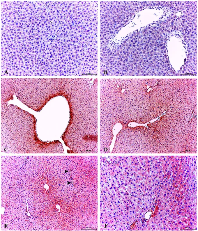
Figure 1. Necrotic/degenerative hepatocytes in centrilobular areas. H&E, Bar, 100 μm (A). Severe edema and necrotic/degenerative hepatocytes in STZ-treated group. H&E, Bar, 100 μm (B).
Şekil 1. Sentrilobüler bölgede nekrotik/dejeneratif hepatositler. H&E, Bar, 100 μm (A). Şiddetli ödem ve nekrotik/dejeneratif hepatositler (Diyabetik grup). H&E, Bar, 100 μm (B).
Figure 2. Comparison of eNOS and iNOS immunopositivity. Statistical difference is indicated as letters. "a" represent values statistically higher than control group. Statistical analysis was performed according to Mann-Whitney U-test. The values represent means ± S.D.
P < 0.05 was considered statistically significant.
Şekil 2. eNOS ve iNOS immünopozitifliklerin karşılaştırılması. İstatistiksel farklar ‘a’ ile gösterilmiştir. ‘a’ istatistiksel olarak kontrol grubundan yüksek olduğunu vurgulamaktadır. Analizler Mann-Whitney U-test ile yapıldı. P < 0.05 istatistiksel olarak anlamlı kabul edildi.
Figure 3. Comparison of GR and SOD1 immunopositivity. Statistical difference is indicated as letters. "a" represent values statistically higher than control group. Statistical analysis was performed according to Mann-Whitney U-test. The values represent means ± S.D. P < 0.05 was considered statistically significant.
Şekil 3. GR ve SOD1 immünopozitifliklerin karşılaştırılması. İstatistiksel farklar ‘a’ ile gösterilmiştir. ‘a’ istatistiksel olarak kontrol grubundan yüksek olduğunu vurgulamaktadır. Analizler Mann-Whitney U-test ile yapıldı. P < 0.05 istatistiksel olarak anlamlı kabul edildi.
Figure 4. Comparison of 8-OHdG immunopositivity. Statistical difference is indicated as letters. "a" represent values statistically higher than control group. Statistical analysis was performed according to Mann-Whitney U-test. The values represent means ± S.D. P < 0.05 was considered statistically significant.
Şekil 4. 8-OHdG immünopozitifliklerin karşılaştırılması. İstatistiksel farklar ‘a’ ile gösterilmiştir. ‘a’ istatistiksel olarak kontrol grubundan yüksek olduğunu vurgulamaktadır. Analizler Mann-Whitney U-test ile yapıldı. P < 0.05 istatistiksel olarak anlamlı kabul edildi.
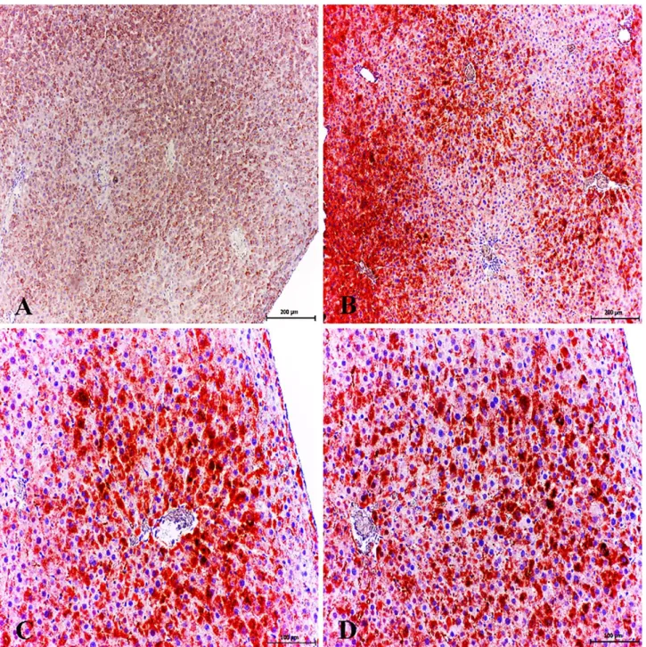
Figure 5. Healthy control group; low expression of 8-OHdG in hepatocytes, Kupffer cells and and pericentral area. ABC technique (anti-8-OHdG), Mayer's hematoxylin counterstain, Bar, 100 μm (A, B). Increased cytoplasmic expression of 8-OHdG in pericentral area and necrotic/degenerative hepatocytes. Diabetic group, ABC technique (anti-8-OHdG), Mayer's hematoxylin counterstain, Bar, 200 μm (C). Increased expression of 8-OHdG in pericentral area, portal vein, hepatic arters and bile duct. Diabetic group, ABC technique (anti-8-OHdG), Mayer's hematoxylin counterstain, Bar, 200 μm (D). Increased expression of 8-OHdG in pericentral area, portal vein (arrowheads), hepatic arteries and bile duct. Diabetic group, ABC technique (anti-8-OHdG), Mayer's hematoxylin counterstain, Bar, 200 μm (E). Increased cytoplasmic expression of 8-OHdG in some periportal hepatocytes, portal vein and bile duct. Diabetic group, ABC technique (anti-8-OHdG), Mayer's hematoxylin counterstain, Bar, 100 μm (F).
Şekil 5. Sağlıklı control grup; hepatositlerde, Kupffer hücrelerinde perisentral bölgede zayıf 8-OHdG sunumları. ABC teknik (anti-8-OHdG), Mayer's hematoksilin arka plan boyaması, Bar, 100 μm (A, B). Nekrotik/dejeneratif hepatositler ve perisentral bölgede şiddetli sitoplazmik 8-OHdG sunumları. Diyabetik grup, ABC teknik (anti-8-OHdG), Mayer's hematoksilin arka plan boyaması, Bar, 200 μm (C). Perisentral bölge, portal ven, hepatik arter ve safra kanalında şiddetli 8-OHdG sunumları. Diyabetik grup, ABC teknik (anti-8-OHdG), Mayer's hematoksilin arka plan boyaması, Bar, 200 μm (D). Perisentral bölge, portal ven (ok başları), hepatik arter ve safra kanalında şiddetli 8-OHdG sunumları. Diyabetik grup, ABC teknik (anti-8-OHdG), Mayer's hematoksilin arka plan boyaması, Bar, 200 μm (E). Bazı periportal hepatositlerde, portal ven ve safra kanalında şiddetli sitoplazmik 8-OHdG sunumları. Diyabetik grup, ABC teknik (anti-8-OHdG), Mayer's hematoksilin arka plan boyaması, Bar, 100 μm (F).
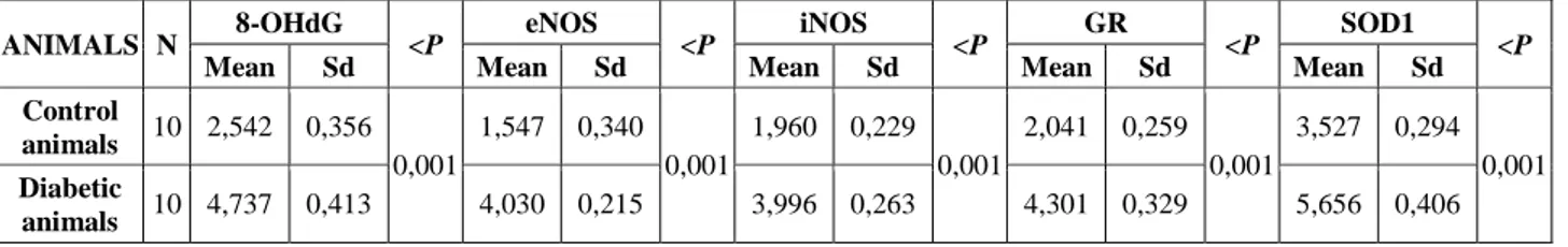
Table 1. Immunoperoxidase technique results and statistical data.
Tablo 1. İmmunoperoksidaz boyama tekniği sonuçları ve istatistiksel veriler.
ANIMALS N 8-OHdG <P eNOS <P iNOS <P GR <P SOD1 <P
Mean Sd Mean Sd Mean Sd Mean Sd Mean Sd
Control animals 10 2,542 0,356 0,001 1,547 0,340 0,001 1,960 0,229 0,001 2,041 0,259 0,001 3,527 0,294 0,001 Diabetic animals 10 4,737 0,413 4,030 0,215 3,996 0,263 4,301 0,329 5,656 0,406
hepatopathy group than in healthy control animals (P<0.001). Statistical analysis of the data on eNOS, iNOS, GR, SOD1 and 8-OHdG expressions in the liver, measured by immunostaining in all the groups, are listed in Table 1.
8-hydroxy-2'-deoxyguanosine (8-OHdG) expressions:
Weak immunoreactivity for 8-hydroxy-2'-deoxyguanosine (8-OHdG) was observed in some hepatocytes (Figure 5A) and no immunostaining around the periacinary areas were observed (Figure 5B) in healthy control group.
Increased 8-OHdG expression was observed only in the cytoplasms of the hepatocytes and in Kupffer cells (Figures 5C, D). In addition, cytoplasmic immunoreaction was localized in some degenerative/necrotic hepatocytes. The most conspicuous finding of the present study was that 8-OHdG expression was markedly increased periacinary area, bile duct and endothelial cells (Figures 5E, F).
8-OHdG expressions in the liver were statistically higher in diabetic hepatopathy group when compared with healthy control animals (P<0.001) (Figure 4).
Endothelial nitric oxide synthase (eNOS) and inducible nitric oxide synthase (iNOS) expressions: Fairly
weak immunoreactivities for eNOS (Fig. 6A) and iNOS (Figure 7A) were observed in hepatocytes in healthy control group.
Immunohistochemical staining demonstrating that iNOS and eNOS immunoreactivity was dramatically intense in the hepatocytes and sinusoidal endothelial cells. Another conspicuous finding of the present study was that eNOS expression markedly increased in veins in hepatic portal area, degenerative/necrotic hepatocyte and endothelial cells (Figures 6B, C, D) and increased iNOS expression in periacinar area. In addition, iNOS expression was found in endothelial cells (Figure 7B) and expression of eNOS was significantly increased in sinusoidal endothelia and especially in degenerative hepatocytes (Figures 6B, C).
eNOS and iNOS expressions in the liver were statistically higher in diabetic hepatopathy group when compared with healthy control animals (P<0.001) (Figure 2).
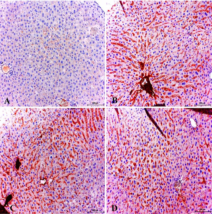
Cu/Zn superoxide dismutases (SOD1) and glutathione reductase (GR) expressions: Fairly weak
immunoreactivity for SOD1 and GR expressions were
observed in hepatocytes and sinusoidal area (Figures 8A, 9A) in healthy control group.
Increased SOD1 expression was observed periacinar area but not periportal area. Moreover, strong SOD1 expression was detected in hepatocyte and endothelia (Figures 8 B, C, D). Another conspicuous finding of the present study was that GR expression markedly increased in sinusoidal endothelial cells and central vein. Importantly, soluble GR antigen staining was observed in the lumens of central vein (Figures 9B, C, D).
SOD1 and GR expressions in the liver were statistically higher in diabetic hepatopathy group when compared with healthy control animals (P<0.001) (Figure 3).
Discussion and Conclusion
Previous studies shown that in diabetic animals, hyperglycemia related OS and NO production at pathological levels cause severe complications in the kidney and brain tissues (8, 9). In this study we have first shown that in the early stages of diabetes an active antioxidant enzyme system steps in. We suggest that this is caused by increasing levels of H2O2 and superoxide
radicals due to hyperglycaemia. Secondarily, we have shown that iNOS and eNOS are responsible for NO production at pathological levels versus healthy control group. We suggest that NO production has an essential level of immunopathological role, providing the largest contribution to the hyperglycaemia-mediated tissue damage in the early stages of diabetes. Finally, we have shown the DNA damage caused by OS in the liver tissue. We suggest that the DNA damage that we observed is caused largely by RNS.
Oxidative stress was described to undertake an important role in the pathogenesis of subchronic and/or chronic diabetic hepatopathology (28, 33, 36, 40). The reason of the pathology related to OS that occur in the liver was described to be due to the decrease in the activities of antioxidant enzymes. In fact, it was shown that activities of antioxidant enzymes are much lower than the physiological limit in diabetic hepatopathology (5, 14, 39, 44). The most striking finding of this study is the fact that there is a statistically significant increase in the antioxidant enzyme expression. We believe that the most
Figure 6. Healthy control group; very slight expression of eNOS in liver. ABC technique (anti-eNOS), Mayer's hematoxylin counterstain, Bar, 100 μm (A). Strong expression of eNOS in sinusoidal area and endothelial cells. Diabetic group, ABC technique (anti-eNOS), Mayer's hematoxylin counterstain, Bar, 100 μm (B). Strong expression of eNOS in portal veins (arrowheads) and sinusoidal endothelial cells. Diabetic group, ABC technique (anti-eNOS), Mayer's hematoxylin counterstain, Bar, 200 μm (C). Strong expression of eNOS in portal vein and sinusoidal endothelial cells. Diabetic group, ABC technique (anti-eNOS), Mayer's hematoxylin counterstain, Bar, 50 μm (D).
Şekil 6. Sağlıklı kontrol grup; karaciğerde zayıf eNOS sunumları. ABC teknik (anti-eNOS), Mayer's hematoksilin arka plan boyaması, Bar, 100 μm (A). Sinüzoidal bölgelerde ve endotel hücrelerde güçlü eNOS sunumları. Diyabetik grup, ABC teknik (anti-eNOS), Mayer's hematoksilin arka plan boyaması, Bar, 100 μm (B). Portal ven (ok başları) ve sinüzoidal endotel hücrelerde şiddetli eNOS sunumları. Diyabetik grup, ABC teknik (anti-eNOS), Mayer's hematoksilin arka plan boyaması, Bar, 200 μm (C). Portal ven ve sinüzoidal endotel hücrelerde şiddetli eNOS sunumları. Diyabetik grup, ABC teknik (anti-eNOS), Mayer's hematoksilin arka plan boyaması, Bar, 50 μm (D).
Figure 7. Healthy control group; very slight expression of iNOS liver. ABC technique (anti-iNOS), Mayer's hematoxylin counterstain, Bar, 100 μm (A). Strong expression of iNOS in endothelial cells (arrowheads) and pericentral area. Diabetic group, ABC technique (anti-iNOS), Mayer's hematoxylin counterstain, Bar, 200 μm (B). Strong expression of iNOS in sinusoidal endothelial cells and pericentral area. Diabetic group, ABC technique (anti-iNOS), Mayer's hematoxylin counterstain, Bar, 200 μm (C). Strong expression of iNOS in hepatocytes and Kupffer cells. Diabetic group, ABC technique (anti-iNOS), Mayer's hematoxylin counterstain, Bar, 50 μm (D).
Şekil 7. Sağlıklı kontrol grup; karaciğerde hafif iNOS sunumları. ABC teknik (anti-iNOS), Mayer's hematoksilin arka plan boyaması, Bar, 100 μm (A). Endotel hücrelerde (ok başları) ve perisentral bölgede şiddetli iNOS sunumları. Diyabetik grup, ABC teknik (anti-iNOS), Mayer's hematoksilin arka plan boyaması, Bar, 200 μm (B). Sinüzoidal endotel hücrelerde ve perisentral bölgede şiddetli iNOS sunumları. Diyabetik grup, ABC teknik (anti-iNOS), Mayer's hematoksilin arka plan boyaması, Bar, 200 μm (C). Hepatosit ve Kupffer hücrelerinde şiddetli iNOS sunumları. Diyabetik grup, ABC teknik (anti-iNOS), Mayer's hematoksilin arka plan boyaması, Bar, 100 μm (D).
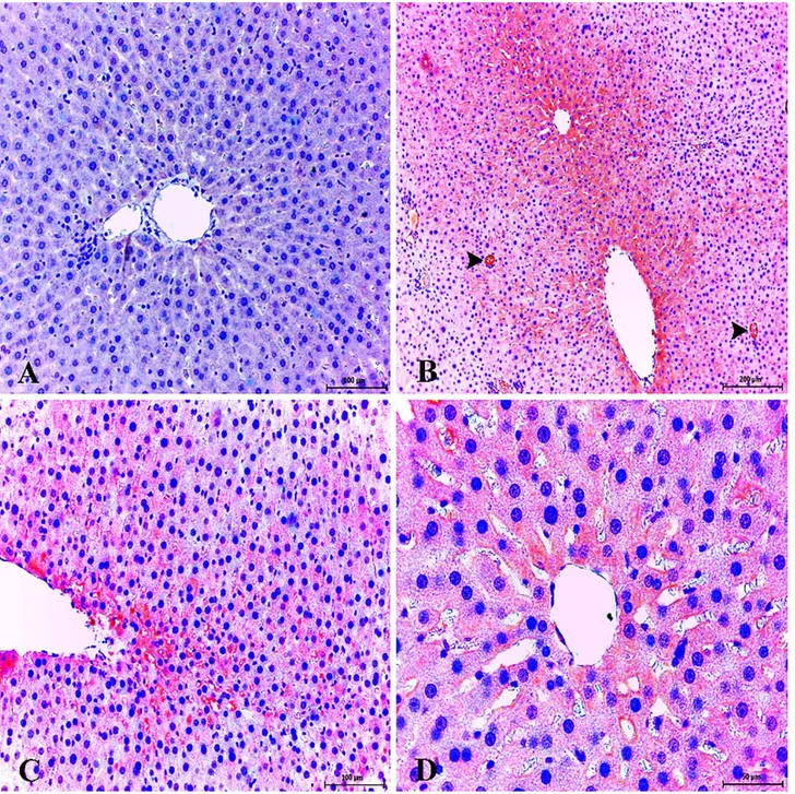
Figure 8. Healthy control group; mild/moderate expression of SOD1 in liver. ABC technique (anti-SOD1), Mayer's hematoxylin counterstain, Bar, 200 μm (A). Very strong expression of SOD1 in pericentral area. Diabetic group, ABC technique (anti-SOD1), Mayer's hematoxylin counterstain, Bar, 200 μm (B). Very strong expression of SOD1 in hepatocytes in pericentral area. Diabetic group, ABC technique (anti-SOD1), Mayer's hematoxylin counterstain, Bar, 100 μm (C). Very strong expression of SOD1 in central vein and hepatocytes. Diabetic group, ABC technique (anti-SOD1), Mayer's hematoxylin counterstain, Bar, 100 μm (D).
Şekil 8. Sağlıklı kontrol grup; karaciğerde hafif/orta şiddette SOD1 sunumları. ABC teknik (anti-SOD1), Mayer's hematoksilin arka plan boyaması, Bar, 200 μm (A). Perisentral bölgede şiddetli SOD1 sunumları. Diyabetik grup, ABC teknik (anti-SOD1), Mayer's hematoksilin arka plan boyaması, Bar, 200 μm (B). Perisentral hepatositlerde şiddetli SOD1 sunumları. Diyabetik grup, ABC teknik (anti-SOD1), Mayer's hematoksilin arka plan boyaması, Bar, 100 μm (C). Sentral ven ve hepatositlerde şiddetli SOD1 sunumları. Diyabetik grup, ABC teknik (anti-SOD1), Mayer's hematoksilin arka plan boyaması, Bar, 100 μm (D).
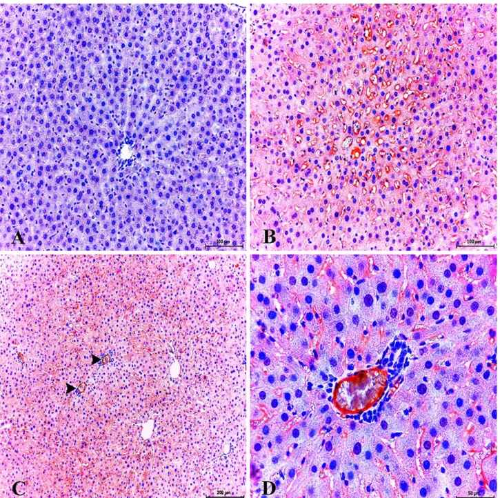
Figure 9. Healthy control group; very slight expression of GR in portal vein and sinusoidal area. ABC technique (anti-GR), Mayer's hematoxylin counterstain, Bar, 100 μm (A). Strong expression of GR in sinusoidal area and endothelial cells. Diabetic group, ABC technique (anti-GR), Mayer's hematoxylin counterstain, Bar, 100 μm (B). Strong expression of GR in sinusoidal area, endothelial cells and portal vein. Soluble GR antigen staining in the lumens of central vein. Diabetic group, ABC technique (anti-GR), Mayer's hematoxylin counterstain, Bar, 100 μm (C). Strong expression of GR in sinusoidal area, endothelial and Kupffer cells. Diabetic group, ABC technique (anti-GR), Mayer's hematoxylin counterstain, Bar, 100 μm (D).
Şekil 9. Sağlıklı kontrol grup; portal ven ve sinüzoidal bölgelerde zayıf GR sunumları. ABC teknik (anti-GR), Mayer's hematoksilin arka plan boyaması, Bar, 100 μm (A). Sinüzoidal bölge ve endotel hücrelerde şiddetli GR sunumları. Diyabetik grup, ABC teknik (anti-GR), Mayer's hematoksilin arka plan boyaması, Bar, 100 μm (B). Sinüzoidal bölge ve endotel hücrelerde şiddetli GR sunumları. Sentral ven lümeninde çözünmüş GR antijenleri. Diyabetik grup, ABC teknik (anti-GR), Mayer's hematoksilin arka plan boyaması, Bar, 100 μm (C). Sinüzoidal bölge, endotel ve Kupffer hücrelerinde şiddetli GR sunumları. Diyabetik grup, ABC teknik (anti-GR), Mayer's hematoksilin arka plan boyaması, Bar, 100 μm (B).
elimination of the free radicals that are excessively produced due to hyperglycemia during the early stages of diabetes. In later stages, it is seen that it may also result from the reduction of the activity of these enzymes. In short, this study confirms that there is an active antioxidant protection mechanism that starts functioning in the early stages but this process is affected adversely and in time the expression of these enzymes become much lower than the physiological limits. There is a significant increase in SOD activity as a compensation mechanism due to the excessive superoxide production (47). It was described that the increase in these radicals may cause problems in the vital functioning of the cells (24, 37). The significant increase in the expression of SOD indicates that there is a significant hyperglycemia dependent increase in the concentration of superoxide radicals in the liver. Therefore, this increase in SOD activity that is induced by the increased concentration of superoxide radicals may occur as a defensive mechanism. This suggests that it is very probable that in the later stages of diabetes depending on SOD depletion there may be a potential superoxide toxicity in liver. Glutathione reductase is involved in the activation of the glutathione peroxidase (GPx) and plays a role in the removal of H2O2 from the environment (30, 46).
Another important finding in this study is the detection of a significant increase in GR expression. This indicates that there is a severe increase in hyperglycemia induced H2O2
production in the liver. Moreover, it was previously shown that in the liver there is a lipid peroxidation dependent increase in GR expression, above the physiological range (26, 42). This shows that in subchronic and/or chronic diabetes mellitus, the increase in H2O2 and lipid
peroxidations due to the decreased GR activity has an important place among the causes of diabetic hepatopathology.
It was previously shown that RNS significantly contributes to the DNA damage and there is a strong positive correlation between them (29). Severe cytoplasmic 8-OHdG expression that was detected in this study indicates the presence of a significant oxidative DNA damage. The damage observed is in the mitochondrial and nuclear DNA. It was also previously shown that NO targets mitochondria and induces successive losses in mitochondrial membrane potential, causes cytochrome c release to the cytosol and thus triggers internal apoptotic pathways (2, 21, 34). It is quite clear that eNOS and iNOS dependent NO production at pathological levels is among the causes of oxidative DNA damage that occurs in the diabetic liver.
There are studies showing that NO inactivates some antioxidants like catalase, glutathione peroxidase and superoxide dismutase (1, 15). Thus, we think that eNOS
the chronic and subchronic stages of diabetes. Therefore, it is clear that revealing the roles of NO in the pathogenesis of diabetic hepatopathy and its molecular mechanisms will also help to develop therapeutic methods to reduce the severity of complications.
NO increases fluid shear stress and the largest contributor to this is provided by eNOS (7, 27). Along with this, there are also studies showing that OS increases fluid shear stress (16, 41). Oxidative DNA damage and severe expression of eNOS and iNOS in diabetic liver, that are among the most important findings of this study, indicate that there is severely triggered fluid shear stress. Therefore, in order to avoid fluid shear stress, anti-oxidant and NO inhibition therapies can be administered in combination. It is likely that it will play a key role in the prevention of hepatopathology that may occur.
This study showed that 8-OHdG, pivotal marker for measuring the effect of endogenous oxidative damage to DNA, was a good biomarker for risk assessment of this disease. In addition to this biomarker might be used to estimate the DNA damage in humans/animals after exposure to hyperglisemia. STZ-induced diabetic animal livers showed enhanced levels of eNOS and iNOS, and prolonged release of NO, which may contribute to hepatotoxicity and parenchyma degeneration.
The research on diabetes suggests that the diabetic hepatopathology that occur in the liver is an extremely complex process. In this study it was seen that the diabetes induced degeneration that occurs in liver does not only originate from hyperglycemia but also from NO that is produced above physiological limits by iNOS and eNOS in parenchymal and endothelial cells. It is also thought that there is a severe nitrositive and fluid shear stress related to the NO production in the liver. Thus, it is quite clear that nitrositive stress is also the reason behind the in the aetiology of oxidative DNA damage.
Acknowledgements
This work was funded and supported by the Scientific Research Projects Commission of the Gümüşhane University, Turkey (Project Code: 15.B0421.02.2). This study was presented as an oral presentation in the 32nd World Veterinary Congress, 13-17
September 2015, Istanbul.
Conflict of Interests
The authors report no conflict of interests.References
1. Asahi M, Fujii J, Suzuki K, et al. (1995): Inactivation of
glutathione peroxidase by nitric oxide. Implication for cytotoxicity. J Biol Chem, 270, 21035-21039.
2. Brookes PS, Salinas EP, Darley-Usmar K, et al. (2000):
Concentration-dependent effects of nitric oxide on mitochondrial permeability transition and cytochrome c release. J Biol Chem, 275, 20474-20479.
3. Brüne B (2003): Nitric oxide: NO apoptosis or turning it
ON? Cell Death Differ, 10, 864-869.
4. Brüne B, von Knethen A, Sandau KB (1999): Nitric oxide
(NO): An effector of apoptosis. Cell Death Differ, 10,
969-975.
5. Calabrese V, Cornelius C, Leso V et al. (2012): Oxidative
stress, glutathione status, sirtuin and cellular stress response in type 2 diabetes. Biochimica et Biophysica Acta,
5, 729-736.
6. Chen T, Zamora R, Zuckerbraun B, et al. (2003): Role of
nitric oxide in liver injury. Curr Mol Med, 6, 519-26.
7. Corson MA, James NL, Latta SE, et al. (1996):
Phosphorylation of endothelial nitric oxide synthase in response to fluid shear stress. Circ Res, 5, 984-991.
8. Dinçel GC, Yıldırım S (2016): Overexpression of ADAMTS-13 and neuronal nitric oxide synthase relates with neuropathology in streptozotocin-induced type 1 diabetic rats. Int J Clin Exp Pathol, 9, 4761-4778
9. Dinçel GC, Yıldırım S (2016): Increased expressions of
eNOS and iNOS correlate with apoptosis of diabetic nephropathy in streptozotocin-induced type 1 diabetic rats.
Kafkas Univ Vet Fak Derg, 22, 381-390
10. Dinçel GC, Atmaca HT (2015): Nitric oxide production
increases during Toxoplasma gondii encephalitis in mice.
Exp Parasitol, 156, 104-112.
11. Dinçel GC, Atmaca HT (2016): Increased expressions of
ADAMTS-13 and apoptosis contribute to neuropathology
during Toxoplasma gondii encephalitis in mice.
Neuropathology, 36, 211-226
12. Dinçel GC, Kul O (2015): Increased expressions of
ADAMTS-13, neuronal nitric oxide synthase, and neurofilament correlate with severity of neuropathology in border disease virus-infected small ruminants. PLoS One,
10, e0120005.
13. Dinçel GC, Kul O (2015): eNOS and iNOS trigger
apoptosis in the brains of sheep and goats naturally infected with the border disease virus. Histol Histopathol, 10,
1233-1242.
14. Dinçer Y, Akçay T, Alademir Z, et al. (2002): Assessment
of DNA base oxidation and glutathione level in patients with type 2 diabetes. Mutat Res, 505, 75-81.
15. Dobashi K, Pahan K, Chahal A, et al. (1997): Modulation
of endogenous antioxidant enzymes by nitric oxide in rat C6 glial cells. J Neurochem, 68, 1896-1903.
16. Edirisinghe I, Rahman I. (2010): Cigarette
smoke-mediated oxidative stress, shear stress, and endothelial dysfunction: Role of VEGFR2. Ann N Y Acad Sci, 1203,
66-72.
17. Frances DE, Ronco MT, Monti JA, et al. (2010):
Hyperglycemia induces apoptosis in rat liver through the increase of hydroxyl radical: New insights into the insulin effect. J Endocrinol, 205, 187-200.
18. Goth L (2000): Lipid and carbohydrate metabolism in
acatalasemia. Clin Chem, 46, 564-566.
19. Halliwell B (1999): Oxygen and nitrogen are
pro-carcinogens. Damage to DNA by reactive oxygen, chlorine and nitrogen species: Measurement, mechanism and the effects of nutrition. Mutat Res, 443, 37-52.
20. Harrison SA (2006): Liver disease in patients with diabetes
mellitus. J Clin Gastroenterol, 40, 68-76.
21. Heneka MT, Loschmann PA, Gleichmann M, et al. (1998): Induction of nitric oxide synthase and nitric
oxide-mediated apoptosis in neuronal PC12 cells after stimulation with tumor necrosis factor-alpha/lipopolysaccharide. J
Neurochem, 71, 88-94.
22. Hon WM, Lee KH, Khoo HE (2002): Nitric oxide in liver
diseases: Friend, foe, or just passerby? Ann N Y Acad Sci,
962, 275-95.
23. Hunt JV, Dean RT, Wolff SP (1988): Hydroxyl radical
production and autoxidative glycosylation. Glucose autoxidation as the cause of protein damage in the experimental glycation model of diabetes mellitus and ageing. Biochem J, 256, 205-212.
24. Hunt JV, Smith CCT, Wolff SP. (1990): Autoxidative
glycosylation and possible involvement of peroxides and free radicals in LDL modification by glucose. Diabetes, 39,
1420-1424.
25. Ignarro LJ, Buga GM, Wood KS, et al. (1987):
Endothelium-derived relaxing factor produced and released from artery and vein is nitric oxide. Proc Natl Acad Sci
USA, 84, 9265-9269.
26. Kakkar R, Mantha SV, Radhi J, et al. (1998): Increased
oxidative stress in rat liver and pancreas during progression of streptozotocin-induced diabetes. Clin Sci
(Lond), 6, 623-632.
27. Kemeny SF, Figueroa DS, Clyne AM (2013): Hypo- and
hyperglycemia impair endothelial cell actin alignment and nitric oxide synthase activation in response shear stress.
PLoS One, 8, e66176.
28. Kon K, Ikejima K, Okumura K, et al. (2010): Diabetic
KK-A(y) mice are highly susceptible to oxidative hepatocellular damage induced by acetaminophen. Am J
Physiol Gastrointest Liver Physiol, 299, 329-337.
29. Kumar A, Pant MC, Singh HS, et al. (2012):
Determinants of oxidative stress and DNA damage (8-OhdG) in squamous cell carcinoma of head and neck.
Indian J Cancer, 3, 309-315.
30. Liu J, Hinkhouse MM, Sun W, et al. (2004): Redox
regulation of pancreatic cancer cell growth: Role of glutathione peroxidase in the suppression of the malignant phenotype. Hum Gene Ther, 3, 239-250.
31. Manna P, Das J, Ghosh J, et al. (2010): Contribution of
type 1 diabetes to rat liver dysfunction and cellular damage via activation of NOS, PARP, IkappaBalpha/NF-kappaB,
MAPKs, and mitochondria-dependent pathways:
Prophylactic role of arjunolic acid. Free Radic Biol Med,
48, 1465-1484.
32. Mariani E, Polidori MC, Cherubini A, et al. (2005):
Oxidative stress in brain aging, neurodegenerative and vascular diseases: An overview. J Chromatogr B Analyt
Technol, 1, 65-75.
33. Maritim AC, Sanders RA, Watkins JB (2003): Diabetes,
oxidative stress, and antioxidants: A review. J Biochem Mol
Toxicol, 1, 24.
34. Moriya R, Uehara T, Nomura Y (2000): Mechanism of
nitric oxide induced apoptosis in human neuroblastoma SH-SY5Y cells. FEBS Lett, 484, 253-260.
35. Nathan C (1992): Nitric oxide as a secretory product of
37. Pacher P, Beckman JS, Liaudet L (2007): Nitric oxide
and peroxynitrite in health and disease. Physiol Rev, 87,
315-424.
38. Poulson HE, Prieme H, Loft S (1998): Role of oxidative
DNA damage in cancer initiation and promotion. Eur J
Cancer Prev, 1, 9-16.
39. Rahigude A, Bhutada P, Kaulaskar S, et al. (2012):
Participation of antioxidant and cholinergic system in protective effect of naringenin against type-2 diabetes-induced memory dysfunction in rats. Neuroscience, 226,
62-72.
40. Rains JL, Jain SK (2011): Oxidative stress, insulin
signaling, and diabetes. Free Radic Biol Med, 5, 567-575.
41. Rouhanizadeh M, Takabe W, Ai L, et al. (2008):
Monitoring oxidative stress in vascular endothelial cells in response to fluid shear stress: from biochemical analyses to micro- and nanotechnologies. Methods Enzymol, 441,
111-150.
42. Shull S, Heintz NH, Periasamy M, et al. (1991):
Differential regulation of antioxidant enzymes in response to oxidants. J Biol Chem, 36, 24398-24403.
43. Uday B, Dipak D, Ranajit BK (1990): Reactive oxygen
species: Oxidative damage and pathogenesis. Curr Sci, 77,
658-666.
45. Wang RN, Bouwens L, Klöppel G (1994): Beta-cell
proliferation in normal and streptozotocin-treated newborn rats: Site, dynamics and capacity. Diabetologia, 11,
1088-1096.
46. Weydert CJ, Cullen JJ (2010): Measurement of
superoxide dismutase, catalase and glutathione peroxidase in cultured cells and tissue. Nat Protoc, 1, 51-66.
47. Wiseman H, Halliwell (1996): Damage to DNA by reactive
oxygen and nitrogen species: Role in inflammatory disease and progression to cancer. Biochem J, 313, 17-29.
48. Yun-Zhong F, Sheng Y, Guoyao Wu (2002): Free
radicals, antioxidants, and nutrition. Nutrition, 18,
872-879.
Geliş tarihi: 26.06.2016 / Kabul tarihi: 22.12.2016
Address for correspondence:
Güngör Çağdaş DİNÇEL Aksaray University, Eskil Vocational School,
Laboratory and Veterinary Health Program. 68800 Eskil/Aksaray, Turkey.