Araştırma Makalesi / Research Article
Amaç: Pankreatikobilyer semptomları olan hastaların MRCP incelemelerinde insidental olarak
sapta-nan 24 jukstapapiller divertikülün (JPD) MRCP bulgularının sunulması ve bunların pankreatik kanal, safra yolları ve pankreas üzerine olan etkilerinin belirlenmesi.
Hastalar ve Yöntemler: 1-T MR ünitesinde elde edilen ve arşivlenmiş MRCP görüntüleri iki radyolog
tarafından tekrar değerlendirilmiştir. Saptanan JPD’lerin yerleşimi ve boyutları, safra kesesinde veya koledokta taş varlığı, safra kanallarında veya pankreatik kanalda dilatasyon ve ana safra kanalında JPD’ye bağlı olarak oluşan deviasyon not edilmiştir. İntrahepatik safra yolları, koledok, pankreatik kanal, safra kesesi ve pankreas parankimi eşlik eden pankreatikobilyer hastalıklar yönünden değer-lendirilmiştir.
Bulgular: Saptanan tüm JPD’ler (n=24) duodenum 2. bölümünün medial kesiminde yerleşimliydi.
JPD’lerin ortalama çapı 2.25 cm ölçülmüştür. Aksiyel T2-ağırlıklı FSE görüntülerde divertiküllerin %95.8’inde (n=23/24) hava-sıvı seviyesi tespit edilmiştir. Kolesistektomi hikayesi olan 6 hasta de-ğerlendirmeden çıkartıldığında hastaların %44.4’ünde (n=8/18) safra kesesinde taş tespit edilmiştir. Hastaların %45.8’inde (n=11/24) koledokta dilatasyon, %58.3’ünde (n=14/24) intrahepatik safra yol-larında dilatasyon, %45.8’inde de (n=11/24) pankreatik kanalda dilatasyon saptanmıştır. Hastaların %12.5’inde (n=3/24) koledokta deviasyon izlenmiştir. Üç olguda (%12.5, n=3/24) koledokolitiyazis saptanmıştır.
Sonuç: JPD’lerin saptanmasında ve pankreatikobilyer sistem üzerine olan etkilerinin
değerlendiril-mesinde MRCP faydalı bir radyolojik yöntemdir. Çalışmamızdaki olgu sayımız fazla olmasa da JPD’nin pankreatikobilyer semptomlara yol açtığını öne sürmekteyiz.
Anahtar Sözcükler: Kolanjiyopankreatografi, Manyetik Rezonans, Duodenum, Divertikül,
Juksta-papiller Divertikül, Pankreatikobilyer Semptomlar
Aim: To describe the MRCP imaging features of 24 juxtapapillary diverticula (JPD) which were
inci-dentally found on the MRCP studies of the patients with pancreaticobiliary symptoms, and to deter-mine whether they effect pancreatic or biliary ducts, gall bladder, and pancreas.
Materials and Methods: Archived MRCP images which were obtained by a 1-T MR unit, were
re-evaluated by two radiologists. The location and size of the JPD were noted. Intrahepatic biliary ducts, common bile duct (CBD), main pancreatic duct, gallbladder, and pancreatic parenchyma were evalu-ated to reveal any associevalu-ated pancreatobiliary disease. Presence of gallbladder stones or choledo-cholithiasis, dilatation of bile ducts or pancreatic duct, deviation of the CBD caused by the JPD were noted.
Results: All of the JPD (n: 24) were located medially at the second part of the duodenum. The mean
diameter of JPD was 2,25 cm. Axial T2-weighted FSE images demonstrated air-fluid levels in 95.8% (n=23/24) of the diverticula. Excluding the six patients with previous cholecystectomy, gallbladder stones were detected in 44.4% (n=8/18) of the patients. CBD was dilated in 45.8% (n=11/24), in-trahepatic biliary ducts were dilated in 58.3% (n=14/24), and pancreatic duct was dilated in 45.8% (n=11/24) of the patients. CBD deviation was observed in 12,5% (n=3/24) of the patients. Three pa-tients (12,5%, n=3/24) had choledocholitiasis.
Conclusion: MRCP is a useful radiological method in determining the JPD, as well as their effects on
the pancreatobiliary system. Even though this is study with a small number of patients, we can still postulate that the JPD can cause changes leading to pancreaticobiliary symptoms.
Key Words: Cholangiopancreatography, Magnetic Resonance, duodenum; diverticulum;
juxta-papillary diverticulum; pancreaticobiliary symptoms
1 Şanlıurfa Eğitim ve Araştırma Hastanesi Radyodiagnostik Bölümü 2 Ankara Üniversitesi Tıp Fakültesi, Radyodiagnostik Anabilim Dalı
Pankreatikobilyer Semptomları Olan Hastalarda İnsidental
Olarak Saptanan Jukstapapiller Divertiküllerin MRCP Bulguları
MRCP Findings of Incidentally Detected Juxtapapillary Diverticula in Patients With Pancreaticobiliary Symptoms
Elif Peker
1, Esra Özkavukcu
2, Nuray Haliloğlu
2, Ayşe Erden
2Duodenal diverticula are very common extraluminal mucosal outpouchings of the duodenal wall which are usually located at the second or third parts of the duodenum (1, 2). They are
usu-ally found incidentusu-ally during vari-ous radiological procedures and rarely become complicated (3). Duodenal diverticula located within a 2-3 cm ra-dius of the ampulla of Vater are called Received: 19.07.2010 • Accepted: 25.10.2010
Corresponding author Uz.Dr. Elif Peker
Şanlıurfa Eğitim ve Araştırma Hastanesi Radyodiagnostik Anabilim Dalı
Phone : +90 414 31 43 / 299 Gsm : +90 505 319 11 99 E-mail Address : elifozyurek0@yahoo.com
juxtapapillary diverticula (JPD). They may cause mass effect on the distal part of the common bile duct and lead to pancreaticobiliary symptoms (3). Magnetic resonance
cholangiopancreatog-raphy (MRCP) can be used as a non-invasive method for the visualization of JPD, as well as their effects on the biliary system. Herein, we present the MRCP findings of 24 cases with JPD which were incidentally found on the MRCP studies of the patients referring with pancreaticobiliary symptoms, and try to determine whether they cause any effect on pancreaticobiliary system.
Materials and Methods
Study Patients
A total of 25 patients with JPD (18 female, 7 male; age range 40–86 years, mean 72 years) who underwent MRCP ex-aminations in our radiology depart-ment between January 2002 and May 2009 were enrolled. One patient who had a large tumor of the pancreatic head was excluded from the study. The study patients were identified from the archive of MRCP reports. All the patients were referred to the MR unit for the evaluation of pancreaticobili-ary disease (i.e. they had elevated liver enzymes or cholestatic parameters like alkaline phosphatase [ALP] or gamma-glutamyl transferase [GGT]).
MRCP Technique
All MRCP examinations were performed using a torso phased-array coil, on a 1-T MRI unit (Signa LX Horizon, GE Medical Systems, Milwaukee, WI, USA). Heavily T2-weighted MRCP slabs were obtained in the coronal or coronal oblique plane using a breath-hold technique. The breath-breath-hold pe-riod for each slab was 2 sec. An average of 13 MRCP sections of 20-70 mm thickness were obtained for each pa-tient. Parameters for the thick MRCP slabs were as follows: TR, 1,700-15,000 msec; TE, 900-1,100 msec; bandwidth, 25-31.2 kHz; matrix size, 256 x 224-256; number of excitations (NEX), 0.5-1.0; field of view (FOV), 35-40 cm. Also, axial T2-weighted fast spin-echo (FSE) MR images of the up-per abdomen were obtained, for better anatomic orientation. The parameters for the FSE sequence were as follows: TR, variable; TE, 102 msec; echo-train length, 4-18; slice thickness, 8 mm; in-terslice gap, 1.5 mm; bandwidth, 41.7 kHz; matrix, 320 x 192–224; NEX, 4; FOV, 36 x 27 cm; acquisition time, 2.05–3.44 minutes. All patients had a fasting period of at least six hours be-fore the MRCP examination.
Review of Data
Archived hardcopy MRCP images were re-evaluated by two radiologists,
to-gether retrospectively. Once the JPD was seen, the location and the size of the JPD were noted. Only the largest measurement of the diameter of the diverticulum was taken into account, regardless of the imaging plane (coro-nal or axial) that the measurement was made. The intrahepatic biliary ducts, CBD, main pancreatic duct, gallbladder, pancreatic parenchyma were evaluated to reveal any associated pancreatobiliary disease. The presence of gallbladder stones or choledocholi-thiasis, dilatation of the bile ducts or the pancreatic duct, deviation of the CBD caused by the JPD were noted. Pancreatic duct diameter larger than 2 mm was accepted as pancreatic duct dilatation. The upper limit for the cali-ber of the CBD was accepted as 7 mm for patients at or under the age of 60. For the patients older than 60 years, one milimeter each for a decade was added to this upper limit. Six of the patients (n=6/24, 25%) had under-gone cholecystectomy operation. For patients with a history of cholecystec-tomy, 9 mm was considered to be the upper limit of CBD.
Results
All of the JPD (n: 24) were located medi-ally at the second part of the duode-num. The diameters of the diverticula ranged between 1–4,5 cm (mean di-Figure 1. Axial T2-weighted FSE image shows air-fluid level within the periampullary diverticulum (D).
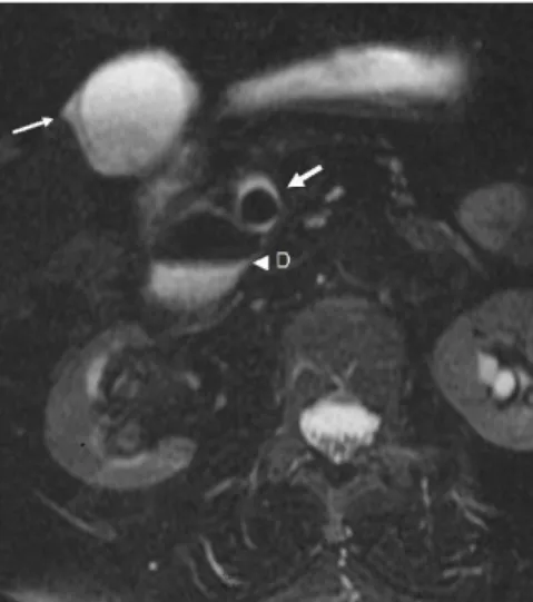
Figure 2. Axial T2-weighted FSE image
reveals gallbladder wall thickening, pericho-lecystic fluid (thin arrow), choledocholithiasis (thick arrow) and periampullary diverticulum (D) with air-fluid level.
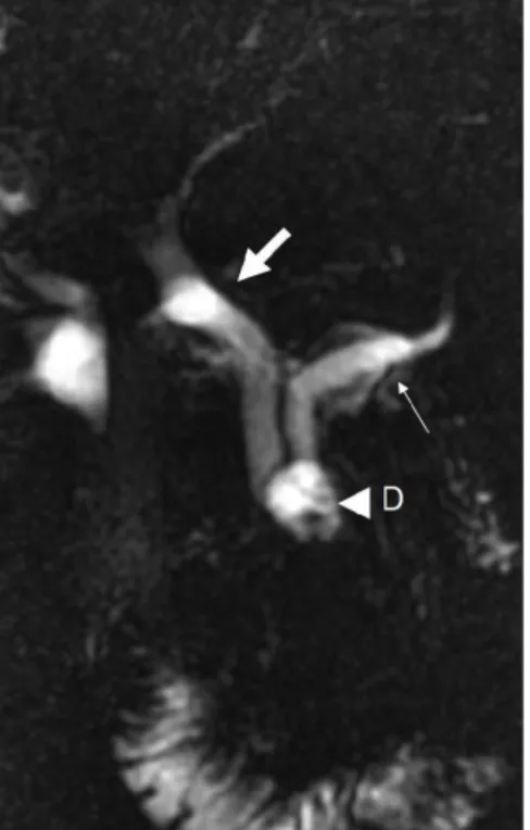
ameter: 2,25 cm). Axial T2-weighted FSE images demonstrated air-fluid levels in 95.8% (n=23/24) of the di-verticula (Fig. 1). Gallbladder stones were detected in 44.4% (n=8/18) of the patients without cholecystecto-my. Gallbladder wall thickening and pericholecystic fluid was present in 22.2% (n=4/18) of the patients (Fig. 2). One patient (5.5%, n=1/18) had diffuse wall thickening of gallbladder with calcifications (chronic cholecysti-tis and porcelain gallbladder) (Fig. 3) and 2 patients (11.1%, n=2/18) had findings consistent with fundal adeno-myomatosis. The CBD was dilated in 45.8% (n=11/24) of all the study patients (Fig. 4). Mild intrahepatic biliary ductal dilatation was detected in 58.3% (n=14/24) of the patients. Pancreatic duct was mildly dilated in 45.8% (n=11/24) of the patients. Pancreatic atrophy was noted in only one patient (4.1%, n=1/24). Four pa-tients (16.6%, n=4/24) had pancreatic side branch dilatations. Deviation of the distal CBD by the diverticulum was observed in 12,5% (n=3/24) of the patients (Fig. 3, 5). Three patients one of which had previous cholecystec-tomy had choledocholitiasis (12,5%, n=3/24).
Discussion
Duodenum is the second most common location for the gastrointestinal di-verticula, after colon. It is difficult to estimate the true prevalence of the duodenal diverticula in the general population; the prevalence on barium meal examination ranges from 0.16 to 6%, prevalence at endoscopic ret-rograde cholangiopancreatography (ERCP) ranges from 5–32.8% and a rate of 23% has been reported at au-topsy (1, 4).
JPD are the extraluminal mucosal her-niations of the duodenal wall from a potentially weak spot, arising within a radius of 2–3 cm from the papilla of Vater (1, 2). They are usually asymp-tomatic and incidentally detected (2). Only 10% of the JPD cases undergo cross-sectional imaging due to pancre-aticobiliary symptoms (2).
The etiology of JPD is postulated to be the disordered duodenal motility (1). Progressive weakening of the intestinal wall with the advancing age, and an
in-crease in the intraluminal pressure may result in diverticula formation (5). Histopathologically, they are similar to the pulsion diverticula of the gastro-intestinal system, and the walls of the diverticula are composed of mucosa and submucosa with scattered smooth muscle (1).
The prevalence of JPD increases with age. They are rarely seen before 40 years of age (4, 6) and usually diagnosed between the ages of 56–70 (1). Be-ing consistent with the literature, we had no patients under 40 years in our study group. The mean age of our pa-tients was 72. There is a slight women dominance among JPD patients (6). We also found a greater rate of fe-males among our patients with JPD (n=18/24).
JPD may cause biliary stasis by compress-ing the distal end of the CBD or may induce reflux of gut bacteria into the bile ducts (7). Lotveit et al. (8) sug-gested that the pressure of the Oddi sphincter may be decreased in asso-ciation with the duodenal diverticula, leading to a reflux of gut bacteria. De-spite the manometric data of Lotveit (8), Kennedy et al. (9) insisted on that there is still a functional stasis within Figure 5. Thick slab MRCP image shows a
diverticulum (D) located in the peripapillary region. There is marked dilatation of the CBD with abrupt deviation at the distal part (thick arrow). Mild intrahepatic biliary duct dilata-tion is also revealed. Pancreatic duct (P) is normal (CBD: Common biliary duct).
Figure 3. Thick slab MRCP image reveals
deviation of the distal CBD due to the peri-ampullary diverticulum (D). The lumen of the gallbladder (G) is filled with stones and the fluid content and volume of the gallbladder is diminished secondary to chronic cholecys-titis (porcelain gallbladder) (CBD: Common
bile duct). Figure 4. Thick slab MRCP image shows marked dilatation of the CBD (thick arrow) and the pancreatic duct (thin arrow) with an abrupt stenosis at the distal end of the ducts due to the presence of a juxtapapillary diverticulum (D).
the bile duct; and infection of a stag-nant system may be easier than an incompetent system (9). B-glucuron-idase-producing bacteria are much likely to be depicted in the bile of the patients with JPD and CBD stones, than the patients with CBD stones but without diverticula (10). Bacte-rial derived B-glucuronidase converts conjugated bilirubin back into its un-conjugated state, which can precipitate as calcium bilirubinate (11) explain-ing the relative likelihood of pigment stones in patients with JPD (12). Pri-mary and recurrent choledocholithia-sis seem to be much more frequent in the patients with JPD (6, 9, 13-17). It has been shown that 40% of the pa-tients who have CBD stones, have also JPD (18). Patients with choledocholi-thiasis were found to be 2,6 times more likely to have a JPD than the patients without stones (9). Similarly, when a JPD is seen at duodenoscopy, it is es-timated that there is a chance for over than 50% for a bile duct stone to be present (9). In our study group, three of the patients (12,5%, n=3/24) had choledocholithiasis, one of them with a history of cholecystectomy. One of these three patients had cholecycstoli-thiasis, whereas the gallbladder of the third patient was normal.
Sitouridis et al. found that JPD may cause deviation at the distal part of the CBD (3). We have observed a deviation of the distal CBD by JPD in 12,5% (n=3/24) of the study patients. None of these patients had choledocholi-tiasis, but one of them had dilatation of CBD, intrahepatic bile ducts, and pancreatic duct without any history of cholecystectomy. The diameter of the JPD of the patients with deviation of the CBD was larger (2.5 cm, 4.5 cm, and 4.5 cm) than the mean diameter (2,25 cm) of JPD in our study group. Several authors have suggested that there
is a relation between JPD and chole-cystolithiasis (6, 13, 14, 19-23). On the contrary, some authors did not support this finding (24, 18). Egawa et al. (25) found that cholecystolithia-sis was significantly more common
with the JPD with a diameter of 20 mm or more, than with the smaller ones. Gallbladder stones were detected in 44.4% (n=8/18) of the patients without cholecystectomy in our study group. Considering that the patients who had previous cholecystectomy probably had had cholecystolithiasis, the real cholecystolithiasis rate among JPD patients must be even higher. The relation between JPD and pancreatitis
is not clear. There are publications in the literature pointing out the effect of JPD in the pathogenesis of pancreatitis (26, 27, 28, 21). But it is not clear whether pancreatitis is caused by the JPD, or associated bile duct stones (1). It is proposed that the compression of the pancreatic duct by the diverticu-lum itself, or ampullary dysfunction secondary to JPD may cause pancre-atitis (1). Uomo et al. (21) have found that idiopathic acute pancreatitis is sig-nificantly more common in JPD pa-tients than in controls, and suggested that the presence of JPD should be searched especially in elderly patients, before defining an acute pancreatitis episode as idiopathic. Also, Leivonen et al. (22) found that the patients with JPD had idiopathic pancreatitis twice as often as controls, but this difference was not statistically significant. Kirk et al. (19) and Lobo et al. (1) were un-able to show that the diverticula were the cause of the pancreatic obstruction and they both commented that there is not enough evidence to propound that JPD contribute significantly to the pathogenesis of pancreatitis. Zoepf et al. (6) also did not support a cor-relation for JPD with acute or chronic pancreatitis. We have observed mild pancreatic ductal dilatation in 45.8% (n=11/24) of our cases. Four patients (16.6%, n=4/24) had pancreatic side branch dilatations. Pancreatic atrophy was noted in only one patient (4.1%, n=1/24). No patient showed any sign of acute pancreatitis. Considering that our findings about pancreas or pancre-atic duct can also be associated with the advanced age of our patients, it is really hard to say that the JPD can cause dilatation of the pancreatic duct.
This subject needs further studies with greater number of patients, and a con-trol group consisting of patients in the same age group.
ERCP is a technically difficult procedure for JPD patients, thus the overall suc-cess rate of ERCP in patients with JPD is usually lower than the patients with-out JPD (1, 4, 6). MRCP can be use-ful in patients with JPD, especially for whom ERCP is technically difficult. It can demonstrate the biliary and pan-creatic ducts and is proven to be useful in the detection of pancreatic disor-ders (29). Therefore, the indications of diagnostic ERCP can be reduced by using MRCP in these patients, and ERCP can be preserved for therapeutic purposes, or for problematic cases to confirm the MRCP findings.
The detection rate of the JPD by MRCP depends on the size and fluid con-tent of the diverticulum (3). Routine MRCP studies without secretin injec-tion has a low sensitivity for the de-tection of JPD. Secretin stimulation can be helpful because the secreted pancreatic fluid fills the diverticulum and makes it more visible (3, 30). Nevertheless, duodenal diverticula are usually easily recognized on MRI stud-ies when completely or partially filled with gas (2). Because of their close re-lationship with the pancreatic head, duodenal diverticula can be potentially misinterpreted as cystic pancreatic le-sions, if their content is purely fluid (31). In addition, it can be difficult to distinguish the diverticulum from the bowel lumen if it is filled with fluid. In our study, we noticed that air-fluid level was the most common feature that helped us to notice the duode-nal diverticula and to differentiate it from a cystic pancreatic mass. Axial T2-weighted FSE sequences were the most useful sequences to demonstrate the air-fluid levels of the diverticula (95.8%, n=23/24). Thus, axial T2-weighted FSE images were the best images to show JPD and to differenti-ate them from pancreatic lesions. If the diagnosis of JPD is in doubt, careful examination of images for the
pres-ence of air-fluid level, or repeating the examination after oral administration of a cup of water or changing the pa-tient’s position may be helpful. Also, some authors recommend follow-up imaging or an upper gastrointestinal barium examination to confirm the diagnosis of a duodenal diverticulum (31). Maziotti et al. (2) suggested oral administration of a supermagnetic iron oxide contrast agent when pre-contrast images show superimposition of bowel-loop fluids.
There are some limitations of our study. One of them is the small number of
cases. In addition, we did not have a control group and due to the retro-spective nature of our study we could not correlate the presence of JPD with another imaging modality, or with pathology. To determine the exact re-lationship between JPD and pancrea-tobiliary diseases, further studies with larger number of patients, including control groups are needed.
In conclusion, MRCP can be a useful ra-diological method to determine the presence, location, and size of the JPD, as well as their effects on the CBD and any associated pancreatobiliary
dis-eases. Axial T2-weighted images are the most useful sequence in demon-strating the air-fluid levels within the diverticula, which we think is the most common and important feature of JPD. JPD may cause deviation of the distal CBD by mass effect which may be a predisposing factor to pancreatico-biliary symptoms. Gallbladder stones, biliary or pancreatic ductal dilatations are quite common in patients with JPD referring with pancreticobiliary symptoms. CBD deviation and cho-ledocholithiasis are the other possible findings, but with a lesser probability.
REFERENCES
1. Lobo DN, Balfour TW, Iftikhar SY, Row-lands BJ. Periampullary diverticula and pancreaticobiliary disease. Br J Surg 1999; 86:588-597.
2. Mazziotti S, Costa C, Ascenti G, Gaeta M, Pandolfo A, Blandino A. MR Cholangio-pancreatography Diagnosis of Juxtapapil-lary Duodenal Diverticulum Simulating a Cystic Lesion of the Pancreas: Usefulness of an Oral Negative Contrast Agent. Am J Roentgenol 2005; 185:432-435.
3. Tsitouridis I, Emmanouilidou M, Gout-saridou F, Kokozidis G, Kalambakas A, Pa-pastergiou C, Tsantiridis C. MR cholangi-ography in the evaluation of patients with duodenal periampullary diverticulum. Eur J Radiol 2003; 47:154-160.
4. Boix J, Lorenzo-Zuniga V, Ananos F, Domenech E, Morillas RM, Gassull MA. Impact of Periampullary Duodenal Diverticula at Endoscopic Retrograde Cholangiopancreatography: A Proposed Classification of Periampullary Duodenal Diverticula. Surg Laparosc Endosc Percu-tan Tech 2006; 16:208-211.
5. Ackermann W. Diverticula and varia-tions of the duodenum. Ann Surg 1943; 117:403-413.
6. Zoepf T, Zoepf DS, Arnold JC, Benz C, Riemann JF. The relationship between jux-tapapillary duodenal diverticula and disor-ders of the biliopancreatic system: analysis of 350 patients. Gastrointest Endosc 2001; 54:56-61.
7. Tham TCK, Kelly M. Association of Peri-ampullary Duodenal Diverticula with Bile Duct Stones and with Technical Success of Endoscopic Retrograde Cholangiopan-creatography. Endoscopy 2004; 36: 1050-1053.
8. Lotveit T, Osnes M, Aune S, Larsen. Stud-ies of the choledochoduodenal sphincter in patients with and without juxta-papillary duodenal diverticula. Scand J Gastroen-terol 1980; 15:875-880.
9. Kennedy RH, Thompson MH. Are duo-denal diverticula associated with choledo-cholitiasis? Gut 1988; 29:1003-1006. 10. Skar V, Skar AG, Bratlie J, Osnes M.
Beta-glucuronidase activity in the bile of gall-stone patients both with and without duo-denal diverticula. Scand J Gastroenterol 1989; 24:205-212 (abstract).
11. Tazuma S. Epidemiology, pathogenesis and classification of biliary stones (Com-mon bile duct and intrahepatic). Best Pract Res Clin Gastroenterol 2006; 20:1075-83.
12. Lotveit T, Foss OP, Osnes M. Biliary pig-ment and cholesterol calculi in patients with and without juxtapapillary duodenal diverticula. Scand J Gastroenterol 1981; 16:241-244 (abstract).
13. Kim M-H, Myung SJ, Seo DW, Lee SK, Kim YS, Lee MH et al. Association of periampullary diverticula with primary choledocholitiasis but not with second-ary choledocholithiasis. Endoscopy 1998; 30:601-604 (abstract).
14. De Koster E, Denis P, Mante M, Otero J, Nyst J, Jonas C, et al. Juxtapapillary duo-denal diverticula: association with biliary stone disease. Acta Gastroenterol Belg 1990; 53:338-343 (abstract).
15. Miyazawa Y, Okinaga K, Nishida K, Okano T. Recurrent common bile duct stones associated with periampullary duo-denal diverticula and calcium bilirubinate stones. Int Surg 1995; 80:120-124 (ab-stract).
16. Lotveit T, Osnes M, Larsen S. Recur-rent biliary calculi: duodenal diverticula as a predisposing factor. Ann Surg 1982; 196:30-32.
17. Hall RI, Ingoldby CJH, Denyer ME. Periampullary diverticula predisposing to primary rather than secondary stones in the common bile duct. Endoscopy 1990; 22:127-128 (abstract).
18. Hagege H, Berson A, Pelletier G, Fritsch J, Choury A, Liguory C, Etienne JP. As-sociation of juxtapapillary diverticula with choledocholitiasis but not with cholecys-tolithiasis. Endoscopy 1992; 24:248-251 (abstract).
19. Kirk AP, Summerfield JA. Incidence and significance of juxtapapillary diverticula at endoscopic retrograde cholangiopancrea-tography. Digestion 1980; 20:31-35 (ab-stract).
20. Leinkram C, Roberts-Thomson IC, Kune GA. Juxtapapillary duodenal diverticula. Association with gallstones and pancreati-tis. Med J Aust 1980; 1:209-210 (abstract). 21. Uomo G, Manes G, Ragozzino A, Ca-vallera A, Rabitti PG. Periampullary ex-traluminal duodenal diverticula and acute pancreatitis: an underestimated etiologi-cal association. Am J Gastroenterol 1996; 91:1186-1188 (abstract).
22. Leivonen MK, Halttunen JAA, Kivilaakso EO. Duodenal diverticulum at endoscopic retrograde cholangiopancreaticography: analysis of 123 patients. Hepatogastroen-terology 1996; 43:961-966 (abstract).
23. Kimura W, Nagai H, Kuroda A, Muto T. No significant correlation between histo-logic changes of the papilla of Vater and juxtapapillary diverticulum. Special ref-erence to the pathogenesis of gallstones. Scand J Gastroenterol 1992; 27:951-956. 24. Novacek G, Walgram M, Bauer P, Schofl
R, Gangl A, Potzi R. The relationship be-tween juxtapapillary duodenal diverticula and biliary stone disease. Eur J Gastroen-terol Hepatol 1997; 9:375-379 (abstract). 25. Egawa N, Kamisawa T, Tu Y, Sakaki N,
Tsuruta K, Okamoto A. The role of juxta-papillary duodenal diverticulum in the for-mation of gallbladder stones. Hepatogas-troenterology 1998 ;45:917-20 (abstract). 26. Shemesh E, Friedman E, Czerniak A, Bat
L. The association of biliary and pancreatic anomalies with periampullary duodenal di-verticula. Correlation with clinic presenta-tions. Arch Surg 1987; 122:1055-1057. 27. Osnes M, Lotveit T. Juxtaminopapillary
diverticulum associated with chronic pan-creatitis. Endoscopy 1977; 8:106-108 (ab-stract).
28. Carey LC. Pathophysiologic factors in re-current acute pancreatitis. Jpn J Surg 1985; 15:333-340 (abstract).
29. Mortelé KJ, Wiesner W, Zou KH, Ros PR, Silverman SG. Asymptomatic nonspesific serum hyperamilasemia and hyperlipas-emia: spectrum of MRCP findings and clinical implications. Abdom Imaging 2004 ;29:109-14.
30. Ono M, Kamisawa T, Tu Y, Egawa N. MRCP and ERCP in Lemmel Syndrome. J Pancreas 2005; 6:277-278.
31. Macari M, Lazarus D, Israel G, Megibow A. Duodenal diverticula mimicking cystic neoplasm of the pancreas: CT and MR imaging findings in seven patients. Am J Roentgenol 2003; 180:195-199.

