Na
+
/I
−
Symporter and Type 3 Iodothyronine Deiodinase
Gene Expression in Amniotic Membrane and Placenta
and Its Relationship to Maternal Thyroid Hormones
Mujde Akturk&Ayla Sargin Oruc&Nuri Danisman&
Serap Erkek&Umran Buyukkagnici&Elmas Unlu&
Uygar Halis Tazebay
Received: 24 May 2013 / Accepted: 1 July 2013 / Published online: 16 July 2013 # Springer Science+Business Media New York 2013
Abstract Placental type 3 iodothyronine deiodinase (D3) potentially protects the fetus from the elevated maternal thyroid hormones. Na+/I−symporter (NIS) is a plasma mem-brane glycoprotein, which mediates active iodide uptake. Our objectives were to establish the distribution of NIS and D3 gene expressions in the placenta and the amniotic mem-brane and to investigate the relationship between placental D3 and NIS gene expressions and maternal iodine, selenium, and thyroid hormone status. Thyroid hormones, urinary io-dine concentration (UIC), and selenium levels were mea-sured in 49 healthy term pregnant women. NIS and D3 gene expressions were studied with the total mRNA RT-PCR method in tissues from maternal placenta (n=49), fetal
placenta (n=9), and amniotic membrane (n=9). NIS and D3 gene expressions were shown in the fetal and maternal sides of the placenta and amniotic membrane. Mean blood selenium level was 66±26.5 μg/l, and median UIC was 143μg/l. We could not demonstrate any statistically signif-icant relationship of spot UIC and blood selenium with NIS and D3 expression (p>0.05). Positive correlations were found between NIS and thyroxine-binding globulin (TBG) (r=0.3, p=0.042) and between D3 and preoperative glucose levels (r=0.4, p=0.006). D3 and NIS genes are expressed in term placenta and amniotic membrane; thus, in addition to placenta, amniotic membrane contributes to regulation of maternofetal iodine and thyroid hormone transmission. Fur-ther studies are needed to clarify the relationship between maternal glucose levels and placental D3 expression and between TBG and placental NIS expression.
Keywords Iodine . Selenium . Na+/I−symporter . Type 3 iodothyronine deiodinase . Placenta . Amniotic membrane
Introduction
Placental type 3 iodothyronine deiodinase (D3) potentially protects the fetus against high thyroid hormone levels of the mother [1]. Na+/I−symporter (NIS) is a plasma membrane glycoprotein, which mediates the active uptake of iodide into cells with the purpose of channeling it to the developing fetus for its proper thyroid hormone synthesis [2]. It is believed that D3 and NIS contribute to regulation of the maternofetal thyroid hormone metabolism.
NIS expression has been shown in the thyroid gland as well as in other tissues such as the salivary gland and milk-producing breast tissue [3–5]. NIS expression has also been shown in placenta, but at lower levels than those in the
M. Akturk (*)
Department of Endocrinology and Metabolism, Faculty of Medicine, Gazi University, Besevler, 06500 Ankara, Turkey e-mail: [email protected]
A. S. Oruc
:
N. DanismanDepartment of Obstetrics, Zekai Tahir Burak Women’s Health Education and Research Hospital, Ankara, Turkey
S. Erkek
:
U. H. TazebayDepartment of Molecular Biology and Genetics, Bilkent University, Ankara, Turkey
U. Buyukkagnici
Division of Biochemistry, Zekai Tahir Burak Women’s Health Education and Research Hospital, Ankara, Turkey
E. Unlu
Division of Radiology, Zekai Tahir Burak Women’s Health Education and Research Hospital, Ankara, Turkey U. H. Tazebay
Gebze Institute of Technology,
Department of Molecular Biology and Genetics, Gebze, 41400 Kocaeli, Turkey
thyroid [6,7]. Thyroid-stimulating hormone (TSH) and io-dide are two main regulators of NIS expression in the thyroid gland [5,8]. The mechanism of placental NIS expression is not yet clear, but iodide and human chorionic gonadotropin have been claimed to have a role [2,9].
Iodothyronine deiodinases are selenoenzymes, which contribute to the regulation of thyroid hormone activity in various tissues [1]. There are three types of iodothyronine deiodinases, type 1 iodothyronine deiodinase (D1), type 2 iodothyronine deiodinase (D2), and D3. The latter (D3) is responsible for inner ring or 5 deiodination of T4 and T3 and produces reverse triiodothyronine (rT3) and 3,3-T2, which are inactive hormone products.
The uterus, placenta, and other maternofetal interfaces such as the fetal skin, respiratory and digestive system epi-thelium, and umbilical arteries and vein express D3 in order to protect the fetus from high thyroid hormone levels and to ensure that the thyroid hormones of the fetus are significantly lower than those of the mother or other adults [1,10–13]. The mechanism of the placental D3 expression is not well understood. It has been claimed that estradiol and progester-one may regulate D3 expression in the uterus [14,15].
Selenium is a required micronutrient for normal develop-ment, metabolism, and growth. All three iodothyronine deiodinases (D1, D2, and D3) contain selenocysteine protein in their active sides. According to a few animal studies, placental D3 activity may not be affected by selenium deficiency [16,17]. To the best of our knowledge, no human studies yet exist about the effects of the iodide, selenium and thyroid hor-mone levels of pregnant women on placenta NIS mRNA expression. Our aim is to assess the distribution of NIS and D3 expression in the fetal side of the placenta (FP), maternal side of the placenta (MP), and the amniotic membrane and to investigate the association between NIS and D3 expression levels in MP and iodine, selenium, and thyroid hormone status in human term pregnancy.
Materials and Methods
Study Protocol
A total of 49 healthy term pregnant women, who were scheduled for a morning elective cesarean section, were assigned to the study. Participants were aged between 22 and 43 years and at 37.5–41 weeks of gestation. Exclusion criteria included multiple pregnancies, diagnosis of pre-eclampsia or pre-eclampsia, chronic diseases (DM, uremia, etc.), previous thyroid operation, and use of thyroid hormone preparation or antithyroid drugs. Onset of labor was another criterion for exclusion. The study protocol was approved by the local ethics board prior to the study, and written informed consent of all cases was obtained.
Gestational age was determined according to the last menstrual period and ultrasonographic biometric measure-ments. Preoperative morning urine and blood samples were drawn. Fasting blood glucose (FBG), insulin, antithyroid peroxidase (anti-TPO), and antithyroglobulin (anti-TG) levels were measured. For other analyses, blood samples were centrifuged and kept at−80°C in a freezer.
During the operation, immediately after removal of the placenta, samples of 2×1×1 cm were obtained from the maternal and fetal sides of the periumbilical zone of the placenta (by dissecting the fetal membrane), and samples of 2 cc were obtained from the amniotic membrane and stored in liquid nitrogen. Samples from the placenta and amniotic membrane were kept at−80°C in a freezer. Methods of Analysis
Urinary iodine concentration (UIC) was analyzed from spot urine samples obtained during morning fast and frozen at−80°C. The analysis was performed via the Sandell–Kolhoff reaction (color-imetric method, with serik ion arsenic acid wet ash method) by using Fisher reagent and Spectronic Genesys 20 autoanalyzer.
Total triiodothyronine (TT3), total thyroxine (TT4), free tri-iodothyronine (FT3), free thyroxine (FT4), TSH, thyroxine-binding globulin (TBG), transthyretin, and TG levels were studied from serum samples and reverse triiodothyronine (rT3) levels from plasma samples. FT3, FT4, TT3, TT4, TSH, and TG levels were studied in the Roche Modular Analytics E170 device with Cobas Elecsys kits manufactured by Roche Diagnostics (Roche Diagnostics GmbH, D-68298 Mannheim). Transthyretin (prealbumin) levels were measured in the Cobas Integra 400 device with Cobas kits manufactured by Roche Diagnostics (Roche Diagnostics GmbH, D-68298 Mannheim). rT3 levels were measured with the RIA method by using the IDS (Belgium) kit. TBG levels were measured with the RIA method by using the B.R.A.H.M.S. (Berlin, Germany) kit. Serum anti-TPO and anti-TG levels were studied by using the solid-phase, enzyme-labeled chemiluminescent sequential immunometric as-say and the Immulite 2000 immunoanalyzer, with Immulite 2000 anti-TPO Ab and anti-TG Siemens kits. Glucose levels were measured with standard enzymatic method (Roche Diag-nostics GmBH, Mannheim, Germany). Serum insulin levels were measured by immunoradiometric assay (sandwich-type assay) using an insulin IRMA kit (Immunotech, Prague, Czech Republic). Selenium was analyzed from serum samples by using an atomic absorption spectrometer (AAS). As samples were prepared for analysis, isoformation was performed to thaw the samples and to transform all selenium content into Se4+. The analyses were performed by using the PerkinElmer AAnalyst 800 Model HG-AAS instrument and the standard water addition technique.
The thyroid ultrasonography of the 38 term pregnant cases were done by a radiology specialist using Aloka
5500 (Tokyo, Japan) and 7.5-MHz linear probe. Length, width, and depth were separately measured for each thyroid lobe, and presence of nodules or heterogeneity characteristics were recorded. Thyroid gland volume (TV) was calculated [18].
RT-PCR Analysis of NIS, D3, and TBP Gene Transcripts
Expression of NIS, D3, and TBP were analyzed by RT-PCR methods followed by agarose gel electrophoresis and densito-metric analysis of relevant bands. Human placental samples were frozen and ground to powder under a continuous supply of liquid nitrogen without allowing the tissue to soften. Sub-sequently, 30 mg of placental tissue powder is taken in a 1.5-ml Eppendorf tube, and total RNA was extracted using RNeasy Protect kit (Qiagen, Hilden, Germany) following the protocol described by the producer. Briefly, tissue was pipetted in 3 ml lysis buffer and taken in a 10-ml glass tube for homogenization by a Becker homogenizer (Palo Alto, CA) at a speed of 1.5 for 40 strokes. Then, following the kit’s protocol, 3μg total RNA is used for cDNA synthesis by using reverse transcriptase enzyme provided in Fermentas (St. Leon-Rot, Germany) First Strand cDNA synthesis kit. The detection and quantification of transcripts corresponding to NIS and D3 genes were done by PCR analysis using the following primer pairs and reaction steps: NIS forward 5′ CTCATCCTGAACCAAGTGAC 3′/NIS reverse 5′ TACATG GAGAGCCACACCA 3′; denaturation for 5 min at 95 °C and then 95 °C for 30 s/60 °C for 30 s/72 °C for 30 s, altogether 40 cycles, with the last extension at 72 °C for 5 min; D3 forward 5′ CCTGCTGCTTCACTCCTTG 3′/D3 reverse 5′ GCGTAG TCGAGGATGTGCT 3′; denaturation at 95 °C for 5 min and then 95 °C for 30 s, 60.3 °C for 30 s, and 72 °C for 30 s, altogether 35 cycles, with the last extension at 72 °C for 5 min. Amounts of these two transcripts were normalized by using the levels of the general transcription factor-associated TBP, and semiquantification was completed. PCR conditions for TBP were as follows: TBP forward 5′ TGCACAGGAGCC AAGAGTGAA 3′/TBP reverse 5′ CACATCACAGCTCCC CACCA 3′; denaturation for 5 min at 95 °C and then 95 °C for 30 s/60 °C for 30 s/72 °C for 30 s, altogether 30 cycles, with the last extension at 72 °C for 5 min. Typically, 50 pg of cDNA was used in all PCRs. After agarose gel electrophore-sis, density of each band is quantified using the BioRad Gel-Doc XR-Plus software platform (BioRad, Turkey).
Statistical Method
Statistical analyses were performed by using the SPSS 16 statistical package. Data were given as mean ± standard deviation or median (25th, 75th percentile) according to their distribution characteristics. Descriptive statistics and corre-lation analyses (Pearson and Spearman) were used, as well as
t test and Mann Whitney U test to show differences between groups. D3 and NIS expression were below the detection limits of our technique in a number of the tissue samples, even though the genetic material was obtained. These cases were excluded from statistical analyses. In samples obtained from MP, FP, and amniotic membrane of the same pregnant women, the difference between the NIS and D3 measure-ments was determined with the nonparametric Friedman test. Post hoc analyses were performed. In the statistical analyses, ap value of <0.05 was considered statistically meaningful. Results
NIS, D3, and TBP gene expression levels were determined with the RT-PCR method in FP, MP, and amniotic membrane tissue samples of nine term pregnant cases in the first part of the study (Fig.1). The medians for D3 in the MP, FP, and amniotic membrane (min–max) were 1.1 (0.59–1.19), 1.08 (0.95–1.28), and 0.58 (0.40–0.96), respectively. The Fried-man test revealed a meaningful difference (p=0.03). When the post hoc analysis was performed, similar D3 levels were found in MP and FP (p>0.05), but D3 levels in the amniotic membrane were lower than those in both sides of the placen-ta (p<0.05) (Fig.2). The median values of NIS expression (min–max) for the MP, FP, and amniotic membrane were 0.40 (0.25–0.45), 0.39 (0.24–0.69), and 0.27 (0.18–0.46), respectively. No statistically significant difference was ob-served between the groups (p=0.17). NIS expression was detected in the FP (n=6), MP (n=7), and amniotic membrane samples (n=6). D3 expression was detected in all the MP and FP samples, and it was found in the amniotic membrane samples of seven cases.
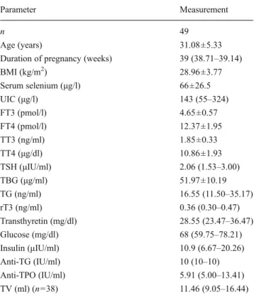
In the second part of the study, D3 and NIS expression levels were studied in the MP in the 49 term pregnant women (NIS gene expression was below our detection limit in two cases), and their relationship to maternal thyroid hormone, UIC, and serum selenium level was investigated. The mean age of the term pregnant women in the study was 31.08±5.3 years, and their mean gestational age was 39 (38.71–39.14)weeks. The thyroid hormone, serum selenium, UIC measurements, and thyroid volumes of the cases are represented in Table1.
Median UIC was 143μg/l (minimum 8 μg/l, maximum 450 μg/l). NIS gene expression was 0.37±0.14. When the cutoff value for UIC was taken as 150 μg/l, the NIS gene expression levels of the cases that had UIC were below (0.39±0.15,n=25) or above (0.36±0.13, n=22). NIS levels of the groups were not different (p=0.4). Similarly, there were no differences in D3 levels between the groups with UIC levels lower or higher than 150μg/l (0.96 (0.79–1.25), 0.91 (0.70–1.1), respectively, p=0.4).
In 49 cases, the median TSH level was found to be 2.06 (minimum 0.72, maximum 7.93)μIU/ml. Median D3 gene
expression was 0.96 (0.76–1.14). When divided into two groups based on the TSH levels of the women (<3 and ≥3 μIU/ml), placental D3 expression was slightly de-creased in the TSH≥3 μIU/ml group (0.81(0.64–1.09), n=12) compared with the TSH <3μIU/ml group (0.96 (0.78–1.17), n=37), but the difference was not statistically significant
(p=0.18) [19]. There was only one woman with overt hypo-thyroidism (D3=0.82). Expression of the NIS gene was not significantly different between the groups with TSH levels lower or higher than 3μIU/ml (p=0.8).
Four term pregnant women had high anti-TPO levels (≥34 IU/ml). The anti-TG levels of 37 cases were at the
A)
1 2 3 4 D3 NIS TBPB)
D3 NIS TBP 1 2 3 4C)
1 2 3 D3 NIS TBPFig. 1 a Maternal side of the placenta (n=4), b fetal side of the placenta (n=4), c amniotic membrane (n=3). The number of the images shows the analysis results of samples obtained from different women. The numbers on the images (1–4 or 1–3) show the analysis results of samples obtained from different patients.NIS Na+/I− symporter,D3
type 3 iodothyronine deiodinase,TBP TATAbinding protein
D3 Gene Expression P = 0.03
Fig. 2 The graphics represent D3 gene expression levels in the mater-nal and fetal side of the placenta and the amniotic membrane by using the RT-PCR method (n=7)
Table 1 Some characteristics and laboratory measurement values of term pregnant women
Parameter Measurement
n 49
Age (years) 31.08±5.33
Duration of pregnancy (weeks) 39 (38.71–39.14)
BMI (kg/m2) 28.96±3.77 Serum selenium (μg/l) 66±26.5 UIC (μg/l) 143 (55–324) FT3 (pmol/l) 4.65±0.57 FT4 (pmol/l) 12.37±1.95 TT3 (ng/ml) 1.85±0.33 TT4 (μg/dl) 10.86±1.93 TSH (μIU/ml) 2.06 (1.53–3.00) TBG (μg/ml) 51.97±10.19 TG (ng/ml) 16.55 (11.50–35.17) rT3 (ng/ml) 0.36 (0.30–0.47) Transthyretin (mg/dl) 28.55 (23.47–36.47) Glucose (mg/dl) 68 (59.75–78.21) Insulin (μIU/ml) 10.9 (6.67–20.26) Anti-TG (IU/ml) 10 (10–10) Anti-TPO (IU/ml) 5.91 (5.00–13.41) TV (ml) (n=38) 11.46 (9.05–16.44)
Data were expressed as mean ± standard deviation or median (25th, 75th percentile)
BMI body mass index, UIC urinary iodine concentration, FT3 free triiodo-thyronine,FT4 free thyroxine, TT3 total triiodothyronine, TT4 total thyrox-ine,TSH thyroid-stimulating hormone, TBG thyroxine-binding globulin, TG thyroglobulin,rT3 reverse triiodothyronine, TG antithyroglobulin, Anti-TPO antithyroid peroxidase, TV thyroid volume
minimum measurable level. There were no cases with high anti-TG levels.
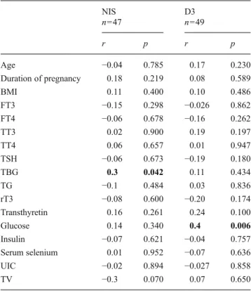
A positive statistically significant correlation was deter-mined between the serum TBG levels of term pregnant women and the placental NIS expression (r=0.3, p=0.042). Correlation of NIS expression with other parameters does not reach a statistically significant level (Table2).
Placental D3 gene expression was positively correlated only with preoperative fasting blood glucose level (min 50– max 111 mg/dl) (r=0.4, p=0.006). As represented in Table2, we could not demonstrate any statistically significant correla-tions between D3 gene expression level and other parameters.
Discussion
In our study, NIS and D3 expression were studied by the RT-PCR methods in the tissue samples obtained from the human
maternal side of placenta, which is in contact with decidua, and the fetal side that is close to the fetal tissues and the amniotic membrane. NIS and D3 gene expressions were found in both sides of the placenta and amniotic membrane, as well. To the best of our knowledge, our study is the first to report the presence of NIS expression in the amniotic mem-brane in the literature. The amniotic fluid is known to contain iodide [20]. A previous study found similar iodine concen-trations in the amniotic fluid in those who did and did not receive iodine supplement. The authors thus claimed that there must be a mechanism that regulates iodine in the amniotic fluid [21]. Our study concluded that the NIS in the amniotic membrane may contribute to iodine transmis-sion from the mother to the fetus. Although the distribution of NIS in the MP and the FP is not yet known in the literature, a previous study conducted on rats found NIS expression in the fetal side of placenta, but, different from our study, not in the maternal side [9]. This discrepancy between the two studies might be related to different meth-odologies used in these works, as immunostaining methods were used by Schroder-van der Elst et al. [9]. As mentioned above, NIS expression was not detected in the different surfaces of the placenta or the amniotic membrane in some cases even though genetic material was obtained. This may have been due to the method used, the characteristics of tissues, or an unidentified mechanism.
Similar to a number of previous studies, we detected D3 expression in human amniotic membrane tissue samples [11,
13,22]. These results suggest that in addition to the placenta and the uterus, the amniotic membrane, which expresses D3, may also regulate the transfer of thyroid hormones from the mother to the fetus in term pregnancy [13]. In another study with rats, D3 was similar in the fetal and maternal sides of the placenta on day 18 of pregnancy, while day 21 had increased D3 activity in the maternal side [23]. However, D3 expression levels in our study were similar in both sides of the placenta, but amniotic membrane expressed lower D3 levels than the placenta consistent with a previous rat study [11]. These findings suggest that placental tissues may contribute to the regulation of maternofetal thyroid hormone transmission more than the amniotic membrane.
One of the most significant findings of our study was the positive correlation between NIS expression and maternal serum TBG. TBG mediates the transfer of thyroid hormones, particu-larly thyroxine. The increasing levels of estrogen leads to in-creased TBG during pregnancy [24,25]. In previous years, it was hypothesized that increased TBG in pregnancy may be“an evolutionary adaptation” to ensure sufficient T4 and/or iodine are provided to the fetus [26,27]. To the best of our knowledge, a relationship between NIS and TBG has not yet been reported in the literature. The possible role of TBG to enhance placental NIS expression and maternofetal iodide transmission as well as its mechanism needs to be investigated in future studies.
Table 2 Correlation between NIS and D3 gene expression levels with demographic characteristics and laboratory measurements
NIS D3 n=47 n=49 r p r p Age −0.04 0.785 0.17 0.230 Duration of pregnancy 0.18 0.219 0.08 0.589 BMI 0.11 0.400 0.10 0.486 FT3 −0.15 0.298 −0.026 0.862 FT4 −0.06 0.678 −0.16 0.262 TT3 0.02 0.900 0.19 0.197 TT4 0.06 0.657 0.01 0.947 TSH −0.06 0.673 −0.19 0.180 TBG 0.3 0.042 0.11 0.434 TG −0.1 0.484 0.03 0.836 rT3 −0.08 0.600 −0.20 0.174 Transthyretin 0.16 0.261 0.24 0.100 Glucose 0.14 0.340 0.4 0.006 Insulin −0.07 0.621 −0.04 0.757 Serum selenium 0.01 0.952 −0.07 0.636 UIC −0.02 0.894 −0.027 0.858 TV −0.3 0.070 0.07 0.650
Ap value of <0.05 was considered as statistically meaningful. The Pearson correlation coefficient was used for the analyses of parameters with normal distribution and the Spearman correlation coefficient for others
NIS Na+/I− symporter/TATA-binding protein,D3 type 3 iodothyronine
deiodinase/TATA-binding protein, BMI body mass index, FT3 free triiodothyronine,FT4 free thyroxine, TT3 total triiodothyronine, TT4 total thyroxine, TSH thyroid-stimulating hormone, TBG thyroxine-binding globulin,TG thyroglobulin, rT3 reverse triiodothyronine, UIC urinary iodine concentration,TV thyroid volume
Interestingly, we found a positive correlation between D3 gene expression levels and preoperative maternal FBG. However, we did not find any correlation between maternal serum insulin and D3 expression. Our subjects were operated on shortly after obtaining their fasting blood glucose, and the blood glucose levels at the time of placental removal were not measured. D3 expression rate or half-life of the placental D3 is not known yet. A previous in vitro study suggested that placental D3 activity is not affected by hunger [28]. The role of iodothyronine deiodinases and thyroid hormones in the carbohydrate metabolism is subject of a recent debate [29,
30]. Recently, Medina et al. [30] reported that fetal and adult pancreas in humans and mice expressed D3 obviously, while D1 and D2 expressions were barely detected. Furthermore, D3KO mice, whose Dio3 gene is disrupted, had impaired insulin secretion and glucose intolerance, because of reduced β cell mass and decreased insulin content. D3 may have a role in regulation of carbohydrate metabolism, but it is not clear yet. Moreover, we investigated the effects of maternal factors such as glucose on the placental NIS and D3, but we did not investigate their impact on the fetus. Therefore, we could not explain the mechanism underlying this correlation with our present knowledge.
We could not demonstrate a statistically significant rela-tionship between spot UIC and NIS expression in term placenta. Even though UIC may be a good indicator of iodine intake of societies, it displays wide variations within and between the days, particularly in pregnant women [31,
32]. In studies conducted on thyroid cell cultures, NIS was reported to be a protein with long half-life (approximately 5 days in the presence of TSH) [8]. The half-life of placental NIS has not been known, but it may be affected by longer-term iodine status than the actual iodine status, which is reflected with the UIC level. Iodine deficiency in pregnancy results in a decrease in serum FT4 levels and an increase in TSH and total molar T3/T4 ratio, a progressive increase in serum TG, and an increase in the thyroid volume [24,32]. Even though UIC in our region is significantly improved than in previous years [33], some of the pregnant women in our study still have low UICs. However, laboratory findings indicate that pregnant women in our study do not have long-term serious iodine deficiency. Moreover, in this study, pla-cental D3 and NIS gene expressions were investigated in the placental samples of healthy term pregnant women without hyperthyroidism and overt hypothyroidism (except one pa-tient) [19]. The lack of a relationship in this study between the thyroid hormone levels and placental D3 and NIS ex-pression levels may be attributed to the fact that the subjects in our study did not suffer from overt thyroid function disorder. Only semiquantitative RT-PCR methods were used in analyses of NIS expression in this preliminary study, as we anticipated that tissue uptake studies using radio-labeled iodide in the tissue samples could hardly be sensitive enough
when NIS expression is not robust. Therefore, further studies are needed.
One additional result of our study has been the informa-tion about the serum selenium levels of pregnant women in the region, which corroborates other studies conducted in Turkish pregnant women [34, 35]. Despite the lack of a recommended borderline value, the whole group of patients seems to have low selenium levels [36]. Even though there is an increased requirement for selenium during pregnancy, decreased selenium levels during this physiological stage have previously been reported [37]. Similar to previous animal studies, our study also showed that the placental D3 expression level was not affected by the maternal serum selenium level [16,17,38].
In conclusion, we showed D3 and NIS expressions in the maternal and fetal sides of the placenta and the amniotic membrane in human term pregnancy by the total mRNA RT-PCR method. In addition to the placental NIS and D3 expressions, the amniotic membrane also contributes to the regulation of maternofetal iodine and thyroid hormone trans-mission by expressing NIS and D3. The placenta and amni-otic membrane express NIS to contribute to providing iodine from mother to fetus and D3 in order to protect the fetus against thyroid hormones in term pregnant women. Howev-er, while D3 expression was probably similar throughout the placenta, it was lower in the amniotic membrane, and thus, the effectiveness of placenta in the regulation of maternofetal thyroid hormone transmission could be higher than that of the amniotic membrane. In healthy term pregnant women without overt thyroid function disorder, we could not find any significant differences of placental D3 and NIS gene expressions with regard to spot UIC and thyroid hormone levels. Our preliminary study demonstrated a positive corre-lation between NIS gene expression and TBG in term preg-nant women. The possible role of TBG in placental NIS expression and the regulation of maternofetal iodide trans-mission needs to be studied. Likewise, the positive correla-tion between preoperative glucose levels and D3 should be investigated.
Acknowledgments This project (106S229/SBAG-3475) was sup-ported by TUBITAK (Scientific and Technical Research Council of Turkey). We gratefully acknowledge Prof. Murat Erdogan for the help in UIC analysis and Elif H. Kamber for the technical assistance of the genetic analysis.
Conflict of Interest The authors declare that they have no conflict of interest.
References
1. St Germain DL, Galton VA, Hernandez A (2009) Minireview: Defining the roles of the iodothyronine deiodinases: current con-cepts and challenges. Endocrinology 150:1097–1107
2. Li H, Richard K, McKinnon B, Mortimer RH (2007) Effect of iodide on human choriogonadotropin, sodium-iodide symporter expression, and iodide uptake in BeWo choriocarcinoma cells. J Clin Endocrinol Metab 92:4046–4051
3. Tazebay UH, Wapnir IL, Levy O, Dohan O, Zuckier LS, Zhao QH, Deng HF, Amenta PS, Fineberg S, Pestell RG, Carrasco N (2000) The mammary gland iodide transporter is expressed during lacta-tion and in breast cancer. Nat Med 6:871–878
4. Riedel C, Dohan O, De la Vieja A, Ginter CS, Carrasco N (2001) Journey of the iodide transporter NIS: from its molecular identifi-cation to its clinical role in cancer. Trends Biochem Sci 26:490–496 5. Bizhanova A, Kopp P (2009) Minireview: The sodium-iodide symporter NIS and pendrin in iodide homeostasis of the thyroid. Endocrinology 150:1084–1090
6. Di Cosmo C, Fanelli G, Tonacchera M, Ferrarini E, Dimida A, Agretti P, De Marco G, Vitti P, Pinchera A, Bevilacqua G, Naccarato AG, Viacava P (2006) The sodium-iodide symporter expression in placental tissue at different gestational age: an immu-nohistochemical study. Clin Endocrinol (Oxf) 65:544–548 7. Bidart JM, Lacroix L, Evain-Brion D, Caillou B, Lazar V, Frydman
R, Bellet D, Filetti S, Schlumberger M (2000) Expression of Na+/I− symporter and Pendred syndrome genes in trophoblast cells. J Clin Endocrinol Metab 85:4367–4472
8. Riedel C, Levy O, Carrasco N (2001) Post-transcriptional regula-tion of the sodium/iodide symporter by thyrotropin. J Biol Chem 15:21458–21463
9. Schroder-van der Elst JP, van der Heide D, Kastelijn J, Rousset B, Obregon MJ (2001) The expression of the sodium/iodide symporter is up-regulated in the thyroid of fetuses of iodine-deficient rats. Endocrinology 142:3736–3741
10. Carvalho DP (2003) Modulation of uterine iodothyronine deiodinases—a critical event for fetal development? Endocrinology 144:4250–4252
11. Galton VA, Martinez E, Hernandez A, St Germain EA, Bates JM, St Germain DL (1999) Pregnant rat uterus expresses high levels of the type 3 iodothyronine deiodinase. J Clin Invest 103:979–987 12. Mortimer R, Galligan JP, Cannell GR, Addison RS, Roberts MS
(1996) Maternal to fetal thyroxine transmission in the human term placenta is limited by inner ring deiodination. J Clin Endocrinol Metab 81:2247–2249
13. Huang SA, Dorfman DM, Genest DR, Salvatore D, Larsen PR (2003) Type 3 iodothyronine deiodinase is highly expressed in the human uteroplacental unit and in fetal epithelium. J Clin Endocrinol Metab 88:1384–1388
14. Wasco EC, Martinez E, Grant KS, St Germain EA, St Germain DL, Galton VA (2003) Determinants of iodothyronine deiodinase activ-ities in rodent uterus. Endocrinology 144:4253–4261
15. Kester MH, Kuiper GG, Versteeg R, Visser TJ (2006) Regulation of type III iodothyronine deiodinase expression in human cell lines. Endocrinology 147:5845–5854
16. Bates JM, Spate VL, Morris JS, St Germain DL, Galton VA (2000) Effects of selenium deficiency on tissue selenium content, deiodinase activity, and thyroid hormone economy in the rat during develop-ment. Endocrinology 141:2490–2500
17. Chanoine JP, Alex S, Stone S, Fang SL, Veronikis I, Leonard JL, Braverman LE (1993) Placental 5-deiodinase activity and fetal thyroid hormone economy are unaffected by selenium deficiency in the rat. Pediatr Res 34:288–292
18. Knudsen N, Bols B, Bülow I, Jørgensen T, Perrild H, Ovesen L, Laurberg P (1999) Validation of ultrasonography of the thyroid gland for epidemiological purposes. Thyroid 9:1069– 1074
19. Stagnaro-Green A, Abalovich M, Alexander E, Azizi F, Mestman J, Negro R, Nixon A, Pearce EN, Soldin OP, Sullivan S, Wiersinga W (2011) Guidelines of the American Thyroid Association for the
diagnosis and management of thyroid disease during pregnancy and postpartum. Thyroid 21:1081–1125
20. Rayburn WF, Robinson A, Braverman LE, He XM, Pino S, Gargas ML, Kinzell JH (2008) Iodide concentrations in matched maternal serum, cord serum, and amniotic fluid from preterm and term human pregnancies. Reprod Toxicol 25:129–132
21. García-Fuentes E, Gallo M, García L, Prieto S, Alcaide-Torres J, Santiago P, Velasco I, Soriguer F (2008) Amniotic fluid iodine concentrations do not vary in pregnant women with varying iodine intake. Br J Nutr 99:1178–1181
22. Roti E, Fang SL, Green K, Braverman LE, Emerson CH (1983) Inner ring deiodination of thyroxine and 3,5,3′-triiodothyronine by human fetal membranes. Am J Obstet Gynecol 147:788–792 23. Schröder-van der Elst JP, van der Heide D, Morreale de Escobar G,
Obregón MJ (1998) Iodothyronine deiodinase activities in fetal rat tissues at several levels of iodine deficiency: a role for the skin in 3,5,3′-triiodothyronine economy? Endocrinology 139:2229–2234 24. Glinoer D (1997) The regulation of thyroid function in pregnancy:
pathways of endocrine adaptation from physiology to pathology. Endocr Rev 18:404–433
25. Glinoer D (2007) The importance of iodine nutrition during preg-nancy. Public Health Nutr 10:1542–1546
26. Schussler GC (2000) The thyroxine-binding proteins. Thyroid 10:141– 149
27. Ekins RP, Sinha AK, Pickard MR, Evans IM, al Yatama F (1994) Transport of thyroid hormones to target tissues. Acta Med Austriaca 21:26–34
28. Emerson CH, Bambini G, Alex S, Castro MI, Roti E, Braverman LE (1988) The effect of thyroid dysfunction and fasting on placenta inner ring deiodinase activity in the rat. Endocrinology 122:809–816 29. Chidakel A, Mentuccia D, Celi FS (2005) Peripheral metabolism of
thyroid hormone and glucose homeostasis. Thyroid 15:899–903 30. Medina MC, Molina J, Gadea Y, Fachado A, Murillo M, Simovic
G, Pileggi A, Hernández A, Edlund H, Bianco AC (2011) The thyroid hormone-inactivating type III deiodinase is expressed in mouse and human {beta}-cells and its targeted inactivation impairs insulin secretion. Endocrinology 152:3717–3727
31. Liberman CS, Pino SC, Fang SL, Braverman LE, Emerson CH (1998) Circulating iodide concentrations during and after pregnan-cy. J Clin Endocrinol Metab 83:3545–3549
32. Abalovich M, Amino N, Barbour LA, Cobin RH, De Groot LJ, Glinoer D, Mandel SJ, Stagnaro-Green A (2007) Management of thyroid dysfunction during pregnancy and postpartum: an Endocrine Society Clinical Practice Guideline. J Clin Endocrinol Metab 92:S1–S47
33. Erdoğan MF, Ağbaht K, Altunsu T, Ozbas S, Yücesan F, Tezel B, Sargın C, Ilbeğ I, Artık N, Köse R, Erdoğan G (2009) Current iodine status in Turkey. J Endocrinol Invest 32:617–622
34. Ozdemir HS, Karadas F, Pappas AC, Cassey P, Oto G, Tuncer O (2008) The selenium levels of mothers and their neonates using hair, breast milk, meconium, and maternal and umbilical cord blood in Van Basin. Biol Trace Elem Res 122:206–215
35. Kilinc M, Guven MA, Ezer M, Ertas IE, Coskun A (2008) Evaluation of serum selenium levels in Turkish women with ges-tational diabetes mellitus, glucose intolerants, and normal controls. Biol Trace Elem Res 123:35–40
36. Rayman MP (2002) The argument for increasing selenium intake. Proc Nutr Soc 61:203–215
37. Izquierdo Alvarez S, Castañón SG, Ruata ML, Aragüés EF, Terraz PB, Irazabal YG, González EG, Rodríguez BG (2007) Updating of normal levels of copper, zinc and selenium in serum of pregnant women. J Trace Elem Med Biol 21:49–52
38. Ramauge M, Pallud S, Esfandiari A, Gavaret J, Lennon A, Pierre M, Courtin F (1996) Evidence that type III iodothyronine deiodinase in rat astrocyte is a selenoprotein. Endocrinology 137:3021–3025