IDENTIFICATION OF CANDIDATE GENES FOR FAMILIAL
ESSENTIAL TREMOR
A THESIS SUBMITTED TO NEUROSCIENCE PROGRAM
OF THE GRADUATE SCHOOL OF ENGINEERING AND SCIENCE OF BILKENT UNIVERSITY
IN PARTIAL FULFILLMENT OF THE REQUIREMENTS FOR THE DEGREE OF
MASTER OF SCIENCE
By
MERVE ŞEN
i
IDENTIFICATION OF CANDIDATE GENES FOR FAMILIAL ESSENTIAL TREMOR
By Merve Şen, September, 2016
We certify that we have read this thesis and that in our opinion it is fully adequate, in scope and in quality, as a dissertation for the degree of Master of Science.
Ayşe Begüm Tekinay (Advisor)
Tayfun Özçelik
Cenk Akbostancı
Approved for the Graduate School of Engineering and Science:
Levent Onural
ii
ABSTRACT
IDENTIFICATION OF CANDIDATE GENES FOR FAMILIAL
ESSENTIAL TREMOR
Merve Şen M.Sc. in Neuroscience
Supervisor: Ayşe Begüm Tekinay, PhD September, 2016
Essential tremor (ET) is one of the most common movement disorders in humans and is characterized by action tremors that occur during voluntary motion. However, due to the strong heterogeneity exhibited by ET patients at etiological, clinical and pathological levels, the genetic architecture and pathophysiology of the disease remain largely unknown. In this thesis, whole exome sequencing and pedigree analysis were performed in 3 ET families with histories consistent with an autosomal dominant pattern of inheritance. In two independent families, we observed a rare variant that cosegregated with the disease and was predicted to affect the function of the protein. In one of these families, a homozygous variant was identified in one affected patient and a heterozygous variant was determined in five affected family members. In a second, four-generation Turkish family, the same heterozygous variant was identified in three ET cases while remaining absent in unaffected family members. In addition, whole exome sequencing allowed us to demonstrate that other missense mutation segregate with essential tremor in a different consanguineous Turkish family. Both variants were observed to involve amino acid substitutions of highly conserved domains. Furthermore, both of the affected genes are expressed in the brain and function as
iii
regulatory elements of the central nervous system. Consequently, we propose that these variants are risk factors involved in the etiology of hereditary ET, and suggest that whole exome sequencing can serve as an effective means of identifying other alleles associated with the disease.
Keywords: Essential tremor, human genetics, whole exome sequencing, movement
iv
ÖZET
AİLESEL ESANSİYEL TREMOR İÇİN ADAY GENLERİN TESPİT
EDİLMESİ
Merve Şen
Nörobilim Yüksek Lisans Danışman: Dr. Ayşe Begüm Tekinay
Eylül, 2016
Esansiyel tremor (ET), insanlarda en sık görülen ve gönüllü hareketlerde aksiyon tremoru ile ortaya çıkan nörolojik bir hareket bozukluğu hastalığıdır. Fakat bu hastalığın genetik nedenleri ve patofizyolojisi bilinmemektedir. Çünkü ET hastaları, etiyolojik, klinik ve patolojik düzeylerde çeşitlilik göstermektedir. Bu çalışmada, ET’nin otozomal dominant kalıtımla aktarıldığı 3 aile, aile ağacı ve ekzon dizilimleri bakımından incelenmiş ve aday mutasyonlar anlatılmıştır. Birbirinden farklı iki ailede nadir bir varyantın proteininin fonksiyonunu etkilediği ve ailede segrege olduğu ortaya çıkarılmıştır. Bu varyant, Türkiye’de yaşayan ve akraba evliliği gözlemlenen 5 kuşaklık
bir ailenin hasta bir bireyinde homozigot ve ailenin diğer bireylerinde heterozigot olarak tespit edilmiştir. Başka bir ailenin tüm hasta bireylerinde ise bu aynı varyant heterozigot olarak gözlemlenmiştir. Ayrıca, başka bir akraba evliliği görülen ve
Türkiye’de yaşayan diğer bir ailede ise farklı bir gendeki mutasyonun ailede hastalıkla beraber kalıtıldığı keşfedilmiştir. Bu varyantların, evrimsel olarak korunmuş amino asit dizilimlerinde değişikliğe neden olduğu ve proteinlerin önemli bölgelerinde olduğu gösterilmiştir. Hastalıkla ilgili bulunan bu genlerin beyinde ifade edildikleri ve merkezi
v
ile alakalı riskli allellerin keşfedilmesi ile ET’nin genetik ve moleküler
mekanizmalarının daha iyi anlaşılmasında önemli bir rol oynayabilir.
Anahtar sözcükler: Esansiyel tremor, insan genetiği, tüm ekzon dizilemesi, hareket
vi
ACKNOWLEDGEMENT
First and foremost I would like to express my gratitude to my advisor Dr. Ayşe Begüm Tekinay, for the continuous support of my Master’s study, for her inspiring guidance, motivation, encouragement and immense knowledge. I consider it as a great opportunity to complete my Master’s under her guidance.
I would like to gratefully and sincerely thank Prof. Dr. Tayfun Özçelik for his continuous support, invaluable guidance and his immense knowledge in the field of human genetics.
I am thankful to Prof. Dr. Cenk Akbostancı and Prof. Dr. Kader Karlı Oğuz for the identification and recruitment of patients, for brain imaging studies and for clinical tests.
I am thankful to my best friends from Zeynep Orhan, Fatih Yergöz, İdil Uyan, Nurcan Haştar and Gökhan Günay from Izmir Institute of Technology and also Hatice Kübra Kara, for their emotional support, encouragement and motivation in my research. I will remember those times that we shared together.
I would like to express my sincere thanks to İdil Uyan and Ahmet Emin Topal for their unforgettable and enjoyable friendship together with laugh forever.
I would like to thank Dr. Seher Yaylacı and İslam Oğuz Tuncay for their friendship and persistent support and excellent collaboration during my research. I would also thank new lab member Neva Yaylacı for her moral support during my thesis.
I would like to acknowledge the financial assistance of The Scientific and Technological Research Council of Turkey (TÜBİTAK) under the BİDEB 2210/C Master Scholarship.
I would like to thank Dr. Emre Onat for his help in my experiments and data analysis and his continuous support during my study.
I would like to thank Alper Devrim Özkan for his support during the thesis writing and editing.
vii
I would like to thank all NBT, BML and UNAM family members and especially Mustafa Beter, Cağla Eren, İbrahim Çelik, Canelif Yılmaz, Özge Uysal, Faruk Okur, Mustafa Fadlelmula, Nuray Gündüz, Tuğçe Önür, Side Selin Su Yirmibeşoğlu, Recep Erdem Ahan and Umut Taşdelen for creating such a warm working environment.
I would like to express my most sincere thanks to Yunus Mermerci and Adile Mermerci Foundation for their help, support and valuable suggestions during my study.
I am thankful to all the Essential tremor patients, relatives and other participants for their cooperation in this study.
I would like to thank my aunts Nuray Özer, Şenay Tatlıcı and Songül Koyutürk and my uncles Nejdet Özer, Sabri Özdemir, Remzi Özdemir and Mustafa Tatlıcı for their support and motivation during my research. I would also thank my all 16 cousins for their support and encouragement during my study.
Finally, I would like to express my deep thanks to my parents Gülay Özdemir and Süleyman Şen and my sisters Duygu Arife Tarım, Nazlı Şen and my spiritual brother Evren Tarım, my grandmother Emine Özdemir and my first nephew Emir Tarım whose support, understanding and love have enabled me to reach the present position in life.
Thank you,
viii CONTENTS
CHAPTER 1 ... 1
Introduction ... 1
1.1 Introduction to Essential Tremor... 1
1.1.1 Tremor in History ... 1
1.1.2 Clinical Features of Essential Tremor... 1
1.1.3 Diagnosis of Essential Tremor ... 4
1.1.4 Pathology of Essential Tremor ... 4
1.1.5 Risk and Etiological Factors for Essential Tremor ... 8
1.1.6 Genetic Heterogeneity in Essential Tremor ... 10
1.1.7 Treatment of Essential Tremor ... 15
1.2 Disease Gene Identification Strategies ... 16
1.2.1 Traditional Methods ... 16
1.2.2 Next Generation Sequencing ... 18
1.2.3 Whole Exome Sequencing Approach ... 19
1.2.4 Identification of Disease-Causing Genes ... 22
1.3 Consanguinity... 25
1.4 Outline of the Thesis ... 27
CHAPTER 2 ... 28
Material and Methods ... 28
2.1 Recruitment of Family Members ... 28
2.2 Clinical Investigations ... 28
2.3 DNA Isolation from Family Members ... 29
2.4 Library construction and whole exome sequencing ... 30
2.4.1 Sample Selection and Preparation ... 30
ix
2.5 Bioinformatics ... 31
2.5.1 Whole Exome Sequencing Data Analysis ... 31
2.5.1 Filtration and Prioritization of Candidate Gene Variants ... 32
2.6 Sanger Sequencing ... 33
2.7 Model 3D Protein Structure of Candidate Variants ... 33
CHAPTER 3 ... 34
Results ... 34
3.1 Matrix Metalloproteinase 19 (MMP19), p.Arg456Gln Missense Mutation Causes Essential Tremor ... 34
3.1.1 Clinical Features of the Families ... 34
3.1.2 Analysis of Whole Exome Sequencing Data ... 41
3.1.3 Variant Annotation and Effect Prediction ... 45
3.1.4 Identification of Disease Causing Determinants... 50
3.1.5 Segregation Analysis of Candidate Variants for ET-5 and ET-49 Families 58 3.2 IMPG1, Interphotoreceptor Matrix Proteoglycan 1, p.Arg580Cys is a Missense Mutation that Causes Essential Tremor in a Consanguineous Family ... 68
3.2.1 Clinical Features of the Family ... 68
3.2.2 Analysis of Whole Exome Sequencing Data ... 73
3.2.3 Functional Annotation of Variants for the ET-31 Family ... 81
3.2.4 Identification of Disease-Causing Determinants ... 81
3.2.5 Segregation Analysis of Candidate Variants for ET-31 Family ... 86
3.2.6 Modelling 3D structure of IMPG1 p.Arg580Cys ... 92
CHAPTER 4 ... 94
Discussion... 94
4.1 MMP19 p.Arg456Gln as the Disease Causing Mutation for Two Different Families with Essential Tremor... 94
x
4.2 IMPG1 p.Arg580Cys as the Disease-Causing Mutation for a Consanguineous
Family with Essential Tremor ... 99
CHAPTER 5 ... 103
Conclusion and Future Perspectives ... 103
BIBLIOGRAPHY ... 105
xi
LIST OF FIGURES
Figure 1.1 Section of the dentate nucleus from an individual with essential tremor ... 7
Figure 1.2 Multiple Lewy bodies are seen in a section of the locus ceruleus from a patient with essential tremor ... 7
Figure 1.3 Segregation of HTRA2 p.G399S in a six-generation consanguineous Turkish family, illustrating essential tremor. ... 14
Figure 1.4 Overview of an exome sequencing pipeline. ... 21
Figure 1.5 Global prevalence of consanguinity. ... 26
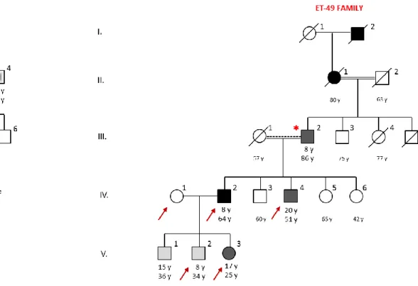
Figure 3.1 Pedigrees of ET-5 and ET-49 families showing the affected and unaffected individuals with essential tremor ... 37
Figure 3.2 Archimedes spiral drawings of ET-5 family members. ... 39
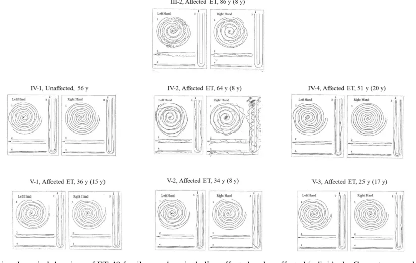
Figure 3.3 Archimedes spiral drawings of ET-49 family members. ... 40
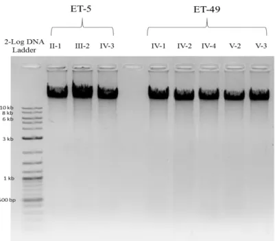
Figure 3.4 Density measurements using agarose gel electrophoresis. ... 42
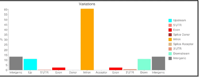
Figure 3.5 Percentage of variants with respect to genomic context for ET-5 and ET-49 families after SnpEff analysis ... 46
Figure 3.6 Filtration and prioritization pipeline for disease causing determinants... 51
Figure 3.7 Pedigree of ET-5 family showing the segregation of essential tremor ……...………...59
Figure 3.8 Segregation analysis results of the ET-5 family……….…... 60
Figure 3.9 Pedigree of ET-49 family showing the segregation of ET with the MMP19 p.Arg456Gln ………...… 61
Figure 3.10 Segregation analysis results of the ET-49 family……….62
Figure 3.11 Predicted protein and domain structure of MMP19……….65
Figure 3.12 Sequence homology of MMP19 protein p.Arg456………..65
Figure 3.13 3D model protein structure of MMP19 protein………... 67
Figure 3.14 Pedigree of ET-31 family showing the affected and unaffected individuals with ET……….………...….70
Figure 3.15 Archimedes spiral drawings of ET-31 family members... 72
Figure 3.16 Density measurements using agarose gel electrophoresis ... 74
Figure 3.17 Percentage of variants with regards to genomic context for the ET-31 family. ... 78
Figure 3.18 Pedigree of ET-31 family segregating essential tremor, with genotypes at IMPG1 p.Arg580Cys. ... 87
xii
Figure 3.19 Segregation analysis result of ET-31 family. ... 88
Figure 3.20 Predicted protein and predicted domain structure of IMPG1 protein ... 91
Figure 3.21 Sequence homology of IMPG1 p.Arg580 ... 91
Figure 3.22 3D model protein structure of IMPG1 protein. ... 93
Figure 4.1.Chromosomal localization of MMP19, protein domain organization of MMP19 and the position of the detected mutation. ... 98
Figure 4.2 Chromosomal localization of IMPG1, protein domain organization of SPACR and position of detected mutation ... 102
xiii
LIST OF TABLES
Table 1.1 Classification of tremors and their characteristics ... 3 Table 1.2 Heritability estimation for selected diseases ... 9 Table 1.3 Mendelian disease gene identifications by exome or genome sequencing .. 24 Table 3.1 Clinical characteristics of tremor in affected and unaffected individuals of families ET-5 and ET-49 ... 38 Table 3.2 Concentrations and qualities of DNA samples isolated from ET-5 and ET-49 families ... 42 Table 3.3 Statistics on the genome sequence produced by the paired-end whole exome sequencing... 44 Table 3.4 Statistics on all variants based on their regions, as identified through whole exome sequencing and SnpEff analysis. ... 47 Table 3.5 Statistics on all variants based on their annotation effects, identified through SnpEff analysis. ... 48 Table 3.6 List of the homozygous damaging variants in proband of the ET-5 family 55 Table 3.7 List of the homozygous variants in proband of the ET-49 family ... 55 Table 3.8 List of the heterozygous variants in the proband of the ET-5 family ...Error!
Bookmark not defined.
Table 3.9 Clinical characteristics of tremor in affected and unaffected individuals of the ET-31 family ... 71 Table 3.10 Concentrations and qualities of DNA samples from the ET-31 family ... 74 Table 3.11 Statistics on the genome sequence produced by the paired-end whole exome sequencing for ET-31 family ... 76 Table 3.12 Statistics on all variants based on their regions, identified through whole exome sequencing and SnpEff analysis for ET-31 family individuals. ... 79 Table 3.13 Statistics of all variants based on their annotation effects, identified through SnpEff analysis for the ET-31 family ... 80 Table 3.14 List of the homozygous damaging variants in proband of the ET-31 Family ... 84 Table 3.15 List of the heterozygous variants in proband of the ET-31 Family ....Error!
xiv
ABBREVIATIONS
BWA Burrows-Wheeler Aligner
CNS Central Nervous System
ESP Exome Sequencing Project
ET Essential Tremor
ExAC Exome Aggregation Consortium
GERP Genomic Evolutionary Rate Profiling
GWAS Genome-Wide Association Study
HTRA2 High Temperature Requirement protein A2
IMPG1 Interphotoreceptor Matrix Proteoglycan 1
ISH In Situ Hybridization
MAF Minor Allele Frequency
MIM Mendelian Inheritance Of Man
MMP19 Matrix Metallopeptidase 19
MRI Magnetic Resonance Imaging
MS Multiple Sclerosis
MSA Multiple Sequence Alignment
NGS Next Generation Sequencing
PD Parkinson's Disease
RT-PCR Real Time Polymerase Chain Reaction
SAMtools Sequence Alignment/Map Tools
xv
Uniprot Universal Protein Resource
VCF Variant Call File
WES Whole Exome Sequencing
WGS Whole Genome Sequencing
URLs
1000 genomes project 1000genomes.org
CADD cadd.gs.washington.edu/
ExAC http://exac.broadinstitute.org/
GATK broadinstitute.org/gatk/
MutationAssesor mutationassesor.org
MutationTaster mutationtaster.org
NHLBI exome sequencing project evs.gs.washington.edu/EVS/
Polyphen-2 genetics.bwh.harvard.edu/pph2/
PROVEAN provean.jcvi.org
SIFT sift.jcvi.org/
1
CHAPTER 1
Introduction
1.1 Introduction to Essential Tremor
1.1.1 Tremor in History
Written accounts of pathological tremors are known from ancient India (5000-3000 BC), Eygpt (700 BC) and Greece (400 BC)1. In addition, records left by physicians such as Galen of Pergamon (130 - 200 A.D.), Sylvius de la Boe (1680), Van Swieten (1745) and Sauvages (1768) contain detailed descriptions of patients exhibiting action and resting tremors, suggesting that the distinction was recognized by these authors1. Action tremors are the characteristic clinical feature of ET and occur during voluntary movement, in contrast to resting tremors that affect stationary extremities2. The first modern description of pathological tremor was provided in 1887 by Dr. Charles Dana, a New York neurologist who identified this condition in several large families1.
1.1.2 Clinical Features of Essential Tremor
Essential tremor (ET [OMIM 190300]) is one of the most common movement disorders, with a frequency of 1% in the population34. The prevalence of ET is also known to be higher among the elderly, with an estimated frequency of 5% in the 65+ age group3. Shaking may occur while patients voluntarily keep a steady posture (postural tremor) or while they move (kinetic tremor)5,2. Kinetic tremor appears during the movement of a body part, such as the rotary movements of the forearm. Postural tremor appears while maintaining a position against gravity, such as holding an outstretched arm in place. Kinetic tremors in the 4-12 Hz range are the characteristic
2
motor feature of ET (Table 1). A trembling voice and slight movements of the chin and head at rest are other symptoms that might be observed in patients. However, ET is not a monosymptomatic disorder and may include a broad range of symptoms, such as parkinsonism, dystonia, sensory abnormalities, cognitive and psychiatric features and sleep disorders6. Recent studies show that certain non-motor disorders, such as cognitive problems, depression, anxiety, social phobia, personal and behavioral changes and hearing and smell disorders are also more frequent in ET patients compared to healthy individuals7.
ET causes serious difficulties in daily activities such as eating, drinking and writing by interfering with motor control8. ET has an effect on upper limbs in at least 95% of
affected individuals. Its effects on other body parts are less frequent: at least 30% of patients have head tremor, at least 20% have voice tremor, at least 10% have chin and face tremor and at least 10% have lower limb tremor9,10. A mild disturbance in gait can also occur in some patients9.
In addition, studies in different populations suggest that essential tremor may cause mild to severe levels of attention deficit, memory loss and impairments in executive functions in patients11.
3
Table 1.1Classification of tremors and their characteristics
Tremor type Frequency Amplitude Feature Disease
Re
stin
g
Low-moderate High (increases with movement)
Muscles are not voluntarily activated and the limb is fully supported
against gravity -Parkinson’s disease -Drug-induced parkinsonism Action Tremor K in etic Tr em o r Intentional
tremor Low (<5Hz) Increases with target directed movement
During any type of movement
-Cerebellar lesions (Stroke, Multiple Sclerosis) -Drug-induced (Lithium,
alcohol) Basic Kinetic
Tremor Variable (3-10 Hz) Does not change with targeted movement Task specific
tremor Variable (4-10 Hz) Variable During specific movement
-Tremor while writing -Tremor in musicians Po stura l Tr em o r
Moderate-High Low, increases with voluntary movement When the limb is positioned against gravity
-Physiological tremor -Essential tremor -Metabolic disorders -After alcohol or drug
4
1.1.3 Diagnosis of Essential Tremor
No specific markers or laboratory tests exist for the diagnosis of ET; medical history and neurological examination are the only criteria for diagnosis. Consequently, the disease is misdiagnosed in approximately 30-50% of cases. The Movement Disorder Society (MDS) has developed a set of consensus criteria in an effort to minimize diagnostic errors12.
According to Deuschl et al., ET can be classified under three groups: sporadic, hereditary and senile. A patient is diagnosed with hereditary ET if they fulfill the consensus criteria of definite (or classical) ET and have at least one other family member that is affected by the disease. In addition, the age of onset should be prior to age 65 for both the patient and at least one affected family member12. In sporadic cases, the patient should fulfill consensus criteria for classical ET at an age of onset lower than 65, and have no other family members with a history of ET. In senile ET, the patient should satisfy the criteria for definite or classical ET with an age of onset after 65 and no family history of the disease10.
1.1.4 Pathology of Essential Tremor
Pathogenesis describes the totality of the changes that occur in an organism following
the onset of disease, including the gradual alterations that accompany its progression. The pathogenesis of ET is not fully understood. According to an initial report with 10 ET cases and 12 controls, pathological findings associated with ET are highly heterogeneous13. These patients could nevertheless be grouped into two categories, based on histopathological examinations of the brain: the first group was characterized by cerebellar degeneration and contained four patients, while the second exhibited Lewy bodies on the brainstem and had six patients13.
5
Cerebellar essential tremor was characterized by large numbers of Bergmann astrocytes and axonal torpedoes in Purkinje cell axons. Bergmann astrocytes form as a non-specific response against neuronal injuries, while axonal torpedoes are swollen regions at the proximal regions of Purkinje cell axons that contain disoriented neurofilaments and may cause Purkinje cell loss13,14. Further cerebellar abnormalities were also identified in ET patients, including neuronal loss, microglial clusters, pallor of the white matter and neuronal atrophy in the dentate nucleus14 (Figure 1.1). These cerebellar
changes are unexpected because it is believed that tremor is mediated by cerebellothalamocortical fibers2. Nevertheless, magnetic resonance imaging studies clearly demonstrate the presence of neuronal loss in the cerebellum15,16.
Instead of cerebellar degradation, a characteristic pattern of Lewy bodies in the brainstem was observed in the remaining six cases13,17. These individuals had numerous Lewy bodies in the locus ceruleus, while fewer bodies were present in the substantia nigra (Figure 1.2). This arrangement of Lewy bodies has not been detected in PD patients and in normal age-matched controls13,17. However, it has been suggested that
ET patients with Lewy bodies in the locus ceruleus are under the risk of developing PD later in life2.
The precise relationship between kinetic tremor and Lewy bodies in the locus ceruleus is not known. However, the locus ceruleus is a well-recognized source of norepinephrine in the central nervous system (CNS) , and axons from this region extend into the cerebellum to form synapses with γ-aminobutyric acid (GABA)-producing Purkinje cells13. The degeneration of the locus ceruleus could compromise this
connection, preventing the stimulation of Purkinje cells and decreasing the production of GABA in the cerebellum13. As such, medical intervention to restore GABA
6
production may serve to ameliorate the effects of ET in patients showing Lewy bodies in the locus ceruleus18,19.
7
Figure 1.1 Section of the dentate nucleus from an individual with essential tremor,
showing neuronal loss. (Copyright 2005 Elsevier. From Louis et al.2 with permission)
Figure 1.1 Multiple Lewy bodies (arrows) are seen in a section of the locus ceruleus
from a patient with essential tremor. (Copyright 2005 Elsevier. From Louis et al.2 with permission)
8
1.1.5 Risk and Etiological Factors for Essential Tremor
Heritability is defined as the degree to which individual genetic variation accounts for phenotypic variation seen in a population. Most heritability estimates have been based on family studies. Epidemiologic evidence shows associations between family history and risk for many diseases (Table). Thus, family history information might aid in assessing risk for a condition, even in the absence of an understanding of the molecular cause of that condition. A family history of ET is a risk factor, and the etiology of ET is often genetic20. The estimates of the proportion of ET patients with a positive family history change between 20% and 90%21.
The frequency of ET increases greatly with age and can be as high as 5.5%22. According
to community-based studies, the prevalence of ET is higher in whites compared to African-Americans23.Nevertheless, there is limited information about the genetic factors responsible for ET. Hereditary ET usually exhibits an autosomal dominant pattern of inheritance; however, a non-Mendelian model of inheritance has also been reported24. Twin studies further support the idea that non-genetic determinants strongly influence the incidence and severity of ET. In one study, pairwise concordance in monozygotic twins was 60%, while another study found this concordance to be 77-93%25,26. β-carboline alkaloids and lead are the main environmental factors identified in case-control studies to potentially produce tremor in patients27,28. β-carboline alkaloids in particular are the most common tremorogenic chemicals in the modern human diet, and Louis et al. showed that the blood concentrations of these chemicals were higher in ET patients than a healthy control group. These chemicals are abundant in meat products that are cooked at high temperatures under elongated times29. In addition, blood concentration of lead is also found to be higher in ET patients than
9
controls, and lead was shown to contribute to cerebellar damage and tremor in ET-diagnosed individuals28,30.
Table 1.2 Heritability estimation for selected diseases
Disease Heritability Estimation
Age-related macular degeneration 49 - 71%31
Alzheimer's disease 58 - 79%32
Autism 30 - 90%33
Bipolar disorder 70%34
Breast cancer 25 - 56%35
Celiac disease 57 - 87%36
Chronic obstructive pulmonary disease 76%37
Colon cancer 13%35
Coronary artery disease 49%38
Crohn's disease 53%39 Epilepsy 70 - 88%40 Essential Tremor 50-90%21 Heart disease 34 - 53%41 Hypertension 30%42 Leukemia 1%35 Lung cancer 8%35 Obesity 70%43 Osteoarthritis 30 - 65%44 Ovarian cancer 40%35 Parkinson's disease 25 - 30%45
Polycystic ovary syndrome 72%46
Prostate cancer 42%35 Rheumatoid arthritis 55%47 Schizophrenia 81%48 Stroke 32%49 Testicular cancer 25%35 Thyroid cancer 53%35 Tourette syndrome 58 - 77%50 Type-1 diabetes 88%51 Type-2 diabetes 26%52
10
1.1.6 Genetic Heterogeneity in Essential Tremor
Despite the widespread occurrence of sporadic ET, twin and family studies have also demonstrated that genetics is an important factor for the disease. According to twin studies, heritability ranges from 45 to 90% for ET25,26. Genome-wide linkage studies performed in North American and Icelandic ET families have identified genetic loci containing ET-associated genes on chromosomes 3q13 (ETM1; OMIM:190300)53, 2p22-p25 (ETM2; OMIM: 602134)54 and 6p23 (ETM3; OMIM: 611456)55.
In 1997, a locus (ETM1, also called FET1) has been identified through the genome-wide scan of 16 Icelandic families, encompassing a total of 75 affected individuals.
FET1 was clearly inherited under a dominant mode of inheritance. The logarithm of
odds (LOD) score was found to be 3.71, but the highest single family LOD score was calculated as 1.29, which is below the threshold of significance for mapping a monogenic disorder to a marker53. In 2006, Jeanneteau et al. identified a Ser9Gly variant in the DRD3 gene at the ETM1 locus in 23 of 30 French families56. This gene codes for the dopamine D3 receptor, which is expressed in Purkinje cells and mediates ERK activation56,57. However, it is not clear whether the Ser9Gly variant is cosegregated with the 16 Icelandic families linked to ETM153. In addition, the
association between ET and the Ser9Gly variant in DRD3 was not observed in other populations58,59.
In 1997, Higgins et al. used linkage analysis to identify the ETM2 locus on chromosome 2p22-p25 in an American family of Czech descent. This gene segregated under autosomal dominant inheritance in this family, with a LOD score of 5.9260. In 2003, this group also identified an ancestral haplotype on chromosome 2p24.2, which was found in 29% of 45 American individuals with a family history of ET60. The HS1-binding protein 3 gene (HS1BP3, OMIM 609359) was also suggested as a gene of
11
interest for ET, and a relationship has been proposed between the disease and an A265G substitution affecting two genes and seven transcripts on a minimal critical region (MRC)61,62. However, the relationship between ET and HS1BP3 has not been confirmed by other studies55,63. In addition, various association and linkage studies have failed to establish correlations between ET and mutations in the ETM2 locus64,65.
The ETM3 locus is located on chromosome 6p23 and segregates with ET in an autosomal dominant pattern. The locus in question was identified through genome-wide non-parametric and parametric linkage analyses in seven North American families including 325 patients. However, no pathologic variants were found following the sequencing of 15 candidate genes in the ETM3 locus55. In addition, linkage to the ETM3
locus was not present in two different linkage studies in Italian families65,66.
Genome-wide association studies have led to the identification of two additional genes. The leucine rich repeat and Ig domain containing 1 (LINGO1, MIM 609791) gene was identified from families of American and European descent67. However, LINGO1 variants that segregate with the disease are found in non-coding intronic regions68. Zhou
et al. suggested that intronic variants, in conjunction with other environmental
components, could disrupt the function of LINGO1 and facilitate the onset of ET69. The
solute carrier family 1 (SLC1A2, MIM 600300) gene was also identified as a potential risk factor for ET in European populations through a genome-wide association study70. However, meta-analysis demonstrated that these variants are not causative21.
FUS/TLS (fused in sarcoma/translated in liposarcoma, MIM137070) is the first
putative ET gene identified through a combination of whole exome sequencing and linkage analysis. One pathogenic variant of FUS is a c.868C>T nonsense mutation that segregated with a large family of French-Canadian origin and was found to cause
12
autosomal dominant ET71. In addition, two rare missense variants (M392I, R377W) were also determined by further screening of ET cases, suggesting that FUS mutations account for a subset of ET cases72,73.
Moreover, Rajput et al. established a connection between the genetic causes of ET and PD in the context of two VPS35 and DNAJC13 variants74. 571 ET patients were genotyped for VPS35 c.1858G>A (p.D620N) and DNAJC13 c.2564A>G (p.N855S) variants and two individuals were observed to harbor the latter mutation, which was previously identified in PD patients74.
Our group has also contributed to the identification of ET-associated genes through exome sequencing. Our recent report showed that a p.G399S missense mutation in HTRA2, which encodes a mitochondrial protease, is responsible for both essential tremor and Parkinson’s disease. Whole exome sequencing of a six-generation
consanguineous Turkish family illustrated that ET is present in both homozygous and heterozygous individuals for this allele, while homozygotes additionally developed Parkinson’s disease later in life (Figure 1.3). As such, homozygous family members for
HTRA2 p.G399S are more severely affected than heterozygotes, even though the presence of a single variant allele is sufficient to cause ET75. HTRA2 produces a serine
protease protein which is released from mitochondria to the cytosol and initiates apoptosis by binding to apoptotic inhibitors76. According to mouse model studies, HTRA2 was also found to be related with Parkinson’s disease. A Mnd2 mouse model study demonstrated that HtrA2 p.S275C causes loss of protease activity and leads to the degeneration of motor neurons, resulting in a phenotype with ataxia, repetitive movements, and akinesis77. In another transgenic mice study, HtrA2 p.G399S was identified as a loss-of-function allele through the behavioral comparison of transgenic mice expressing either wild-type HtrA2 or the variant allele. Mice overexpressing the
13
WT variant showed motor impairments that were not present in HtrA2 p.G399S-overexpressing mice, suggesting that the overexpression of the variant allele can rescue its function78.
Another ET-related exome sequencing study has discovered a pathogenic variant of the
TENM4 (MIM 610084) gene in a Spanish population. In particular, two novel missense
mutations were shown to exhibit a dominant inheritance pattern in one family79. Clark et al. has published a recent study concerning the identification of candidate genes for familial early onset essential tremor. In this study, they identified four genes in five different families following the analysis of 37 early-onset families. In two different families, they determined a heterozygous variant (c.46G4A (p.Gly16Ser)) and a heterozygous variant (c.164C4T (p.Pro55Leu)) in the NOS3 gene. The p.Asp379Glu variant in KCNS2, p.Gly350Arg variant in HAPLN4 and p.Ala133Val variant in USP46 genes were also shown to segregate with ET in three different families80.
14
Figure 1.3 Segregation of HTRA2 p.G399S in a six-generation consanguineous Turkish family, illustrating that essential tremor is present in
subjects both homozygous and heterozygous for this allele. Homozygous family members for HTRA2 p.G399S are more severely affected than heterozygotes. Individuals that were selected for exome sequencing (IV: 3, IV: 4, VI: 5) are indicated with arrows. Black symbol illustrates ET-diagnosed and red symbol illustrated PD-ET-diagnosed individuals. N: non-variant, wild-type allele, glycine; V: the variant allele serine for HTRA2 p.G399S.
15
1.1.7 Treatment of Essential Tremor
Several treatments can alleviate the effects of ET for some patients; however, there is no conclusive cure for ET. Drugs can improve the quality of social life by reducing functional disability or discomposure81. Different β-blockers can be used for treatment, with propanol being the most common drug for this purpose82. Atenolol, sotanol and
nadolol are other β-blockers that are used for the treatment of ET patients82. Primidone is the drug of choice for the treatment of tremor82. Primidone is more effective when taken in combination with propranol, and can reduce tremors in 75% of patients83,84. If the patient suffers from both tremors and anxiety, benzodiazepines such as alprazolam and clonazepam are preferred due to their psychoactive effects85. These drugs enhance
the effect of the neurotransmitter gamma-aminobutyric acid (GABA) on GABA receptors, resulting in sedative, muscle relaxant and anti-anxiety effects86. Patients who do not respond to drugs can instead be treated by surgical approaches such as deep brain stimulation87.
Nevertheless, it should be emphasized that ET treatments would benefit greatly from the identification of new causative genes for essential tremor, which will enable the development of personalized treatment methods depending on the genetic history of each patient.
16
1.2 Disease Gene Identification Strategies
1.2.1 Traditional Methods
Genes associated with Mendelian diseases are typically identified through Sanger sequencing of candidate genes, which are selected based on their similarity with genes related to similar diseases, relevance between predicted protein function and the physiology of the disease, and positional mapping analyses88. However, non-Mendelian diseases require higher scrutiny due to their variability, and a broad range of analysis techniques have been developed for their identification. The most important genetic mapping methods are karyotyping89, linkage analysis90, homozygosity mapping91, copy number variation analysis92 and SNP based association analysis93.
Linkage analysis is the primary tool for the positional mapping of Mendelian genes94. In 1910, Thomas Morgan used Drosophila cultures to determine that traits are co-inherited because of the physical proximity of the factors responsible for their appearance95. Accordingly, genes that are clustered in close proximity on a chromosome tend to co-segregate because they are less likely to experience recombination than distant genes. Based on this theory, distances between trait-encoding genes can be determined through recombination studies95. Consequently, if two traits are consistently co-segregated in a given family, it is likely that they are linked96.
Logarithm-of-odds (LOD) scores can be used to determine the probability that a particular gene is truly associated with the disease of interest rather than co-segregating due to chance97. LOD scores greater than 3 are considered to be suitably robust for suggesting linkage in Mendelian disorders98.
17
Linkage mapping has been highly successful in mapping genes and gene variants affecting Mendelian traits. However, it is relatively inefficient in mapping loci underlying common polygenic disorders99. Genome wide association studies (GWASs) are based upon the principle of linkage disequilibrium (LD) at the population level and focus on the nonrandom associations between alleles at different loci. These association studies have been utilized for the evaluation of many common genetic variants in different individuals, depending on the associations between single nucleotide polymorphisms (SNPs) and common diseases such as diabetes, heart diseases, auto-immune diseases and psychiatric disorders99. Accordingly, if one allele is more frequent in individuals with a specific disease, that variant is evaluated to be associated with that disease99. The first successful GWAS was published in 2005 and described the relationship between age-related macular degeneration and two SNPs that exhibited significantly changed allele frequencies in affected patients compared to healthy controls100. However, GWAS is not a suitable technique for the identification of rare genetic diseases, as it usually identifies hundreds of variants that are common in the population (i.e. with frequencies over 5%), have to establish biological relevance to the disease of interest and explain only a small portion of the total inherited risk. In addition, only a small number of risk alleles identified by GWAS efforts are nonsynonymous SNPs in exons101.
Homozygosity mapping is another successful approach for the identification of recessive disease genes and is done by determining homozygous regions shared by affected family members102. This method depends on genome wide single nucleotide
polymorphism (SNP) arrays of affected and unaffected family members. However, despite recent advances in sequencing technology, the genetic basis of several common
18
disorders remains unknown due to the limited number of family members, locus heterogeneity, variable expressivity and reduced penetrance.
1.2.2 Next Generation Sequencing
The first true sequencing method was described in 1977 by the hallmark paper of Sanger et al., whose approach involved the use of dideoxy-nucleotides for controlled chain termination103. This discovery contributed to the development of Sanger sequencing in 1987104,105 and has been the basis of DNA sequencing for nearly 30 years afterwards. Although technological developments have increased the length of DNA that can be sequenced (to c. 1000 bp) and facilitated the parallel sequencing of DNA strands in capillaries, Sanger sequencing was nevertheless limited by its inability to analyze DNA in a high-throughput manner.
This inability was one of the main impediments to the Human Genome Project (HGP), which was initiated in 1990 with the aim of identifying the 3 billion base pairs that comprise the human genetic code. Despite this rocky start, however, the project eventually finished ahead of schedule through the development of a new approach called shotgun sequencing106. This method involves the production of small DNA fragments through the mechanical or enzymatic breakdown of a DNA isolate, which are subsequently cloned into sequencing vectors107. Cloned DNA sequences can then be sequenced individually, and the entire genome sequence can be determined by the alignment and assembly of fragmented sequences using overlapping regions107. The
10-year project culminated in the publication of first drafts by Lander et al.108 and Venter
et al. in 2001106, followed by a complete publication by Jasny and Roberts et al. after three more years109. The success of HGP also led to the initiation of certain important projects, such as the International HapMap Project and 1000 Genomes Project. Of these, the International Hapmap project aims to produce a haplotype map of the human
19
genome and determine the patterns of common genetic diversity in the genetic code110. This approach attracted great attention to the prospect of identifying the genetic causes behind common human disorders, and was completed in 2005 with the genotyping of 4.6 million SNPs110. The 1000 Genomes project aims to uncover more detailed genetic factors involved in human health and disease111. This project also provides various resources on human genetic variation, including the genomes of at least one thousand volunteers from a broad range of ethnic groups111.
Shotgun sequencing also spurred the development of massive parallel sequencing techniques that would be used in next generation sequencing (NGS) efforts112–114. NGS is performed by breaking the entire genome into smaller chunks and ligating them to pre-designated adapters for rapid, irregular reading during DNA synthesis. Consequently, the technique is also known as sequencing by synthesis115. As NGS produces millions of short, fragmented DNA sequences that only partially overlap, the problem in sequencing moved from the logistics and chemistry of the sequencing reaction to data management and analysis of the resulting fragments. As such, new algorithmic approaches have been developed (and are actively in development) to create a coherent genetic sequence from these disjointed fragments116.
1.2.3 Whole Exome Sequencing Approach
Whole exome sequencing (WES) is a practical application of NGS and limits itself to the variations found in the coding regions (exons) of all currently known genes. Newly developed technologies such as whole genome and whole exome sequencing have replaced traditional methods because of their decreased costs, higher accuracy, capacity for rapid diagnosis and compatibility with a number of user-friendly software for data analysis117,118.
20
The human genome contains around 3 x 10⁹ bases, but only about 3 x 10⁷ base pairs (i.e. 1% of the genome) are responsible for encoding proteins119. The WES approach
involves the sequencing of this comparatively small amount of DNA, using hybridization techniques to separate it from the non-coding “chaff”120,121. Two main approaches exist for the “capture” of exomes: solution-based and array-based applications.
In solution-based WES, DNA samples are broken down and hybridized to target regions in the genome by biotinylated oligonucleotide probes. Magnetic streptavidin beads can then be used to bind to biotinylated probes, thereby retaining the coding regions while the rest of the DNA is washed off. The remaining sample is then amplified through polymerase chain reaction (PCR), resulting in the selective enhancement of the coding regions (Figure 1.4)122. Array-based WES is similar to its solution-based counterpart, except that the probes are bound to a high-density microarray123. It also needs a lower amount of DNA and is more efficient than solution-mediated approaches124,125. NGS has greatly advanced our capacity to determine the genetic factors underlying various disorders. The first report about WES was published by Ng et al. in 2009121. In this report, four individuals that were diagnosed with Freeman-Sheldon syndrome were sequenced by whole exome sequencing, and following the exclusion of common variants, the MYH3 gene was discovered as a previously unknown gene for a rare Mendelian disorder121. The first application of NGS for molecular diagnosis of a patient came from Choi et al., who reported the clinical diagnosis of a suspected Bartter syndrome individual of Turkish origin126. They demonstrated that a well-conserved
recessive mutation in SLC26A3 gene is associated with congenital chloride diarrhea and utilized WES as a clinical tool that is able to facilitate the diagnostic evaluation of patients in addition to disease gene discovery126. Their study therefore allowed the
21
correct diagnosis of individuals who were suffering from congenital chloride diarrhea (CLD) but were misdiagnosed with Bartter syndrome126.
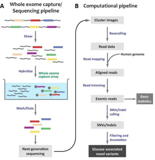
Figure 1.4 Overview of an exome sequencing pipeline. SNV: single nucleotide variant,
Indel: Insertion/Deletion. (A) In sequencing pipeline, isolated genomic DNA is sheared into 200-300 bp fragments. Exomes are hybridized with biotinylated probes and non-targeted regions are washed and eluted. At the end, libraries are sequenced. (B) Flowchart of a computational pipeline for WES, including data analysis. (Copyright 2012 by The Korea Genome Organization. From Choi et al 127 with permission)
22
1.2.4 Identification of Disease-Causing Genes
Family based linkage analysis is usually the first step for disease gene identification, although it may be insufficient for the determination of diseases caused by de novo mutations128. Family based linkage studies have become common place with development of microarrays for GWAS and NGS129.
WES has been performed for the determination of disease causing variants in various rare Mendelian diseases, including spastic paraplegia, brain malformations, cerebellar ataxia, retinitis pigmentosa and nonsyndromic mental retardation.130–134 (Table 1.2). WES has also been performed for complex and common disorders such as Alzheimer’s disease, essential tremor, multiple sclerosis and the familial form of Parkinson’s disease to investigate rare genetic variants related with these diseases 71,135–137.
Determination of disease causing genes starts with the alignment of WES data to the human reference genome, which is obtained from the HGP. A minimum of 30 reads per base is generally considered to be sufficient for accurate sequencing. Certain databases (dbSNP, ExAC, 1000genomes, EVS) are then used to determine whether the identified variants have previously been reported and exclude common variants. After this step, the remaining variants have their functions annotated, and the neutral-coding variants are eliminated based on functional filtering. This step ensures that all identified variants contain protein-damaging mutations such as nonsense mutations and frameshift insertions or deletions, which generate non-functional or truncated proteins.
Several in silico analysis methods (such as SIFT, PROVEAN, PolyPhen2,
MutationAssessor and MutationTaster) are then used to evaluate the deleteriousness of missense mutations and determine how damaging each variant will be to the complete protein138–142.
23
The exome sequencing approach has been successfully used for the identification of disease-causing variants. However, this method still suffers from a number of restrictions. First, our knowledge of protein-coding regions is still incomplete; consequently, it is probable that some “non-coding” regions in fact contain genes.
Second, some sequences fail to be targeted by the capture probe design, resulting in “leaks” in the sequenced exomes. Third, not all sequences can be mapped to the
reference genome. And lastly, it is becoming increasingly evident that other sequences such as microRNAs, promoters and conserved elements also play important roles in the pathogenesis of various disorders, but none of these are considered in exome sequencing94.
Overall, however, exome sequencing is a rapid, powerful and cost effective technique for the identification of new genetic variants for Mendelian and non-Mendelian diseases.
24
Table 1.3 Mendelian disease gene identifications by exome or genome sequencing
(Copyright 2011 BioMed Central Ltd. From Gilissen et al143 with permission)
Disorder Inheritance Gene identified Scope References Congenital chloride diarrhea Recessive SLC26A3 Exome Choi et al. 126
Miller syndrome Recessive DHODH Exome Ng et al.144
Charcot-Marie-Tooth neuropathy Recessive SH3TC2 Genome Lupski et al.145
Metachondromatosis Dominant PTPN11 Genome Sobreira et al.146
Schinzel-Giedion syndrome Dominant SETBP1 Exome Hoischen et al.147
Nonsyndromic hearing loss Recessive GPSM2 Exome Walsh et al. 148
Perrault syndrome Recessive HSD17B4 Exome Pierce et al.149
Hyperphosphatasia mental retardation
syndrome Recessive PIGV Exome Krawitz et al.
150
Sensenbrenner syndrome Recessive WDR35 Exome Gilissen et al.151
Cerebral cortical malformations Recessive WDR62 Exome Bilguvar et al. 130
Kaposi sarcoma Recessive STIM1 Exome Byun et al.152
Spinocerebellar ataxia Dominant TGM6 Exome Wang et al. 131
Combined hypolipidemia Recessive ANGPTL3 Exome Musunuru et al.153
Complex I deficiency Recessive ACAD9 Exome Haack et al. 154
Autoimmune lymphoproliferative syndrome Recessive FADD Exome Bolze et al. 155
Amyotrophic lateral sclerosis Dominant VCP Exome Johnson et al.156
Nonsyndromic mental retardation Dominant Various Exome Vissers et al.157
Kabuki syndrome Dominant MLL2 Exome Ng et al.158
Inflammatory bowel disease Dominant XIAP Exome Worthey et al.159
Nonsyndromic mental retardation Recessive TECR Exome Caliskan et al.160
Retinitis pigmentosa Recessive DHDDS Exome Züchner et al. 133
Osteogenesis imperfecta Recessive SERPINF1 Exome Becker et al. 161
Dilated cardiomyopathy Dominant BAG3 Exome Norton et al. 162
Hajdu-Cheney syndrome Dominant NOTCH2 Exome Simpson et al. 163
Hajdu-Cheney syndrome Dominant NOTCH2 Exome Isidor et al. 164
Skeletal dysplasia Recessive POP1 Exome Glazov et al.165
Amelogenesis Recessive FAM20A Exome O'Sullivan et al.166
Chondrodysplasia and abnormal joint
development Recessive IMPAD1 Exome Vissers et al.
167
Progeroid syndrome Recessive BANF1 Exome Puente et al. 168
Infantile mitochondrial cardiomyopathy Recessive AARS2 Exome Götz et al. 169
Sensory neuropathy with dementia and
hearing loss Dominant DNMT1 Exome Klein et al.
170
25
1.3 Consanguinity
Consanguinity has been identified as a risk factor for rare diseases since the turn of the century. Consanguineous individuals are those who descend from strongly related (i.e. second cousins or closer) ancestors, and share an extraordinarily high amount of genetic material. The first report detailing the detrimental effects of consanguineous marriages was published in 1902 and concerns alkaptonuria patients172. This phenomenon can also be seen in Mendelian inheritance. The co-occurrence of two recessive alleles is much more frequent in these families: for example, assuming that each couple shares 1/8 of their genome, their progeny will be homozygous for 1/16 of all loci.
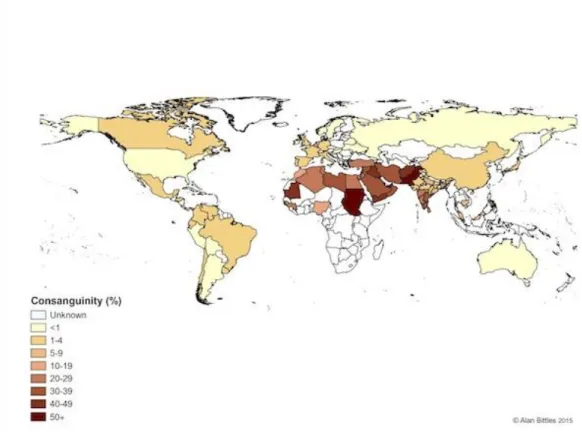
The frequency of recessive diseases strongly correlates with the rate of consanguineous marriages. More than 50% of marriages are consanguineous in certain regions of North Africa, South India, West Asia and eastern and southern rims of the Mediterranean, resulting in strong incidence of genetic disorders in these populations173 (Figure 1.5). As such, the likelihood of recessive disorders creates potentially detrimental health effects in consanguineous marriages, and the disease genes in question can be identified by determining the regions that are shared between family members174. Accordingly, a prospective disease-associated variant should be found in other affected family members, but also be absent from unaffected family members. These regions can be determined by genome-wide SNP genotyping arrays by analyzing shared homozygous regions175. In addition to autosomal recessive disorders, consanguinity is also important
for diseases exhibiting a dominant inheritance pattern because the presence of a second allele often alters the severity of the disease75.
26
Figure 1.5 Global prevalence of consanguinity. (http://www.consang.net with
27
1.4 Outline of the Thesis
In this dissertation, I detail the identification of the candidate genes related with familial essential tremor under five chapters, including this introductory section.
The first chapter provides a general outlook on the identification of genetic disorders in general and ET in particular. The second chapter is a methods section that explains how clinical assessment of families, whole exome sequencing analysis and disease gene identification were performed.
The third chapter covers the identification of two genes related with ET. ET is one of the most frequent movement disorders in humans; however, its genetic causes and pathophysiology are unknown. We identified candidate genes in three ET families based on WES. In two independent Turkish ET families, we identified a rare and potentially damaging variant in the matrix metallopeptidase gene (MMP19) as a causative allele. In a third Turkish family, we determined a rare and damaging missense mutation in IMPG1. Our results suggest that MMP19 and IMPG1 might be responsible for essential tremor in these families.
The fourth chapter contains a discussion of my findings, and the final chapter summarizes my study and offers future perspectives on determining the genetic causes of ET.
28
CHAPTER 2
Material and Methods
2.1 Recruitment of Family Members
Three families; ET-5, ET-31 and ET-49, were investigated in the present study. ET-5 is a family from Bayır Village (Muğla, Turkey) and has four individuals (spanning four
generations) suffering from essential tremor. ET-49 is a consanguineous family from Ilgın (Konya, Turkey), includes 6 patients and a control group, and has a family history
of ET for at least five generations. ET-31 family is a consanguineous family from Corum, Turkey includes 11 patients and 4 control individuals, and has a family history of ET for at least four generations. All individuals are coded by the blood ID. The study was approved by the Institutional Review Board at Bilkent University. Additional cohorts involving patients having similar neurological phenotypes were included in the study to identify other patients with candidate mutation. Before the study, all the participants were asked to sign an enlightened consent form that was prepared according to the guidelines of the Ministry of Health in Turkey.
2.2 Clinical Investigations
Clinical investigations were performed at Ankara University Medical School and Hacettepe University Medical School. All clinical examinations were performed according to the Helsinki Declaration (http://www.wma.net). Each participant was examined for essential tremor consistent with criteria of both the Washington Heights-Inwood Genetic Study of Essential Tremor (WHIGET) and the Consensus Statement of the Movement Disorder Society on Tremor (MDS). After the diagnosis of essential tremor, a pedigree was developed and histories about the distribution of the disease was
29
collected through communication with family members. Each participant’s tremor score was graded for resting and postural tremors according to 0 to +3 tremor ratings. As stated in WHIGET criteria, 0 means no visible tremor, +1 is of low amplitude; +2 tremor is of moderate amplitude and +3 tremor is of large amplitude. Participants were asked to carry through four distinct tasks to evaluate kinetic tremor: finger-to-nose movement, pouring water, drinking water from a cup and drawing. Tremor severity was estimated in the course of each task. To separate essential tremor symptoms from Parkinson Disease, bradykinesia, muscular rigidity and postural instability were also evaluated according to the diagnostic criteria of the UK Parkinson Disease Society Brain Bank.
2.3 DNA Isolation from Family Members
Peripheral blood samples from patients and their relatives were collected in K3-EDTA-containing BD Vacutainer® Blood Collection tubes (Becton Drive, NJ, USA) by a specialist utilizing veinpuncture technique. Blood samples were carried to the laboratory under cold chain conditions and stored in 1.5 ml micro centrifuge tubes containing 1 mL blood samples at -80 ºC.
Each individual's genomic DNA was isolated from 200 µL of peripheral whole blood samples using Nucleospin® Blood kit (Macherey-Nagel Inc., PA, USA) in accordance with the manufacturer’s instructions. To get high DNA concentrations that are required for whole exome sequencing, washing steps were repeated two times and DNA was eluted with double distilled water rather than elution buffer.
Quality of genomic DNA samples was measured by densitometry analysis using horizontal 1% agarose gel electrophoresis with TAE Buffer. To analyze the quantity and purity of DNA samples, spectrophotometric analysis was performed with
30
NanoDropTM ND-1000 Spectrophotometer (NanoDrop Technologies, Inc, DE, USA). DNA concentration, A260/280 and A260/230 values were recorded. A260/280 ratio is used for determining the purity of DNA and a ratio of ~1.8 is generally accepted as “pure”. The 260/230 ratio was used for assessing the purity of nucleic acids and ideal
values are generally in the range of 2.0-2.2.
2.4 Library construction and whole exome sequencing
2.4.1 Sample Selection and Preparation
Two affected individuals (II-1 and IV-3) and one control (III-2) were selected from the ET-5 family for WES. Likewise, four affected (III-2, IV-2, IV-4, V-2 and V-3) and one unaffected (IV-1) individuals were selected from the ET-49 family, and three affected individuals (III-2, III-3 and III-4) and one control (II-13) were chosen from the ET-31 family for exome analysis. High quality DNA were isolated from selected individuals as described.
2.4.2 Library Construction
Library construction was performed for whole exome sequencing using a commercial kit (Illumina TruSeq DNA Sample Prep Kit), based on the manufacturer’s instructions. Accordingly, DNA samples were diluted in suspension buffer to get a final amount 2.5 µg in 55 µL and sheared into 200-300 bp for 120 s at 6 ºC in a sonicator to produce
dsDNA fragments with 3’ or 5’ overhangs. For checking the quality of DNA, sheared
DNA was run on agarose gel. The final amount of sheared DNA was 1 µg. The overhangs were then converted to blunt ends utilizing End Repair Mix for 30 min at 30
ºC. After cleaning DNA fragments with 160 µL of AMPure XP Beads, this mixture was
placed on a magnetic stand. The DNA samples with beads were washed with 80% ethanol and dried, and DNA samples were resuspended in Resuspension Buffer. The
31
DNA fragments were incubated with A-tailing mix for 30 min at 30 ºC, allowing single ‘A’ nucleotides to be added to 3’ ends of DNA fragments and preventing them from
ligating to one another. The ligation reaction was performed by adding multiple paired-end adapters with ‘T’ nucleotides on their 3’ fragments, which were ligated to
complementary dsDNA fragments following incubation for 10 min at 30 ºC. This reaction was inactivated by Stop Ligation Buffer and the fragments were cleaned up with AMPure XP Magnetic Beads and amplified by PCR. Exomes were captured by SeqCap EZ Exome Capture Kit (Roche) and hybridized with biotinylated capture probes at 47 ºC for 72 h. The exomes were cleaned with Qiagen QIAquick PCR Purification Kit and their concentration and quality were determined with ABI system KAPA Illumina Library Quantification Kit with Real Time PCR. Libraries were sequenced on an Illumina HiSeq2500 and converted to basecall (.bcl) file format.
2.5 Bioinformatics
2.5.1 Whole Exome Sequencing Data Analysis
Qualities of sequences were determined using Ilumina Real Time Analysis Software and sequences were converted to .bcl file format. The data included in .bcl files were converted to FASTQ files through Illumina CASAVA software. Burrows-Wheeler Aligner176 (BWA, v0.6.1-r104) was used to align the fragments to the human reference
genome177 (hg19). PCR duplications were removed by Sequence Alignment/Map Tools (SAMtools) software package178 and sequence depth of exonic regions were obtained by BED tools179. SNP and Indel variants and base quality score recalibration were done with Genome Analysis Tool Kit180 (GATK; v3.0–0-g6bad1c6). Variants were positionally and functionally annotated with SnpEff181 (4.2, 2015-12-05) and
32
of the determined variants, regions were analyzed using the Integrative Genomics Viewer (IGV) platform, which allows interactive assessment of genomic datasets183.
2.5.1 Filtration and Prioritization of Candidate Gene Variants
The following criteria was used to obtain a list of variants for ET-affected families: (1) including only protein-coding region variants, (2) excluding all common variants that have MAF bigger than 0.02 in ExAC (Exome Aggregation Consortium), (3) including variants shared between affected individuals within a family. After variants were annotated positionally and functionally with annotation tools, only protein coding region variants were selected based on their annotation impacts, which were determined with SnpEff. ‘HIGH’ and ‘MODERATE’ variants were selected for further analysis. The ‘HIGH’ variants included frameshift, splice acceptor, splice donor, stop loss
variants and ‘MODERATE’ variants are composed of inframe insertion/deletion, missense variants and splice region variants. Common SNPs and variants that are frequently seen in the population (minor allele frequency (MAF) is bigger than 0.02) were filtered using ExAC, the 1000 Genomes Project, the NHLBI Exome Sequencing Project and our in-house database and selected for further analysis. Variants were removed without protein altering effect(synonymous, start-retained and stop-retained) and remained with nonsynonymous effect in coding region (i.e. missense, nonsense) or alteration resulting in frameshifts, premature stop codons, loss of stop codons, coding INDELS, splice site and splice region variants. We filtered a variant if it is also found as homozygous or compound heterozygous in the genome of his control sample. For patient-patient pairs, we selected the variants that are shared by affected individuals within the same family. We also considered hemizygous sequence variants on the X chromosome found in brother–brother pair families as candidate mutations. We filtered heterozygous variants only if the control sibling has the same variation and/or any of
33
the control individual has the same variant in homozygous state. We prioritized as potentially damaging if they generated a premature stop codon or were mutations with the following scores on in silico prediction tools: Provean <-2.5, SIFT =<0.05, Polyphen2 HDIV; D: Probably damaging >=0.957, P: possibly damaging 0.453<=pp2hdiv<=0.956, Polyphen2 Hvar D: Probably damaging >=0.909, P: possibly damaging 0.447<=pp2hvar<=0.909, MutationTaster prediction for A (disease causing automatic) and D (disease causing), MutationAssessor >1.94 (M: Moderate and H: High), CADD >=10, GERP >=3 and PhastCons_Mammalian >=0.90.
2.6 Sanger Sequencing
Segregation analysis of the candidate variants for other informative relatives of ET-5, ET-49 and ET-31 families were performed in order to determine if the candidate genes were co-segregated with autosomal ET. The appropriate primers (Table A1) were designed with the Primer3Plus bioinformatics tool. Segregation analysis was performed with Chromas Lite analysis software (Technelysium Pty Ltd) and Multiple Sequence alignment was done with Clustal Omega.
2.7 Model 3D Protein Structure of Candidate Variants
In order to deduce structural alignments and compare the active sites and other relevant regions of the candidate variants, 3D structures of related protein models were derived from Swiss Model, Raptor X or Phyre2 and analyzed with Swiss-PdbViewer (DeepView, v4.1)184.