Annals of Medical Research
DOI: 10.5455/annalsmedres.2019.12.874
Original Article
Long duration single pulse intense pulsed light application
in erythematotelangiectatic rosacea: An instrumental
retrospective study
Erkin PekmezciIstanbul Medipol University, Faculty of Medicine, Department of Dermatology, Istanbul, Turkey Copyright © 2020 by authors and Annals of Medical Research Publishing Inc.
Abstract
Aim: Erythematotelangiectatic rosacea is a challenging condition due to poor therapeutic options. Treatment with intense pulsed
light is partially effective after numerous sessions. Regarding the ‘thermal relaxation time’ principle and characteristic histopathology of erythematotelangiectatic rosacea, dilated capillaries and venules throughout the dermis, we proposed that directly targeting the largest vessels available by applying a long duration single pulse intense pulsed light mode, should decrease the mean number of treatment sessions to obtain a significant outcome.
Material and Methods: Eighty seven erythematotelangiectatic rosacea patients who completed the proposed one month apart,
four times consequtive intense pulsed light treatments with 25 ms pulse duration, single pulse and 10 J/cm2 fluence in the years of 2017-2019 were recruited retrospectively. Instrumental evaluations and self-assessment scorings were performed during the treatment period.
Results: No adverse effect was recorded. Instrumentally evaluated results revealed a significant erythema quenching effect obtained
at the end of 2nd treatment session (p=0.0011). Considering the decrease in erythema, both the instrumental and self-assessment values were found extremely significant after the 4th session, compared to initial values (p<0.0001, for both).
Conclusion: Considering the fast improvement recorded, intense pulsed light therapy with long pulse duration, single pulse, and low
fluence may present a time saving and effective treatment option in erythematotelangiectatic rosacea patients with varied levels of severity. Also, the satisfactory outcome obtained by directly targeting the largest vessels available beginning from the initiation of therapy period, may be regarded as an indicator of the practicability of an identical mode of intense pulsed light treatment in diverse clinical presentations of erythematotelangiectatic rosacea.
Keywords: Erythema; fast improvement; rosacea; intense pulsed light
Received: 22.12.2019 Accepted: 17.03.2020 Available online: 23.05.2020
Corresponding Author: Erkin Pekmezci, Istanbul Medipol University, Faculty of Medicine, Department of Dermatology, Istanbul, Turkey E-mail: [email protected]
INTRODUCTION
Rosacea is a chronic condition which is manifested by facial flushing, persistent erythema, telangiectasia, papules and pustules. It generally involves mid-facial region. There are four rosacea subtypes; erythematotelangiectatic, papulopustular, phymatous and ocular, but also two or more forms may be together in one patient (1,2). Erythematotelangiectatic rosacea (ETR) is the most frequent one and also the type which has the strongest vascular component among all (1). Generally women are affected more often. Although recent large retrospective database studies have demonstrated prevalence rates ranging from 1.3% to 2.1%, such low percentages may not be standing for the true rates, as the administrative databases generally recorded the patients with severe rosacea forms, potentially missing a substantial amount
of patients with mild illness. Considering these limitations, rosacea as a dermatological disease might be more common than previously reported (3).
The precise causal pathway of rosacea still remains obscure. In the ground of genetic predisposition; bacterial, environmental, psychological aspects and nourishment styles are considered as triggering factors. One leading theory is based on vascular response: Flushing and telangiectasias are major symptoms in rosacea patients. A combination of structural abnormality of cutaneous vasculature on the face, higher blood flow of facial skin and vascular dysregulation via humoral and neural mechanisms may explain the rationale behind this theory (2). Since it is a multifactorial chronic inflammatory ailment that waxes and wanes, the goal of treatment should be
subsiding acute flares with rapid acting treatments and maintaining the results with lifestyle management (3). Although the antibiotics and antiinflammatory therapy control the microbial and inflammatory components, there is no totally effective treatment of persistent facial redness and erythematotelangiectatic lesions (2,4). This challenge can be targeted with specific light and laser therapies to a certain extent (2,5).
Intense pulsed light (IPL) is known to be a rewarding therapeutic device in various diseases of skin including rosacea. Parameter flexibility makes the targeting of both superficial and deep vessels as well as large areas of telangiectasia, erythema, and flushing, but requires clinical expertise for appropriate treatment with minimized complications (3). Although histo-pathologic examination of ETR biopsies typically show nonspecific features, one important characteristic change is the presence of enlarged, dilated capillaries and venules in diverse thicknesses of the dermis (6). Also, according to ‘thermal relaxation time’ (TRT) principle, for larger vessels with longer TRTs, pulse durations can be lengthened to obtain an effective outcome. Even though the treatment of ETR with IPL is mostly significant according to physicians’ objective evaluations (7), in patients’ perspective, it is partially effective after numerous treatment sessions. Therefore we proposed that, whatever the patient’s severity of erythema, directly targeting the largest vessels available in the dermis should shorten the period of therapy and lower the number of treatment sessions, by applying long duration single pulse IPL mode. In this retrospective analysis it is also figured out the effect of a standardized IPL therapy with an identical mode, adjusted for larger vessels, in pure ETR patients with various degrees of severity.
MATERIAL and METHODS
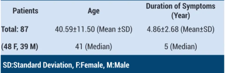
Eighty seven patients with ETR and persistent facial erythema, who referred to our hospital dermatology clinic for treatment, and completed four consecutive monthly IPL sessions in the years of 2017-2019 were retrospectively recruited. For both legal and ethical considerations the signed approval of each patient was received before each treatment session. All of the patients had Fitzpatrick skin types II-III. The demographic data and duration since the onset of symptoms are shown in Table 1.
Table 1. The demographic data and duration of the symptoms
Patients Age Duration of Symptoms (Year) Total: 87 40.59±11.50 (Mean ±SD) 4.86±2.68 (Mean±SD)
(48 F, 39 M) 41 (Median) 5 (Median)
SD:Standard Deviation, F:Female, M:Male
The erythema severity values of each patient were measured instrumentally by using the erythema probe of Multi Skin Test Center® MC 1000 (Courage+Khazaka
electronic GmbH, Cologne, Germany) immediately before the first and third sessions and one month after the fourth session. So the initial, 2nd and 4th session values were obtained. The measurements were taken from three points as the left and right malar regions, and the dorsum of the nose. The arythmetic mean of three values was considered as the erythema index (EI) of the patient. Also each patient was asked to fulfill a self-assessment scale immediately before the first and one month after the fourth sessions. Patient’s self-assessment scale was comprised of 5 semiquantitative erythema values as; 0: Null, 1: Mild, 2: Moderate, 3: Marked, and 4: Severe erythema. The patients were told to assess the 1-4 values as approximate 25%-100% quantities of a red hue. The value selected was considered as the patient’s self-assessment score (PSAS).
IPL sessions were performed by using 550 nm cut-off filter and 8.3 cm2 handpiece of the IPL module of Synchro
HP Platform™ (DEKA M.E.L.A. S.r.l., Florence, Italy). The identical mode in the form of single pulse with 25 ms duration and 10 J/cm2 fluence, was applied to all patients
in all sessions. Here, although the longest pulse duration available was selected, the fluence was kept relatively low. Only one pass was applied on mid-facial region with the rectangular 8.3 cm2 hand piece vertically, beginning
from the left and ending on the right preauricular regions. The patients were cautioned to use a low-potent topical steroid for 5 days b.i.d. to preclude inflammation after each session and a 50+ SPF sun proof three times during daylight throughout the therapy and assessment periods. Each patient was photographed on the same days which the instrumental evaluations were performed. Summary of the study outline is presented in Table 2. Student’s t test (two-tailed, paired) was used for statistical analysis.
Table 2. Summary of the study outline
T0 T1 T2 T3 T4 Instrumental evaluation X X X
Self assessment evaluation X X
IPL session X X X X
Photograph X X X
T:Time in Months
RESULTS
None of the patients presented adverse effects such as purpura or pigment alterations and the expected mild increase in the erythema after the IPL sessions waned in 1-3 days. The mean and standard deviation values of instrumental (EI) and self-assessment (PSAS) evaluations are presented in Table 3. A gradual decrease in the mean values of evaluations can be seen through the progression of treatment sessions.
Table 3. The mean and standard deviation (SD) values of the instrumental and self assessment evaluations
Initial (T0) 2nd Session (T2) 4th Session (T4) Instrumental (EI) 63.78±4.80 (SD) 61.48±4.30 (SD) 60.07±4.03 (SD) Self assessment (PSAS) 2.87±0.69 (SD) NA 2.45±0.62 (SD)
T:Time in Months, NA: Not Applicable
The comperative p values of the evaluations are shown in Table 4. As it can be seen in the table, the significance of instrumental p value after the first two sessions (2nd session vs. initial) is greater compared to the last two (4th vs. 2nd session). Both the instrumental (EI) and self-assessment (PSAS) values are found extremely significant after the 4th sessions compared to initial values.
Table 4. The comperative p values of the instrumental and self assessment evaluations 2nd Session vs. Initial (T2 vs. T0) 4th vs. 2nd Session (T4 vs. T2) 4th Session vs. Initial (T4 vs. T0) Instrumental (EI) =0.0011 =0.0275 <0.0001 Self assessment
(PSAS) N.A. N.A. <0.0001
T:Time in Months, NA: Not Applicable
Figure 1. Improvement curve. The line shows the improvement
in instrumental evaluations (EI) after the 2nd and 4th treatment sessions compared to initial values
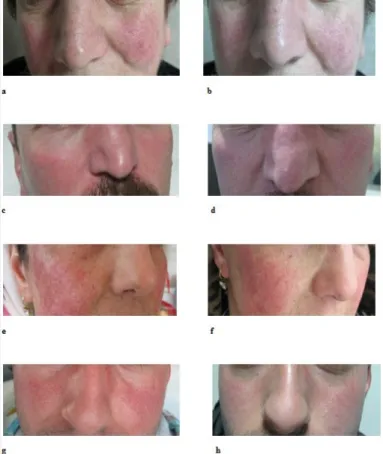
It is also formed an improvement curve (Figure 1) by taking the decreases in instrumental evaluation (EI) means after the 2nd and 4th sessions from the initial mean which are demonstrated in Table 3. In Figure 1 it can be seen that approximately two-thirds (i.e. 2.3/3.71) of total improvement was achieved after the 2nd session. In Figure 2, various degrees of clinical improvement after the 2nd session are demonstrated as before (a,c,e,g,) and after (b,d,f,h) photographs.
Figure 2. Various degrees of clinical improvement after the 2nd
treatment session. Before (a,c,e,g) and after (b,d,f,h) photographs
DISCUSSION
IPL has long been shown to be pertinent in various clinical frameworks with the ease of parameter flexibility (3). By using the desired cut-off filter it can be utilized the theory of selective photothermolysis with the non-coherent, polychromatic light the device emits (7). Also, most IPL systems provide a relatively large spot size increasing the efficacy of treatment and enabling deeper light penetration. IPL treatments are clinically effective on the local cutaneous vasculature, which is the main cause of the symptomatology seen in patients with rosacea (3). An important characteristic change in histo-pathology of ETR is the presence of enlarged, dilated capillaries and venules in varied thicknesses of the dermis. These vessels generally have bizarre shapes and are sometimes defined as ‘tortuous’. Mild to moderate edema, which is rarely visible to the naked eye, but is almost always present on histology, is responsible for the clear aspect of the upper dermis. Edema in patients with ETR may be due to an increased number of vessels and defective permeability (6).
Filters are used to limit the wavelengths emitted by the IPL device in order to improve dermal penetration and minimize absorption of energy by other chromophores. The longer wavelength filters allow the deeper penetration of the light (8). As the shorter wavelengths emitted by IPL devices are highly absorbed by melanin, patients with darker skin phototypes should be treated with a longer
wavelength filter (8,9). IPL energy is delivered as a series of single, double, or triple pulse sequences with total pulse durations of 2–25 ms and interpulse delays of 10-500 ms. Longer pulse durations are used to heat deeper vessels more effectively. This way of application also reduces the risk of purpura and post inflammatory pigment alterations (10).
The aim of treating vascular lesions with IPL is to raise the blood vessel temperature high enough to cause its coagulation, leading to its destruction and replacement by fibrous granulation tissue. Because of its polychromaticity, IPL can target oxyhemoglobin and deoxyhemoglobin which are predominantly found in clinically red and blue lesions respectively. The diameter of the targeted vessel is an important consideration when choosing the treatment settings of the IPL device (11). Selection of pulse duration is mainly guided by TRT, which is related to the size of the target. As a general rule, larger the chromophore, longer is the TRT since large objects take long time to cool (12). For small vessels in the papillary dermis with a diameter of 0.1 mm, the TRT is about 10 ms, but for larger and deeper vessels of 0.3 mm in diameter, it increases approximately nine-fold (11) as TRT is proportional to the square of the vessel’s diameter (8). Therefore for larger vessels with longer TRTs, pulse durations can be lengthened. This is especially important in telangiectasias, as when the vessel seems to be resistant to treatment, increasing the pulse duration can result in an effective clinical response (7). More difficulty is encountered while lasing the smaller targets, because the shorter pulse duration required makes the epidermis more vulnerable due to its melanin content (12).
Although IPL has a proven high grade efficacy in the treatment of telangiectasias (13), the effective number of treatment sessions and parameter adjustments vary to a great extent for each individual patient (14-16), and sometimes it is a matter of trial and error. With the classical shorter but multiple pulse applications, generally four or more IPL sessions are needed for vascular lesions (7). In our study, the adjusted identical mode for all patients, revealed a significant result, the two-thirds of total improvement, at the end of 2nd session. In Figure 1, it can be seen the marginal decrease in improvement as the number of sessions increase. It is quite likely that the long duration single pulse application primarily affected the majority of the larger and dilated vessels with longer TRTs in the dermis, rather than the smaller superficial ones, resulting the early erythema quenching effect. These preliminary data are notable due to both displaying the applicability of an identical mode of IPL treatment targeted for larger vessels in different clinical presentations of ETR, and revealing a significant efficacy both objectively (i.e. instrumental evaluation) and subjectively (i.e. self-assessment scoring).
CONCLUSION
Regarding the fast improvement recorded without any adverse effect, IPL application with long duration, single
pulse, and low fluence may be a time saving and effective treatment in ETR patients with varied levels of severity. Competing interests: The authors declare that they have no competing interest.
Financial Disclosure: There are no financial supports.
Ethical approval: This article that has been written regarding the three year retrospective data obtained at our routine dermatology outpatient clinic. The relevant and applicable ethical principles (i.e. Declaration of Helsinki) are considered during the writing process.
REFERENCES
1. Lim HS, Lee SC, Won YH, et al. The efficacy of intense pulsed light for treating erythematotelangiectatic rosacea is related to severity and age. Ann Dermatol 2014;26:491-5.
2. Saghari S, Keri J, Shanler S, et al. Rosacea (type 2 sensitive skin). In: Bauman L, ed. Cosmetic Dermatology. 2nd edition. New York: McGrawHill 2009;128-32.
3. Weinkle AP, Doktor V, Emer J. Update on the management of rosacea. Clin Cosmet Investig Dermatol 2015;8:159-77.
4. Lee WJ, Lee YJ, Lee MH, et al. Prognosis of 234 rosacea patients according to clinical subtype: the significance of central facial erythema in the prognosis of rosacea. J Dermatol 2016;43:526-31.
5. Hoffmann MA, Lehmann P. Physical modalities for the treatment of rosacea. J Dtsch Dermatol Ges 2016;14::38-43.
6. Micali G, Gerber PA, Lacarrubba F, et al. Improving treatment of erythematotelangiectatic rosacea with laser and/or topical therapy through enhanced discrimination of its clinical features. J Clin Aesthet Dermatol 2016;9:30-9.
7. Kaufman J. Lasers and light devices. In: Bauman L, ed. Cosmetic Dermatology. 2nd edition. New York: McGrawHill 2009;212-20.
8. 8. Vallee JA, Kelly KM, Rohrer TE, Arndt KA, Dover JS. Lasers in the treatment of vascular lesions. In: Kaminer MS, Arndt KA, Dover JS, Rohrer TE, Zachary CB, eds. Atlas of Cosmetic Surgery. 2nd edition. Philadelphia: Saunders Elsevier 2009.p.135-53.
9. Rohrer TE, Ort RJ, Arndt KA, et al. Lasers in the treatment of pigmented lesions. In: Kaminer MS, Arndt KA, Dover JS, Rohrer TE, Zachary CB, eds. Atlas of Cosmetic Surgery. 2nd edition. Philadelphia: Saunders Elsevier 2009;155-77.
10. Husain Z, Alster TS. The role of lasers and intense pulsed light technology in dermatology. Clin Cosmet Investig Dermatol 2016;9:29-40.
11. Goldberg DJ. Current trends in intense pulsed light. J Clin Aesthet Dermatol 2012;5:45-53.
12. Patil UA, Dhami LD. Overview of lasers. Indian J Plast Surg 2008;41:101-13.
13. Wat H, Wu DC, Rao J, et al. Application of intense pulsed light in the treatment of dermatologic disease: a systematic review. Dematol Surg 2014;40:359-77. 14. Schroeter CA, Haaf-von Below S, Neumann HA.
Effective treatment of rosacea using intense pulsed light systems. Dermatol Surg 2005;31:1285-9.
15. Kassir R, Kolluru A, Kassir M. Intense pulsed light for the treatment of rosacea and telangiectasias. J Cosmet Laser Ther 2011;13:216-22.
16. Campolmi P, Bonan P, Cannarozzo G, et al. Intense
pulsed light in the treatment of non-aesthetic facial and neck vascular lesions of 85 cases. J Eur Acad Dermatol Venereol 2011;25:68-73.
![Ofloxacin plus Rifampicin versus Doxycycline plus Rifampicin in the treatment of brucellosis: a randomized clinical trial [ISRCTN11871179]](data:image/gif;base64,R0lGODlhAQABAIAAAP///wAAACH5BAEAAAAALAAAAAABAAEAAAICRAEAOw==)