1
CELL PENETRATING PEPTIDE AMPHIPHILE INTEGRATED LIPOSOMAL SYSTEMS FOR ENHANCED DELIVERY OF CARGO TO TUMOR CELLS
A THESIS
SUBMITTED TO THE MATERIALS SCIENCE AND NANOTECHNOLOGY PROGRAM OF GRADUATE SCHOOL OF ENGINEERING AND SCIENCE OF
BILKENT UNIVERSITY
IN PARTIAL FULFILLMENT OF THE REQUIREMENTS FOR THE DEGREE OF
MASTER OF SCIENCE
By
MURAT KILINÇ August, 2013
2
I certify that I have read this thesis and that in my opinion it is fully adequate, in scope and in quality, as a thesis of the degree of Master of Science.
...
Assoc. Prof. Dr. Ayşe Begüm Tekinay (Advisor)
I certify that I have read this thesis and that in my opinion it is fully adequate, in scope and in quality, as a thesis of the degree of Master of Science.
...
Assoc. Prof. Dr. Mustafa Özgur Güler
I certify that I have read this thesis and that in my opinion it is fully adequate, in scope and in quality, as a thesis of the degree of Master of Science.
... Assoc. Prof. Dr. Fatih Büyükserin
Approved for the graduate school of engineering and science: ...
Prof. Dr. Levent Onural
3 ABSTRACT
CELL PENETRATING PEPTIDE AMPHIPHILE INTEGRATED LIPOSOMAL SYSTEMS FOR ENHANCED DELIVERY OF CARGO TO TUMOR CELLS
Murat Kılınç
M.S. in Materials Science and Nanotechnology Supervisor: Assoc. Prof. Ayşe Begüm Tekinay
August, 2013
Liposomes have been extensively utilized as effective nanocarriers due to their enhanced solubility, higher stability and greater ability to facilitate the slow release of encapsulated drugs compared to free drug administrations. Liposomes are also preferred as drug vectors due to their non-toxic nature, biodegradability and structural resemblance to the cell membrane. However, their low internalization efficiencies pose an important challenge for their use in drug delivery applications. Internalization issues inherent in many liposomal systems can be circumvented by the use of cell penetrating peptides, which non-covalently attach on the liposome surface and greatly enhance liposomal uptake in a receptor- and charge-dependent manner.
In this study, we examined the liposomal dynamics effected through the integration of an amphiphilic cell penetrating peptide into a simple liposome system. Peptide amphiphiles with a cell penetrating arginine-rich domain were incorporated into liposomal membranes formed by negatively charged dioleoylphosphoglycerol (DOPG) phospholipids in the presence of cholesterol. Throughout the present study, we sought to analyze the effect of peptide incorporation on (a) the physical characteristics, such as size, surface potential
4
and membrane polarity, of the liposomal membrane, (b) the alterations in the encapsulation and delivery mechanisms of hydrophilic (Rhodamine B) and hydrophobic (Nile Red) drug models and (c) the enhancement of therapeutic capability in liposomes loaded with the drugs Doxorubicin-HCl and Paclitaxel. Our results revealed that the modification of liposomes by cell penetrating peptide amphiphiles results in the improvement of cargo delivery and the enhancement of the therapeutic effects of the anti-cancer drugs Doxurubicin and Paclitaxel.
5 ÖZET
LİPOZOMLARIN HÜCRE DELİCİ PEPTİTLERLE MODİFİYE EDİLİLEREK ANTİKANSER İLAÇ GÖNDERİMİNİN GELİŞTİRİLMESİ
Murat Kılınç
Malzeme Bilimi ve Nanoteknoloji Programı, Yüksek Lisans Tez Yöneticisi: Assoc. Prof. Ayşe Begüm Tekinay
Ağustos, 2013
İlaçlara kattığı yüksek çözünürlük, stabilite ve zaman salınımı gibi özellikleri olması itibariyle liposozmlar etkili nano taşıyıcılar olarak yaygınca kullanılmaktadır. Toksik etki göstermemeleri, biyobozunurluğu ve hücre zarına benzer yapıda olmaları da lipozomların tercih edilmesindeki diğer sebeplerdir. Buna rağmen lipozomların hücre içine alınımının kısıtlı olması, lipozomların ilaç taşıyıcısı olarak kullanımında önemli sorunlar teşkil etmektedir. Bu durumun üstesinden gelme amacıyla lipozomları reseptör ve yük bağıntılı işlev gören hücre delici peptitlerle modifiye edip lipozomların hücre içine girişini desteklemek kayda değer bir stratejidir.
Bu çalışmada, liposomları tek adımda modifiye eden bir sistem geliştirilmiş olup, entegre olan amfifilik hücre delici peptitlerin lipozomların etkinliği üzerindeki rolü test edilmiştir. Arjinin miktarı itibariyle zengin hücre delici bölümü ve hidrokarbon zincirine sahip hücre delici peptitler DOPG ve kolesterolden oluşan eksi yüklü lipozomlara entegre edilmiştir. Çalışmada modifiye edilmiş lipozomların (a) boyut, yüzey potansiyeli ve zar polaritesi gibi fiziksel özellikleri, (b) hidrofilik Rhodamin B ve hidrofobik Nile Red gibi ilaç modellerinin enkapsülasyon ve iletim etkinliği, (c) Paclitaxel ve Doxurobicin-HCl gibi
6
anti kanser ilaçlarla yüklenmesi sonrası terapütik etkinliği araştırılmıştır. Sonuçlar ışığında amfifilik hücre delici peptitlerle kovalent bağ olmaksızın modifiye edilmiş lipozomların ilaç iletimi yönünden daha iyi olup, anti kanser ilaçlarla yüklendiğinde terapütik etkinliğini arttığını göstermiştir.
7
ACKNOWLEDGEMENTS
I have spent two years in this laboratory, as a part of the NBT and BML research groups, and I was blessed with many an invaluable experience during my stay.
I would like to thank my advisor Dr. Ayşe Begüm Tekinay for her support and guidance. I learned a lot from her, and I would like to express my gratitude for her contributions to my academic and personal development. Her guidance was beyond academic subjects, and she prepared me for future by all means. I also would like to thank Dr. Mustafa Özgür Güler for his guidance and support throughout the research we have conducted. This work would not be possible without Dr. Güler.
It was great to work with Dr. Rükan Genç and Melis Şardan. This project could not be accomplished without them. I am grateful for their collaboration and friendship.
When I first started to my master’s study, I have been working with Seher Üstün, Ayşegül Tombuloğlu and Hilal Ünal in various projects. I appreciated to work with them and I would like to thank for their cooperation and companionship.
I would like to thank Aref Khalily, Ruslan Garrifulin and Fatih Genişel for their companionship and assistance. By dealing with my endless questions, they taught me what I know about the peptide synthesis.
Ceren Garip and Nuray Gündüz were my companions in muscle regeneration projects. I am grateful for their friendship, endurance and sincere efforts. They worked really hard and did not loosen support either in good and bad times of ongoing projects.
And lastly, I would like to thank my dear brother Rashad Mammadov for his companionship. He enlightened my path in a lifelong mental quest and provided me insights to become
8
invincible, fearless and without boundaries. I shall consider myself most fortunate, should I have opportunity to work with him in future.
9 TABLE OF CONTENTS ABSTRACT ... 3 ÖZET ... 5 ACKNOWLEDGEMENTS ... 7 TABLE OF CONTENTS ... 9
LIST OF TABLES & FIGURES ... 12
CHAPTER 1 INTRODUCTION ... 15
1.1CANCER ... 15
1.2STRATEGIESFORCANCERTREATMENT ... 15
1.2.1 Cytoreductive Surgery ... 16
1.2.2 Radiotherapy ... 17
1.2.3 Immunotherapy ... 17
1.2.4 Chemotherapy ... 18
1.3NANOSCALEDELIVERYOFANTICANCERAGENTS ... 20
1.3.1 Polymer Based Nanocarriers ... 22
1.3.2 Inorganic Nanospheres ... 23
1.3.3 Liposomes ... 24
1.3.3.1 Physical Properties of Liposomes ... 25
1.3.3.3 Characterization of Liposomes ... 29
Determination of Lamellarity: ... 30
Size Measurement: ... 30
Determination of Surface Charge: ... 31
Encapsulation Efficiency: ... 32
10
1.3.3.4 Functionalization of Liposomes ... 33
1.3.4 Liposome Surface Modifications ... 36
1.3.4.1 Antibodies ... 36
1.3.4.2 Small Molecules ... 37
1.3.4.3 Proteins ... 38
1.3.4.4 Sugars ... 39
1.3.4.5 Peptides ... 39
1.3.4.6 Cell Penetrating Peptides ... 40
1.4MOTIVATIONANDGOALS ... 43
CHAPTER 2 CELL PENETRATING PEPTIDE MODIFICATION FOR IMPROVED LIPOZOMAL DELIVERY OF ANTI CANCER THERAPEUTICS ... 44
2.1INTRODUCTUON ... 44
2.2MATERIALS&METHODS ... 47
2.2.1 Chemicals and Solutions ... 47
2.2.2 Peptide Synthesis and Characterization ... 48
2.2.3 Liposomes ... 48
2.2.3.1 Liposome Preparation ... 48
2.2.3.2 Characterization of Liposomes ... 49
Particle size and Zeta-Potential by Dynamic Light Scattering: ... 49
Transmission Electron Microscopy: ... 49
Nile Red Polarization Studies: ... 49
Quantification of the number of membrane integrated peptide amphiphile: ... 50
Determining encapsulation capacities of liposomes: ... 50
2.2.4 Cell Culture ... 51
11
2.2.4.2 Liposome Uptake Quantification ... 52
2.2.2.4 Imaging ... 53
2.3RESULTS&DISCUSSION ... 54
2.3.1 Synthesis and Characterization of Peptides and Liposomes ... 54
2.3.1.1 Peptides ... 54
2.3.1.2 Liposomes ... 58
2.3.2 Safety of Liposomal Carriers ... 64
2.3.3 Enhanced Uptake via CPP Modification ... 66
2.3.3.1 Liposomes with Hydrophilic Content ... 66
2.3.3.2 Liposomes with Hydrophobic Content ... 70
2.3.3 Release Dynamics of Liposomes ... 76
2.3.4 Therapeutic Effects of CPP Modified Liposomes ... 78
2.3.4.1 Encapsulation of Doxorubicin and Paclitaxel ... 78
2.3.4.2 Therapeutic Effects of Doxorubicin Loaded Liposomes ... 80
2.3.4.3 Therapeutic Effects of Paclitaxel Loaded Liposomes ... 84
2.4CONLUSION&FUTUREPERSPECTIVES ... 88
12
LIST OF FIGURES & TABLES
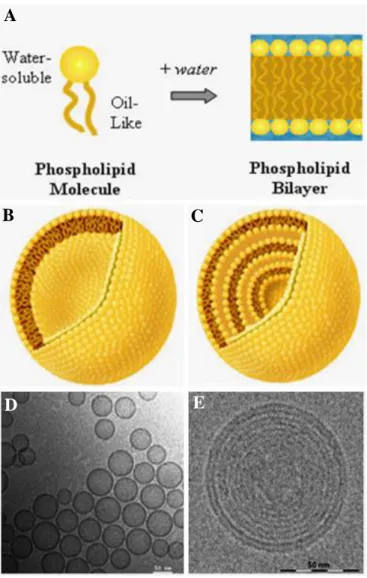
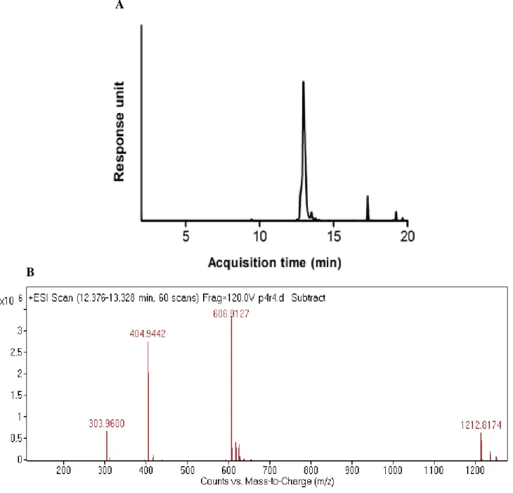
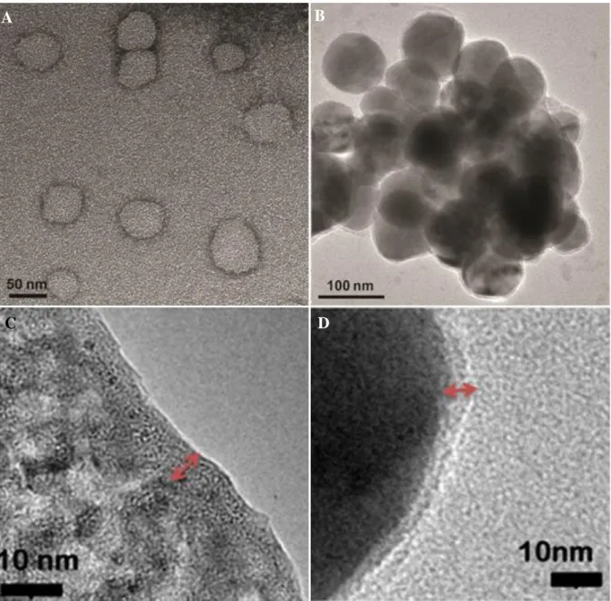
Figure 1 Basic physical properties of unilamellar and multilamellar lipsomes. A. Schematics of basic lipids and bilayer. B. Schematic of unilamellar liposomes. C. Schematic of multilamellar liposomes. D. Cryo-TEM image of unilamellar liposomes. Adopted from Battersby et al.[20] E. Cryo-TEM image of a multilamellar liposome. Image acquired from vironova.com. Scale bars identify 50 nm distances. All schematics adopted from encapsula.com. ... 27 Figure 2 Strategies for liposome modification. (Top) Liposome Modification with hydrophilic polymer without bioactivity, frequently used for passive tumor targeting. (Bottom) Modification with cell penetrating peptides improves cellular uptake and drug delivery. (Left) Modification with antibodies utilized to achieve specific active targeting. (Right) Modification with bioactive small molecules to achieve ligand specific active targeting. Image adopted form Paliwal et al. [27] ... 35 Figure 3 Chemical representation of C12 (Lauryl)-PPPPRRRR-NH2, cell penetrating peptide amphiphile. Peptide consists of 3 main domains. C12 forms hydrophobic domain and facilitates incorporation with bilayer. Poly-Proline at linker domain prevents b-sheet formation and allows Poly-Arginine tip to reach bilayer surface. Poly-Arginine sequence at cell penetrating domain improves internalization and endosomal escape. ... 55 Figure 4 A. Liquid chromatogram of Lauryl-PPPPRRRR-NH2. B. Mass spectrum of corresponding peptide molecule. Mass data [M+H]+ (calculated) = 1212.54, [M+H]+ (observed) = 1212.82 (observed [(M+2H)/2]+= 606.91, [(M+3H)/3]+= 404.94, [(M+4H)/4]+= 303.96) ... 57 Figure 5 Transmission electron microscope images of liposomes. A & C. DOPG:Chol B & D. DOPG:Chol:PA. C & D are close section of single liposomes and red arrows point the unilamellar membrane. Scale bars = 10 nm, 50 nm and 100 nm, respectively. ... 61
13
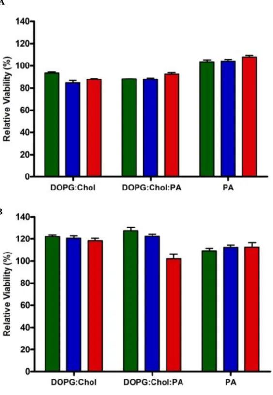
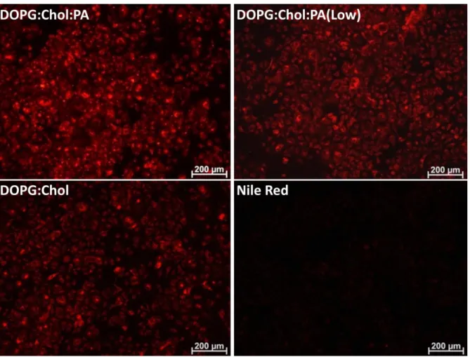
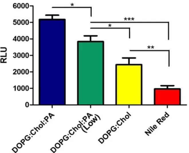
Figure 6 Polarity change in DOPG:Chol liposomes from polar towards nonpolar when positively charged cell penetrating peptide amphiphile was integrated to their membrane. Black DOPG:Chol liposomes. Red DOPG:Chol:PA liposomes. ... 63 Figure 7 Viability of MCF7 cells against drug free liposomes after cell exposure to liposomes w/o PA and only PA in free form for A. 4 h and B. 24 h. Samples were optimized to final peptide concentrations of 250 (green), 25 (blue) and 12.5 (red) µM. Results were normalized to untreated cells in PBS. (n=4) ... 65 Figure 8 Uptake of 4.5 µM Rhodamine B within DOPG:Chol and DOPG:Chol:PA liposomes by MCF7 breast cancer cells after 3 h of treatment. Free Rhodamine B was used as control. Acquired signals were normalized to protein concentration of samples to calculate relative uptake value. (*** stands for p < 0.0001) (n=4) ... 69 Figure 9 Uptake of 10 µM Nile Red by MCF7 cells. NR was administrated in free or liposome encapsulated form for 3 h. Fluorescent microscopy images of cells following liposomal DOPG:Chol:PA, DOPG:Chol:PA (Low), DOPG:Chol, and free Nile Red administration. ... 73 Figure 10 Uptake quantification of 10 µM Nile Red by MCF7 cells. NR was administrated in free or liposome encapsulated form for 3 h. DOPG:Chol:PA and DOPG:Chol:PA(Low) differs in terms of amount of CPP incorporated. Uptake levels quantified via ethanol lysis method prior to fluorescence spectroscopy. ... 75 Figure 11 In vitro release profile of DOPG:Chol and DOPG:Chol:PA liposomes at pH 7.4 and pH 5.5. Both liposome formulations are stable at physiological condition while they both show slow release slightly triggered by acidic pH. ... 77 Figure 12 Dose response of MCF7 cells against free Doxorubicin-HCl and Doxorubicin-HCl loaded DOPG:Chol and DOPG:Chol:PA liposomes. After 24 h of exposure to Doxorubicin-HCl, viability of cells was measured by Alamar Blue. Results were normalized to untreated cells in PBS. (*** stands for p < 0.001, ** stands for p < 0.01, * stands for p < 0.05) (n=4) . 81
14
Figure 13 Time response of MCF7 cells to 1, 3 and 6 h of 10 µM free or liposomal Doxorubicin-HCl treatment. Following administration, cells were incubated in fresh media for further 24 h and viability of cells was measured. (*** stands for p < 0.001, ** stands for p
< 0.01, * stands for p < 0.05) (n=4)... 83
Figure 14 Dose response of MCF7 cells against free Paclitaxel and Paclitaxel loaded DOPG:Chol and DOPG:Chol:PA liposomes within spectrum of concentrations ranging from 0.2 nM to 10 µM final concentration. Subsequent to 24 h of exposure to Paclitaxel, viability of cells was measured. Cell proliferation in the presence of DOPG:Chol:PA liposomes was significantly lower (p < 0.001) than both DOPG:Chol liposome and free Paclitaxel at all Paclitaxel concentrations except 10 μM. At 10 μM Paclitaxel concentration, free Paclitaxel showed significantly lower effect compared to both DOPG:Chol and DOPG:Chol:PA liposomes (p < 0.001). (n=4) ... 85
Figure 15 Time response of MCF7 cells to 1, 3 and 6 h of 30 µM free or liposomal Paclitaxel exposure. Cytotoxic effects of Paclitaxel loaded DOPG:Chol and DOPG:Chol:PA liposomes. All results were normalized to viability level of nontreated cells. (*** stands for p < 0.001, ** stands for p < 0.01, * stands for p < 0.05) (n=4) ... 87
TABLE 1 PHYSICAL PROPERTIES OF LIPOSOMES ... 59
TABLE 2 ENCAPSULATION OF MODEL DYES ... 67
TABLE 3 ENCAPSULATION OF TRACKING DYES BY LIPOSOMES ... 71
TABLE 4 DOX ENCAPSULATION CAPACITIES ... 79
15 CHAPTER 1
INTRODUCTION
1.1 CANCER
The World Health Organization (WHO) lists cancer as the second most prevalent cause of death in developed countries, after cardiovascular diseases. In the developing world, cancer is ranked as the third most common lethal medical condition. In addition to being a terminal illness, cancer poses an immense economic and psychological burden on the affected and their families. Factors like aging, unhealthy lifestyles (malnutrition, smoking etc.) and pollution increase the incidence and mortality rates of cancer. By 2020, 16 million new cases are estimated to emerge around the world. Due to the frequency of incidence, high mortality rates and difficulty of treatment associated with many cancers, efficient treatment strategies against this disease are urgently necessary.
1.2 STRATEGIES FOR CANCER TREATMENT
Cancer can be managed with variety of options, such as cytoreductive surgery, radiotherapy, chemotherapy and immunotherapy. The selection among these therapies depends on parameters such as the type (e.g. whether the tumor in question is metastatic), location, size and structure of the malignant tumor. In all treatments, the primary goal is the removal of all cancerous cells with minimum collateral damage to the native, healthy body tissue. However, current treatment technologies are rarely capable of eliminating all malignant cells while keeping normal cells perfectly healthy. When tumors are structurally distinct and less invasive, or located in non vital organs, it may be possible to
16
remove all cancer tissue from body by surgery. But this is not a common case, since many types of malignancies either occur in vital organs or have a natural tendency to invade vital tissues and form malignant structures that cannot be separated from native tissue by mechanical means. In addition, cancer cells can spread to very distant tissues via metastasis, or may recur in the same location and necessitate the resumption of treatment. Chemotherapeutic agents and radiotherapeutic applications are often capable of eliminating cancer cells to a greater extent than invasive surgery, but also cause systemic side effects and result in the death of healthy cells. Immunotherapy is not associated with particularly severe side effects, yet it is also slow to act and provides enough time for tumor cells to evolve defensive mechanisms.
A variety of cancer treatment options, each with their distinct sets of advantages and disadvantages, are available for specific types of cancers in specific degrees of progress. We will discuss these treatment options briefly to develop a broad understanding of cancer treatment before detailing advanced anticancer drug delivery strategies.
1.2.1 Cytoreductive Surgery
Local tumors with non-invasive characteristics can be ectomized by basic surgery. This procedure involves the removal of either the tumor tissue or the entire organ in which the tumor is nested. Removal of excessive prostate tissue in prostate cancer and mastectomy in breast cancer are examples of such surgical procedures. The main concern in anti-cancer surgeries is the elimination of leftover anti-cancer cells on the tumor site. Since anti-cancer cells reproduce at rates much greater than their healthy equivalents, even a single remaining malignant cell may well end up forming a new tumor.
17
Against metastatic cancers, surgery can only provide short term relief and cannot serve as a conclusive treatment. Even if the majority of the tumor mass is removed from its initial location, surviving cancer cells frequently establish metastatic tumors throughout the body by traveling through blood and lymph streams. The development of these new tumors almost inevitably necessitates further treatment.
1.2.2 Radiotherapy
Cancer cell irradiation is an effective way to kill target cells. In this procedure, radio waves are focused on target tissues and cancer cells are exposed to ionizing radiation. The main rationale of this treatment is to disrupt cell metabolism via radiation-induced DNA damage. When the cell physiology is disrupted, survival and division rates decrease and the target tissue shrinks in mass. The main problem associated with radiotherapy is the exposure of healthy cells to ionizing radiation. Compared to tumor cells, healthy cells have more efficient DNA repair mechanisms, and are much more resistant to radiation. However, the failure to repair a single incident of oncogenic mutation may result in the occurrence of cancer in any tissue exposed to radiation therapy. Furthermore, as with surgery, radiotherapy alone is not a sufficient treatment method against metastatic tumors. Local tumors can be destroyed efficiently, yet new treatments will be necessary when metastasized cancer cells form new tumor masses throughout the body.
1.2.3 Immunotherapy
Cancer cells can be eliminated by manipulating the immune system to fight against tumor cells. For this purpose, cytokines and interferons are frequently utilized to enhance the immune alertness of body. Since cancer cells divide often and have weak DNA repair mechanisms, they are likely to produce mutated antigens that can be recognized when
18
immune alertness is high. When these tumor antigens are recognized by T cells and natural killer cells, the tumor tissue is rapidly invaded by the immune system and apoptotic signals are produced to initiate cell death in tumor cells.
Cell based therapies, which involve the recruitment or priming of immune cells against malignant tumors, are also among available methods. Perhaps the most striking example of such a treatment approach is dendritic cell therapy, which utilizes the priming of these cells against tumor antigens.[1-2] In this procedure, isolated dendritic cells are co-cultured with tumor cells and primed against tumor antigens. When dendritic cells are transplanted to the patient, they activate the immune system against cancer cells, which eventually leads tumor shrinkage. This procedure is capable of increasing life expectancies by up to several years, even with conditions normally associated with very poor prognoses, such as pancreas cancer.
Overall, immunotherapy is an efficient way to manage cancer, yet there are several weaknesses associated with it. First, a boost to the immune system may trigger the development of autoimmune disorders, which will harm healthy tissue along with cancer cells. Further, cancer cells are capable of rapidly adapting to hostile conditions and surviving cells may be able to escape from enhanced immune surveillance by producing new antigens. These survivors can then continue to divide and restore the tumor to its original size.
1.2.4 Chemotherapy
Chemotherapy is use of cytotoxic chemical substances against cancer cells, and primarily relies on drugs that display more pronounced effects against frequently dividing cells. By
19
interfering with specific cellular processes (e.g. DNA replication or microtubule dynamics), these drugs are able to cause cytotoxic damage or trigger growth inhibition. For example, drugs like Doxorubicin, Cisplatin and Daunomycin intercalate with DNA to impair transcription and replication.[3-4] Other drugs, like Paclitaxel, Taxanes and Epothilones, inhibit the movement of microtubules, which is essential for cell division.[5-6] These drugs share the common ability to block the cell cycle at a certain point, triggering a cell cycle arrest that will eventually result in cell death. Since such drugs are administered directly into the bloodstream, they can reach every part of body, which renders them highly effective against metastatic tumors.
While chemotherapeutics are effective against fast-dividing cancer cells, they are also associated with severely detrimental side effects due to the considerable cytotoxicity they exhibit against healthy cells. Since chemotherapeutics are widely distributed throughout the body, they are capable of significantly altering the physiology of systems with high cell turnovers, usually for the worse. Disruption of hematopoietic stem cell division and adult neurogenesis are examples of side effects caused by these treatments, which are much more severe than the hair and nail losses associated with other treatments. In order to reduce side effects and increase treatment efficiency, sophisticated drug administration methods need to be developed. These strategies must focus on delivery vehicles with accurate tumor targeting and effective internalization capacities. As the focus of the present work is the delivery of chemotherapeutic agents using advanced nanocarrier systems, the next section will be devoted to an in-depth discussion of nanocarrier-based drug delivery systems to further detail the opportunities and restrictions this novel nanomedical approach presents.
20
1.3 NANOSCALE DELIVERY OF ANTICANCER AGENTS
The uptake inefficiency, misdistribution and metabolic interference of therapeutic agents such as pharmaceuticals, peptides or nucleic acids, continue to represent major challenges in their medical applications. The cell membrane acts as an impermeable barrier to a wide range of these therapeutic substances and, by blocking their entry into cells, prevents them from manifesting their physiological impact. For many years it has been a great challenge to increase the transportation rate of therapeutics through the cell membrane. To this end, the discovery of liposomes around half a century ago served as an attractive opportunity for pharmaceutical applications. Later still, natural and synthetic cell penetrating peptides, which enhance the translocation of materials across the cell membrane, were discovered, and this finding opened new possibilities in biomedical research.
Integration of nanotechnological applications into medical research revolutionized the area of cancer therapeutics. A variety of nanocarrier systems can be designed to accurately deliver drugs into target cells with little to no loss of efficiency. The ideal nanoscale delivery agent ensures that the anticancer drug is accurately localized in the tissue(s) of interest without loss of function or efficiency.[7] In order to achieve the greatest therapeutic efficiency, nanocarriers should be able to a) improve the half-life of the drug cargo by protecting it from degradation b) prevent non-specific localization and action of drugs c) prevent the premature escape of the drug from circulation d) improve the absorption of the drug by the target tissue and/or e) improve the cellular uptake of the drug.[8] An ideal nanocarrier should be capable of fulfilling all of these criteria.
Many parameters may alter the effectiveness of nanocarrier dynamics. The size of nanoparticles may determine their localization patterns in the body: Small particles, for
21
example are capable of crossing biological barriers easily and can be excreted quickly by kidneys.[9] Surface charge is another important criterion, and positively charged nanoparticles in particular may have a higher capacity for cellular uptake due to their affinity for the negatively charged cell membrane.[10] On the other hand, using nanoparticles with negative surface charges can prevent non-specific internalization events and improve selectivity when utilized alongside targeting residues. Another important feature of nanocarriers is surface hydrodynamics. Nanocarriers with hydrophobic surfaces tend to aggregate and are eventually captured by immune cells. Hydrophilic polymer coats therefore improve solubility and circulation times.[11]
In addition to operational requirements, biocompatibility and biodegradability are key properties of any nanoscale agent in internal medicine.[7] The carrier has to be metabolized into smaller nontoxic components that can be cleared from circulation by means of renal excretion or further metabolization. Therefore, both composition and size of nanocarriers are important parameters. For instance, liposomes are perfectly biocompatible since they consist of natural or synthetic lipids that can easily be metabolized. Metal-based carrier systems, such as gold and iron oxide nanoparticles, cannot be metabolized by cells and have a size-dependent tendency to accumulate in various body parts.[12] Thus, in the case of inorganic nanoparticles, it is necessary to adjust particle sizes such that renal excretion is feasible. As stated earlier, the size of the nanocarrier is important in crossing biological barriers, such as the blood brain barrier or the nephrons of the kidney.[9]
Despite displaying significant advantages over free drugs, current nanocarrier systems are far from having ideal features all across the board. There are various types of nanocarrier
22
systems with individual strengths and weaknesses. In this section, we will review these nanocarrier systems to develop a broader understanding of nanomaterial applications on cancer therapeutics.
1.3.1 Polymer Based Nanocarriers
Polymeric nanoparticles are sub-micron level carriers that can be synthesized from natural or synthetic polymers. Depending on the polymer type and synthesis procedure, particles with different sizes, shapes and encapsulation capacities can be obtained. The critical feature of polymer based nanoparticles is that they are suitable for surface modifications to facilitate tumor targeting or controlled release.[13] An abundance of end groups on the nanocarrier surface allows the efficient chemical modification of these nanoparticles with various targeting or penetration-enabling residues. In order to control the release dynamics, drugs can be absorbed or encapsulated within the polymer based structure. Structural changes within the drug-carrier complex may also alter treatment efficiency.[14]
Polymers like PLGA, PLA, chitosan, guar gum and gelatin are commonly used for nanocarrier fabrication. Various methods of synthesis and modification can be utilized to improve drug carriage and facilitate targeted therapy. For instance, a study by Sharma et al., reports guar gum nanoparticles functionalized by folate and methotrexate to specifically target colon cancer tissue.[15] Like most polymer based nanocores, guar gum particles have an abundance of functional groups amenable for modification, which were utilized for the attachment of a great number of targeting residues. In vivo studies done by this group confirmed the preferential uptake of nanoparticles to the large intestine. In another study, paclitaxel loaded gelatin nanoparticles were used for the intravascular
23
treatment of bladder cancer.[16] It is notable that gelatin based nanoparticles displayed targeting abilities without any modification. Since tumors produce high amount of unstructured collagen, gelatin based materials may preferentially attach to cancerous tissue via strand invasion. In this particular study, nanoparticles were administered to the bladder itself and the nanocarriers were thus protected from proteolytic destruction, which would have happened if they were given directly into the bloodstream.
1.3.2 Inorganic Nanospheres
A large range of inorganic nanocarrier systems can be used for the delivery of therapeutics such as DNA, proteins and chemotherapeutic agents. However, inorganic nanoparticles have to be modified with biological (or at least biofunctional) materials to operate safely and efficiently. Additionally, these modifications have to improve the interaction between the nanocarrier and the therapeutic agent in order to increase drug stability and prevent premature leakage.[17] Many inorganic nanocarrier materials are inert, display low solubilities and cannot be efficiently metabolized by body. Surface modification of these particles must be adjusted depending on the material type, therapeutic agent and target tissue. For instance, gold nanoparticles are frequently modified with thiol groups, while carbon based nanostructures are commonly functionalized with carboxyl groups.[17]
In order to produce efficient and clinical grade inorganic nanocarriers, two main problems must be solved. First, a suitable chemical interaction must occur between the nanocarrier and the therapeutic agent. Drug-carrier interactions have to be in optimal levels to provide controlled release. Interactions between nanocarriers and drugs are mainly mediated by hydrophobicity or electrostatic forces. Since inorganic nanocarriers generally have dense cores, they cannot be used for the encapsulation of therapeutics. As such, surface
24
modifications must be performed to facilitate the adsorption of therapeutic agents onto the inorganic core surface. For instance, in a study done by Kneuer et al., silica based nanoparticles were modified with (2-aminoethyl)-3-aminopropyltrimethoxysilane or N-(6-aminohexyl)-3-aminopropyltrimethoxysilane to create a positive surface charge on the silica surface.[18] This surface, due to its high positive charge, was able to adsorb DNA molecules and form a DNA carrier system.
The second challenge associated with inorganic nanospheres is to attach a variety of functional groups to improve therapeutic dynamics of the drug-carrier complex without compromising the core-drug interaction. Improvements on cellular penetration and targeting capabilities are common objectives for the secondary modification of nanocarriers. For example, it has been shown that magnetic nanoparticles modified with polyethylene glycol and folic acid display enhanced internalization by breast cancer cells compared to nanoparticles modified with only polyethylene glycol.[19] In another study, iron oxide nanoparticles were modified with arginine-glycine-aspartate (RGD) peptide and chlorotoxin, which target several types of integrins and matrix metalloproteins that are overexpressed in variety of cancer tissues.[20] By doing so, they were able to achieve cancer-specific targeting capacity and tumor shrinkage.
1.3.3 Liposomes
Liposomes are biomimetic delivery systems that have been used to administer various therapeutic and imaging agents. Compared to free drugs, they are significantly advantageous in terms of pharmacokinetics, efficiency and management of side effects. In addition to their innately superior physicochemical properties, they can be modified for specific purposes such as tissue targeting or improved cell penetration. For these reasons,
25
liposomal chemotherapeutics have been used as advanced anti cancer agents for several decades. There are many forms of liposomal chemotherapeutics that are already being used by clinics, such as Doxil and Marqibo.[21-22] Many more such drugs, like MBP-426 and ThermoDox, are in development and will soon be ready for clinical trials.[23-24] Since liposomes are the main focus in this thesis, the following sections will encompass an extensive review of liposomal drug delivery. We will first detail the physicochemical properties of liposomes, and then continue with their surface modification potential and anti cancer applications.
1.3.3.1 Physical Properties of Liposomes
Liposomes are nanosized vesicular structures artificially produced from natural or synthetic lipids and cholesterol. When combined under suitable conditions, lipids align to form bilayered spherical structures in water, which eventually stabilize as liposomal vesicles. Since lipids have both hydrophobic and hydrophilic sites, they are highly disposed towards a configuration where their hydrophilic regions face outwards and their hydrophobic sections collapse together.
Liposomes can be produced in various forms. Parameters like size, lamellarity and composition significantly influence various features, such as stability, drug containment capacity, and solubility. If need be, synthesis conditions can be altered to adjust these properties. Liposomes can be multilamellar or unilamellar. Briefly, multilamellar liposomes have multiple concentric bilayers within each other while unilamellar liposomes consist of a single lipid bilayer. Unilamellar liposomes are also classified further with respect to their size. Liposomes ranging from 20-40 nm in diameter are classified as small unilamellar vesicles, while these that with 40-80 nm diameter sizes are
26
called medium unilamellar vesicles.[25] Any liposomal vesicle larger than 100 nm is considered a large unilamellar vesicle. Multilamellar liposomes tend to be larger, since larger volumes are necessary to build up their concentric series of bilayers. Figure 1 demonstrates the basic structural differences between unilamellar and multilamellar liposomes.
27
Figure 1 Physical properties of unilamellar and multilamellar liposomes. A. Schematics of basic lipids and a lipid bilayer. B. Schematic of unilamellar liposomes. C. Schematic of multilamellar liposomes. D. Cryo-TEM image of unilamellar liposomes. Adopted from Battersby et al.[26] E. Cryo-TEM image of a multilamellar liposome. Image acquired from vironova.com. Scale bars are 50 nm. All schematics adopted were from encapsula.com.
B D A C E D
28
Phosphatidyl Glycerol (PG), Phosphatidyl Serine (PS) and Phosphatidyl Choline (PC) are natural lipids found in such products as animal fat and eggs, while lipids like Distareol Phosphatidyl Choline (DSPC), Dipalmitoyl Phosphatidyl Choline (DPPC), Dioleoyl Phosphatidyl Glycerol (DOPG) and Dipalmitoyl Phosphatidyl Serine (DPPS) are synthesized artificially.
These lipids differ in size, shape and/or charge, and provide different physicochemical characteristics to the liposomes they form. These characteristics are significant enough to alter the native behavior of these liposomes throughout the body. A study done by Gabizon et al. clearly demonstrates this effect.[27] In this study, researchers prepared a variety of liposomes with similar physical properties yet different lipid compositions. PG-PC, PG-DSPG-PC, DPPG-DSPG-PC, HPI-DSPC and GM-DSPC liposomes were prepared in the presence of cholesterol, and the biodistribution of liposomes were analyzed following intravenous administration. After 24 h of exposure, high accumulations of PG-PC-Chol, GM-DSPC-Chol and DPPG-DSPC-Chol liposomes were observed in liver and spleen. In terms of passive tumor targeting, HPI-DSPC-Chol and GM-DSPC-Chol liposomes were superior compared to others.
In addition to phospholipids, cholesterol is a staple as a liposome constituent. Cholesterol has no capacity to form a bilayer or vesicular structure by itself, and yet has drastic effects on liposome properties when used in conjunction with phospholipids. The addition of cholesterol improves the rigidity and stability of the vesicle membrane and reduces the permeability of water-soluble molecules.[25] The use of cholesterol is therefore clearly advantageous, especially when the drug load is hydrophilic and leaky.
29
Effects of cholesterol content to liposome structure were extensively studied by Kirby and colleagues.[28] In this study, the stability of negatively or positively charged small unilamellar liposomes with varying cholesterol contents was tested both in vivo and in vitro. Cholesterol rich liposomes were found to be capable of remaining stable in both circulation and in vitro environment regardless of their surface charge. In contrast, cholesterol-poor and cholesterol-free liposomes degraded very quickly when exposed to whole blood, plasma or serum. When injected intraperitoneally, high cholesterol, neutral charged liposomes demonstrated the greatest stability.
Beside its ability to provide stability to the supramolecular structure of liposomes, cholesterol is also capable of improving the stability of the cargo. Research by Lee et al. demonstrated that liposomes containing high amounts of cholesterol are more efficient in terms of stabilizing retinol, a lipophilic compound.[29] Two properties of cholesterol allow this molecule to increase drug stability. First, by bridging strong bonds between lipids, cholesterol prevents the leakage and eventual loss of cargo. In addition, by forming strong bonds with hydrophobic molecules, cholesterol further blocks the dissipation of material and acts as a stabilizing barrier against diffusible reactants from outside.
1.3.3.3 Characterization of Liposomes
The development of accurate quality assessment criteria for liposomes is crucial to enable the objective comparison between different liposome formulations. Since many physicochemical properties have vital roles in liposome dynamics, many properties have to be measured accurately to gain a thorough understanding of the liposome’s ability to deliver a given drug. The most important aspects of liposomes are their lamellarity, size, surface charge, encapsulation efficiency and release profile. In this section, we will review
30
why these characteristics are important, and how they can be quantified.
Determination of Lamellarity:
Lamellarity is an important factor on liposomal dynamics due to its influence on the encapsulation and release of drugs.[30] Additionally, after endocytosis, it is likely that the fate of the liposome and its contents is significantly affected by the number of lamellae. As such, an accurate analysis of lamellarity is crucial.
Lamellarity can be measured with a variety of complex methods, such as NMR (by estimating intravesicular lipid content) and small angle x-ray scattering (by Fourier Transformation of scattered light, which will give the a probability distribution for the distances between electrons).[30] More direct and simple methods, such as freeze fractured TEM, can also be used for lamellarity analysis. In this technique, small amounts of sample are loaded into the imaging chamber, and freeze-shock is applied. Liposomes keep their morphology due to the high speed cooling, and can be imaged via electron microscopy. Acquired images can be analyzed and statistically consistent lamellarity rates can be obtained using this technique.
Size Measurement:
Size is an essential aspect of liposomes in clinical use. As mentioned previously, localization, longevity and fate of liposomes are heavily affected by size. There are several techniques available to calculate the exact size of, or make comparative size analyses between, liposomes. These include microscopy techniques and chromatographic methods.
31
Any microscopy technique that allows liposomes to maintain their size and shape can be used for size measurement. The most important concern in image-based size assessment methods is to keep liposomes from size and shape changes, which is usually accomplished by structurally fixing them prior to imaging. When this criterion is fulfilled, any method with proper resolution and magnification power can be used. As with lamellarity analysis, freeze fractured TEM can be used for the size assessment of liposomes. Other variations of electron microscopy systems, such as cryo-EM or EM with negative staining also provide the required information. Other than electron beam dependent systems, atomic force microscopy (AFM) is also used in liposome size assessment.[31-32]
In addition to microscopy based methods, chromatographic techniques also can be applied in order to compare and quantify liposomal size. Size exclusion chromatography is an ideal method for the separation of materials with distinct sizes, including liposomes. HPLC with size exclusion chromatography is commonly utilized in order to separate liposomes and non encapsulated materials.[30] However, it must be kept in mind that liposomes may get stuck in column during the progress of this technique, which can render it difficult to retrieve them. Depending on the type of lipid and surface modification, the interaction between the separation column and liposomes may be strong enough to interfere with both size measurements and liposome retrieval.
Determination of Surface Charge:
Surface charge is one of the most important aspects of liposomes, and plays a crucial role in determining the solubility and cell-liposome interactions of any given drug delivery vesicle. It may be affected from a variety of parameters, such as the type(s) of lipid used, surface modifications and cargo. Particles with low surface charges tend to pull each other
32
and aggregate. When we consider the relatively large initial size of liposomes, in vivo aggregations may result in tremendous damage throughout the body. Particles with high surface charges can repel each other through electromagnetic interactions, and are therefore more likely to dissolve and diffuse than to aggregate.
In order to quantify surface charge, zeta potential measurements can be performed. This approach relies on the detection of minute changes in light scattering patterns following the application of electromagnetic forces on the sample. The scattering pattern fluctuates much more with highly charged materials compared to neutral and low charged materials.[30]
Encapsulation Efficiency:
Liposomes cannot encapsulate all the cargo dissolved in preparation media. A fraction will remain in media, while rest is successfully loaded. The ratio between loaded and unloaded cargo is an important parameter that enables us to estimate the amount of cargo per carrier molecule. By doing so, it is possible to adjust treatment doses to utilize optimal concentrations of drug and carrier. The main principle underlying the calculation of encapsulation efficiency is to separate unloaded material from liposomes and quantifying fractions. This can be achieved through various methods. Liposomes can be filtered through membranes with small pores to obtain a solution that contains only the unloaded material. Liposome solutions can be dialyzed with a membrane of appropriate size. Liposome pellets can be destructed with detergents and the cargo encapsulated by liposomes can be exposed for subsequent measurement. Finally, size exclusion chromatography can be applied in order to separate the unloaded cargo from the liposomes.[33] After separation, the amount of loaded and unloaded material can be
33
measured via absorbance or fluorescence, depending on the nature of the subject material.
Profiling Liposomal Release:
Profiling release dynamics is a crucial effort to estimate the time dependent efficiency of the drug-carrier complex. Vesicles with high release rates are predisposed towards aggressive behavior in shorter periods of time. Liposomes with low release rates tend to provide a sustained long term effect. Release rates, and therefore therapeutic dynamics, are affected by various conditions such as pH and temperature. Considering these parameter in release profiling experiments allows researchers to further understand and predict the behavior of liposomes in the body. Release dynamics of liposomes can be determined using an appropriate dialysis method.[34] Briefly, after separating liposomes from unloaded cargo materials, liposomes are placed in a dialysis bag. As liposomes release their contents, the concentration of the subject material will be increased in the solution outside the membrane. By acquiring small aliquots from this solution, time-based changes in the concentration of the released material can be observed. As stated earlier, the precise procedure for concentration measurements depends on the nature of the subject material. This method can also be applied at different pH or temperature values to understand release dynamics in various environmental conditions.
1.3.3.4 Functionalization of Liposomes
Surface modification is an essential tool for the functionalization of liposomes for specific purposes. Site specific drug delivery is possible by using different combinations of surface modifications. A variety of materials, such as carbohydrates, inorganic polymers, proteins, peptides and metals, can be attached to liposome surfaces to improve liposomal efficiency with respect to a specific purpose. For instance, several polymers (such as PEG) can be
34
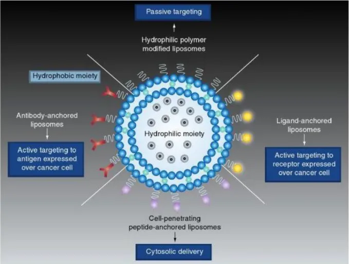
used to improve the stability of liposomes and extend their circulation times. Antibodies can be conjugated to facilitate target-specific attachment. Used alongside a series of magnetic tools, some metals (such as iron oxide) can be used for active targeting to a subject tissue. The use of peptides with specific affinities (such as cell penetrating properties) is another method to improve liposomal dynamics by enhancing cellular uptake. Figure 2 demonstrates schematics of various liposomal surface modifications and functions. In the next section, we will discuss several types of liposome modifications and the therapeutic benefits associated with each modification type.
35
Figure 2 Strategies for liposome modification. (Top) Liposome modification with hydrophilic non bioactive polymers is frequently used for passive tumor targeting. (Bottom) Modification with cell penetrating peptides improves cellular uptake and drug delivery. (Left) Modification with antibodies is utilized to achieve target-specific attachment. (Right) Modification with bioactive small molecules is utilized to achieve ligand specific active targeting. Image adopted form Paliwal et al. [35]
36 1.3.4 Liposome Surface Modifications
1.3.4.1 Antibodies
As mentioned earlier, gene expression profiles of cancer cells are different from their healthy counterparts. In some cases, this difference in gene expression results in alterations in surface antigen productions. Changes in surface antigen populations, allows cancer cells to be targeted via specific antibodies, which can be anchored to the liposome surface. For example, it is known that many types of breast cancer cells overexpress HER2, which is a growth factor receptor located on the cell surface.[36] Overexpression of this antigen provides an appropriate immunologic target for drug delivery applications. Using various forms of immunoliposomes, many studies have reported efficient targeting, accumulation and multiple drug delivery to HER2 over expressing tumors.[37-41] Up to sixfold higher tumor accumulations were observed with liposomes modified with HER2 antibodies compared to their non modified equivalents.
Liposomal targeting by immunological modification can also be applied to other forms of cancer. A study done by Sapre et al. reported that the efficiency of immunoliposomes against human B cell lymphoma.[42] Cancerous human lymphoid B cells overexpressing the surface antigen CD19 were targeted via liposomes modified with anti-CD19. Following modification, researchers observed longer circulation times and higher cytotoxicities against these cancer cells in blood.
Another modification strategy is to target the vascularization of tumors. Vascular Endothelial Growth Factor (VEGF) is the primary regulator of angiogenesis and the expression of VEGF receptor (VEGFR) generally corresponds to the level of vascularization in a specific tissue.[43] As cancer cells grow continuously and tend to
37
form bulk tumors, they must induce angiogenesis to remain connected with blood circulation. Therefore, solid tumors have to overexpress signals capable of inducing the neovascularization required. As stated above, the extent of VGEF signaling corresponds to the vascularization of a given tissue, and the components of this signaling pathway are therefore abundantly expressed in many solid tumors. Thus, it is reasonable to utilize targeting strategies involving materials with an affinity to VEGFR or VEGF itself. Many studies reported improvement in tumor localization and cytotoxicity following the use of VEGF targeting antibodies.[44-45]
1.3.4.2 Small Molecules
Small molecules displaying high affinities to cancer cells can be used for liposome modification and may facilitate tumor specific targeting. For example, folic acid plays a crucial role in DNA biosynthesis, cell division and other metabolic events.[46] As cancer cells divide and replicate continuously, many forms of cancer cells overexpress folate receptors. Since receptor expression patterns for cancer and healthy cells are different, the increased affinity between folate and its receptor in tumor cells can be used to achieve liposomal targeting. Several studies have demonstrated the in vivo and in vitro efficiency of liposomes modified with folic acid-PEG conjugations, and have found that folate-modified liposomes increase cancer cell cytotoxicity while doing little to no damage to healthy tissues.[47-48]
Estrogen is another small molecule that can be used to target some types of cancer cells. 60-80 percent of all breast cancer cells overexpress estrogen receptor (ER).[49] As such conjugating estrogen to liposome surface is a reasonable approach for tumor recognition. Several studies utilized this approach to specifically target breast cancer cells.[50-51] A
38
study by Paliwal et al. reported the improvement of treatment efficiency following the estrogen modification of doxorubicin-loaded stealth liposomes on tumor cytotoxicity.[50] In vitro cytotoxicity of liposomes to ER positive and negative cells was studied comparatively along with in vivo biodistribution and tumor growth inhibition. Estrogen modified liposomes achieved higher cytotoxicity against ER positive cancer cells compared to non modified liposomes. In addition to improved cytotoxicity, estrogen modified liposomes demonstrated distinctively higher drug stability and better tumor accumulation, such that drug half life was 50% higher and tumor accumulation six fold higher in liposomes with estrogen modification compared to non modified ones.
1.3.4.3 Proteins
Several proteins have been found to be useful for the manipulation of anticancer pharmacodynamics in liposomes, antibodies and small molecules. The main principle behind protein-based modifications is similar to previously mentioned approaches. Modification with transferrin (TF) protein can be given as an example. TF is an iron binding glycoprotein that stabilizes ferric iron and facilitates endocytosis via TF receptor (TFR).[52] It has been previously reported to be overexpressed in several cancer types, being associated with metastasis in particular.[53] As such, TF can be used as a targeting moiety in order to improve the therapeutic effect of various drug delivery systems against TFR-overexpressing cancer cells. Yue et al. functionalized Paclitaxel loaded vesicles with TFR-recognition capabilities and characterized the resulting improvements in delivery pharmacodynamics.[54] They have demonstrated that, following TF modification, the functionalized liposomes accumulated earlier in tumor tissue, and facilitated a greater extent of tumor shrinkage. In another study, Wu et al. developed TF modified liposomes encapsulating multiple drugs and reported that TF modification could improve the uptake
39 and cytotoxicity of liposomes.[55]
1.3.4.4 Sugars
Several types of sugars can be used for liposome functionalization. For instance, various cancer types, such as stomach, colon, breast, ovarian cancers and leukemia, overexpress the hyaluronic acid (HA) receptor CD44.[56] Eliaz et al. reported improvements in the targeting capacity of in HA-modified liposomes against CD44-overexpressing cancer cells.[57] In this study, researchers demonstrated that HA modified liposomes could selectively bind to CD44-overexpressing cancer cell lines. When loaded with Doxorubicin, HA modified liposomes achieved significantly better delivery rates, such that IC50 value of nonmodified liposomes was around 175 fold higher compared to HA modified liposomes. Active targeting of CD44 by HA modification was also reported in several other studies.[58-60]
1.3.4.5 Peptides
Peptides can be used to modify liposomesfor a variety of purposes, such as targeting tumors or improving pharmacokinetics. Peptides are small chains of amino acids that display distinct and adjustable chemical properties depending on their sequences. Peptides are advantageous in surface modification applications since they are easy to synthesize, structurally stable and can be tailored to serve specific purposes. By using different peptide sequences, it is possible to achieve targeting or improved cell penetration.
Integrins are cell surface proteins involved in homodimeric cell to cell interactions mediating attachment, migration and division, and have been popular target molecules for drug delivery applications.. It has been previously reported that several types of integrins
40
are overexpressed in cancer cells.[61] This difference in expression can be utilized for active tumor targeting purposes. Peptides with RGD motifs have an affinity for native integrin proteins and are frequently used as tumor targeting moieties. In one study, for instance, liposomes modified with PEG-RGD conjugates were shown to target angiogenic tumors better and enhance tumor shrinkage, demonstrating higher accumulation in the tumor site and greater cytotoxicity compared to non-modified liposomes.[62]
In addition to targeting, a family of peptide sequences can be used to improve cellular uptake. These positively-charged sequences are called cell penetrating peptides (CPP), and display a high capacity to facilitate entry into cells, rendering them particularly useful for the effective delivery liposomes into cytosolic or nuclear compartments.Following the derivation of the CPP from Human Immunodeficiency Virus proteins (HIV), many unifying features of CPPs have been characterized, and both natural and artificial CPP sequences continue to to provide new strategies for drug delivery applications.[63] As cell penetrating peptides are the main focus of this thesis, they will be discussed separately in the following section.
1.3.4.6 Cell Penetrating Peptides
Many different approaches, such as viral vectors, nanoparticles and liposomes, have been used for the delivery of DNA, drugs or other agents into cells. However, these methods all invariably display a number of undesirable properties, ranging from immune reactivity to inefficiency of delivery. The discovery of cell penetrating peptides (CPP), which improve cellular internalization and endosomal escape of macromolecules, has allowed the development of new strategies for drug delivery. Due to their ability to facilitate cellular internalization, CPPs offer a substantial opportunity to improve cargo delivery into target
41
cells. Thus, CPPs are an important focus on gene and drug delivery studies.
CPPs are short peptide sequences that mainly consist of positively charged amino acids. These low molecular weight, highly cationic peptides are rich in basic amino acids like arginine or lysine.[64] In addition to their charge, their penetration capacity is based on their sequence and target cell type, since compatible membrane-CPPs interaction is crucial to achieve efficient penetration.[65]
CPPs are capable of entering into cells via receptor mediated and/or non receptor mediated mechanisms. Although the internalization mechanisms are not fully understood, the coexistence of different uptake types is frequently suggested.[66] Since CPPs mainly consist of cationic amino acids, electrostatic interactions are important for the transduction of CPPs (and their cargo) through the cell membrane.[67] In addition to electrostatic interactions, receptor mediated macropinocytosis and subsequent endosomal release also have primary roles in the accumulation of CPPs in the cytoplasm.[68] Coexistence of various internalization mechanisms may help explain the delivery efficiency of CPPs.
Several different strategies have been developed to integrate CPPs on liposome surfaces. This integration can be facilitated either through the efficient linkage of CPPs with reactive residues on the surface, or via non-covalent integration, as we have achieved in our study. One commonly used approach is the addition of cysteine to the CPP sequence to facilitate the formation of strong disulphide bonds with liposome surface residues.[69] Spacer molecules are also used to connect CPPs and liposomes. In addition to the covalent linkage of CPPs and liposomes, CPPs can be integrated into the lipid bilayer by non
42
covalent interactions. In our study, we modified CPPs with a hydrophobic lauric acid, which is both chemically and structurally similar to fatty acid chains of membrane lipids. By doing so, we allowed the non covalent incorporation of our CPP during liposome formation, such that our modified CPPs were added to the structure of liposomes due to their similarity to the other lipids within the liposome matrix.
43 1.4 MOTIVATION AND GOALS
Peptide amphiphiles (PA) possess both a bioactive “head group” and a lipid-like alkyl tail in one molecule, which renders them highly suitable for the functionalization of liposome-based drug carrier systems. PAs can be designed and chemically synthesized with high yield and specificity and, more importantly, are easy to incorporate into liposomes without experiencing activity loss or requiring laborious chemical functionalization steps, primarily due to the amphipathic properties that they share with lipids.[64, 70] Here, we designed and incorporated a poly-arginine cell penetrating amphiphilic peptide segment into a liposomal system, and examined the integration of this arginine-rich peptide amphiphile into liposomes formed by negatively charged dioleoylphosphoglycerol (DOPG) phospholipid molecules in the presence of cholesterol. We aimed to elucidate the changes in the physical characteristics of the resulting liposomes in terms of size, surface potential and membrane polarity, and characterized their encapsulation capacities using hydrophilic and hydrophobic dyes Rhodamine B and Nile Red. After optimizing the encapsulation efficiencies of liposomes using fluorescent dyes, we then profiled the dynamics of liposome uptake. Furthermore, we investigated the possible augmentation of anti-cancer properties in known cancer drugs, doxorubicin-HCl and paclitaxel, loaded into cell penetrating peptide amphiphile-modified liposomes. This study not only analyzes the effect of peptide amphiphile integration on the physical properties of liposomes, but also discusses the in vitro therapeutic effect of peptide amphiphile integrated liposomes on MCF7 breast cancer cells in terms of cellular uptake and cytotoxicity.
44 CHAPTER 2
CELL PENETRATING PEPTIDE MODIFICATION FOR IMPROVED LIPOSOMAL DELIVERY OF ANTI CANCER THERAPEUTICS
This part of thesis is based on article “Cell Penetrating Peptide Amphiphile Integrated Liposomal Systems for Enhanced Delivery of Anticancer Drugs to Tumor Cells” Published in
RSC Faraday Discussions
2.1 INTRODUCTION
Liposomes are vesicles with varying diameters and contain lipid bilayers surrounding aqueous compartments. The self-organization of lipid molecules into bilayer in aqueous environment through their amphipathic character is responsible for the formation of spherical vesicles. Liposomes have been considered as potential drug delivery agents for several decades due to their biocompatibility, biodegradability and their resemblance to cell membrane. They have a long history of use to improve the delivery of many therapeutics such as vaccines, antibiotics and anticancer drugs.[71] Hydrophobic materials can be incorporated into the lipid bilayer of liposomes, while hydrophilic components can be encapsulated within aqueous lumen.
Liposomes happen to be reliable drug delivery vehicles due to the following properties: They are biocompatible, biodegradable and similar to cell membrane which makes
their administration safe.[72]
They increase the longevity of cargo which will be exposed to metabolic interference of humorous environment unless it is protected.
45 materials.[73]
Phosphatidylcholine (PC), phosphatidylglycerol (PG), phosphatidylethanolamine (PE) and dioleoyltrimethylammonium propane (DOTAP) are commonly used lipids which can be isolated from natural resources or produced synthetically. PC is used for preparation of uncharged liposomes while PG, PE or DOTAP are used to introduce charges. Liposomes ordinarily contain cholesterol to improve stability and prevent leakage of cargo.
Various approaches have been practically used to functionalize liposomes including chemical coupling of lipid molecule and ligand of interest before liposome formation, covalent conjugation of biologically active segments to the liposome surface and non-covalent association of liposome constituents.[74-75] Their versatile nature enables design of various functional liposomal systems decorated with a wide range of bioactive molecules such as antibodies, viral proteins, carbohydrates, peptides, aptamers, and vitamins for therapeutic delivery.[71, 76-77] The easiness of functionalization together with their extensive encapsulation capacity make them attractive tools for the development of systems which deliver the cargo to the target location with enhanced in vivo stability and circulation time. Besides their facile integration, the versatility of modifications enables diverse biofunctionality to the liposomal carrier.
Amphiphilic peptides comprised of a bioactive peptide sequence and a hydrophobic segment have great potential for functionalization of liposomal carriers. They can be designed and chemically synthesized with high yield and specificity and more importantly, they can be easily incorporated into liposomes noncovalently due to their lipid-like amphipathic property with minimized activity loss or without laborious
46
chemical functionalization steps.[78-81] The easiness of functionalization together with their extensive encapsulation capacity make them attractive tools for development of carrier systems, which can deliver cargo to the target with enhanced in vivo stability and circulation time.[82] Besides their facile integration, the versatility of peptide sequences provides diverse biofunctionality to the liposomal carriers.
The importance of cell penetrating peptides including HIV-Tat derived peptides, oligoarginines, and chimeric cell penetrating peptides have been emphasized in several works for delivery of therapeutic agents to target cells.[83-84]Arginine-rich peptides were also synthesized and investigated for enhanced cellular uptake efficiency by conjugating with large molecules such as fatty acids.[85]
Herein, we designed and synthesized an arginine-rich, cell penetrating peptide amphiphile molecule and examined its integration into liposomal formulation of 1,2-dioleoyl-sn-glycero-3-[phosphor-rac-(1-glycerol)] (DOPG) phospholipid in the presence of cholesterol. We studied size, surface potential, and membrane polarity of the resulting liposomes with and without peptide amphiphile incorporation. Encapsulation capacities of these carriers were examined by using hydrophilic and hydrophobic dyes, Rhodamine B and Nile Red, respectively. After optimization of the encapsulation efficiencies of liposomes, in vitro uptake profile and cytotoxicity of cancer drugs including Doxorubicin-HCl and Paclitaxel entrapped in liposomes with and without peptide amphiphile molecules were examined on MCF7 human breast cancer cell line.
47 2.2 MATERIALS & METHODS
2.2.1 Chemicals and Solutions Lipids:
1,2-dioleoyl-sn-glycero-3-[phosphor-rac-(1-glycerol)] (DOPG) was purchased from Avanti Polar Lipids and Cholesterol from AppliChem.
Peptides:
9-Fluorenylmethoxycarbonyl (Fmoc) and tert-butoxycarbonyl (Boc) protected amino acids, [4-[α-(2’,4’-dimethoxyphenyl)Fmoc-aminomethyl]phenoxy]acetamidonorleucyl-MBHA resin (Rink amide [4-[α-(2’,4’-dimethoxyphenyl)Fmoc-aminomethyl]phenoxy]acetamidonorleucyl-MBHA resin), 2-(1H-Benzotriazol-1-yl)-1,1,3,3 tetramethyluronium hexafluorophosphate (HBTU) and Lauric acid were purchased from NovaBiochem, ABCR and Merck. Piperidine, Acetic anhydride, Dichloromethane (DCM) and Dimethylformamide (DMF), N,N-diisopropylethylamine (DIEA), trifluoroacetic acid (TFA) : triisoproplysilane (TIS) were purchased form Sigma.
Cell Culture Reagents:
Dulbecco’s Modified Eagle Medium (DMEM), Penicillin/streptomycin (PS) antibiotic combination and fetal bovine serum (FBS) were purchased from Invitrogen Gibco.
Liposome Contents:
Anticancer drugs Doxorubicin-HCl (DOX) and Paclitaxel (PTX) were acquired from Applichem. Our tracking materials, Nile Red (NR) and Rhodamine B (RHB) were purchased from Alfa Aesar.