CASE REPORT
Jpn J Radiol (2010) 28:369–371 DOI 10.1007/s11604-010-0431-3
Leiomyoma in a female urethral diverticulum
Demet Karadag · Oya Caglar · Ahmet Hakan Haliloglu Omur Ataoglu
Abstract Neoplasms arising within urethral diverticula
are rare. It is important to know if a diverticulum is fi lled by tumor, but traditional diagnostic methods, such as voiding cystourethrography, cannot detect a tumor or diverticulum, as in our case. We report an unusual case of leiomyoma developing in a female urethra diverticu-lum and review the literature.
Key words Leiomyoma · Urethral diverticulum ·
Female · CT · MRI
Introduction
Urethral diverticulum in women is a rare entity, with an incidence ranging from 0.6% to 6.0%.1
The occurrence of a tumor in a urethral diverticulum is rare,2,3
although benign, premalignant, and malignant tumors have been detected there. This article reports an unusual case of a leiomyoma in a female urethral diverticulum.
Case report
A 49-year-old woman presented with a complaint of painless macroscopic hematuria of 7 years’ duration. She also reported that she had noticed, by palpation, the presence of a painless mass along the anterior vaginal wall. Physical examination revealed a nontender mass in the anterior wall of the vagina.
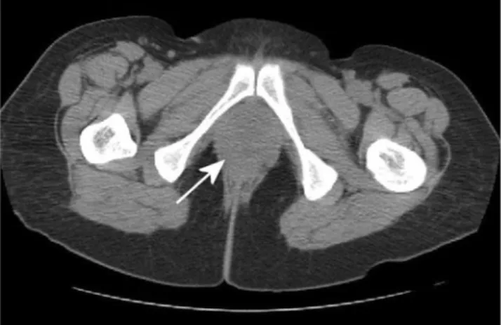
Her urine culture was negative, and routine labora-tory investigations were normal. At cystourethroscopy and voiding cystourethrography (VCUG), there was no evidence of a urethral diverticulum. Transvaginal ultra-sonography (US) at that time showed a 3.5 × 2.5 cm solid mass located along the anterior aspect of the vagina but not attached to the vagina (Fig. 1). Computed tomography (CT) and magnetic resonance imaging (MRI) demonstrated a solitary, solid, well-defi ned mass along the urethra (Figs. 2, 3). On MRI, the mass was isointense to muscle on T1-weighted images and slightly hyperintense on T2-weighted images. After intravenous gadolinium administration, the mass showed diffuse and homogeneous enhancement.
The mass was excised under spinal anesthesia, and the urethral defect was reconstructed with a 4-0 absorbable suture. She had an uneventful postoperative course.
Histopathological evaluation demonstrated a leio-myoma arising in a urethral diverticulum. Examination of sections revealed that the lesion consisted of cells with a blunt nucleus and eosinophilic cytoplasm that formed bundles. Areas of focal hyalinization were observed. There was no marked mitosis or cytological atypia. Diffuse staining with desmin and actin was observed in the immunohistochemical (IHC) examina-tion (Fig. 4).
Received: December 25, 2009 / Accepted: February 17, 2010 © Japan Radiological Society 2010
D. Karadag (*) · O. Caglar
Department of Radiology, Faculty of Medicine, Ufuk University, Ankara, Turkey
Tel. +90-312-204-4000; Fax +90-312-287-2390 e-mail: [email protected]
A.H. Haliloglu
Department of Urology, Faculty of Medicine, Ufuk University, Ankara, Turkey
O. Ataoglu
370 Jpn J Radiol (2010) 28:369–371
Fig. 1. Transvaginal sonogram. The diverticulum is fi lled with the tumor mass
Fig. 2. Computed tomography reveals a periurethral tumor (arrow)
a
b d
c Fig. 3. Axial T1-weighted
(a), axial STIR (b), sagittal T2-weighted (c), and postcontrast sagittal T1-weighted (d) images demonstrate a large T1 isointense, T2 slightly hyperintense, markedly enhanced, well-defi ned mass (arrows) lying along the urethra
a b
Fig. 4. a Histology of the operative specimen shows spindle cells arranged in bundles (H&E, ×100). b High-power view of the spindle cells showing no signifi cant atypia or mitotic fi gures
Jpn J Radiol (2010) 28:369–371 371
Discussion
Urethral diverticula may be congenital or acquired. Whereas most urethral diverticula are congenital in males, those in females are usually acquired. The etiol-ogy is uncertain, although it has been suggested that acquired diverticula may develop as a result of rupture of dilated and infected periurethral glands.1,4
In addition, urethral injury during childbirth and urethral or vaginal surgery may cause a urethral diverticulum. The inci-dence of urethral diverticulum is lower in white women (1.1%) than in black women (6.0%),3
who account for 78%–86% of female urethral diverticulum cases.4
Ure-thral diverticula are usually located in the posterolateral wall of the middle third of the urethra.1
Calculi may occur in 1.5%–10.0% of female urethral diverticula.5
Approximately 25% of the lesions are multiloculated.6
The symptoms of the urethral diverticula are not spe-cifi c, although patients may complain of dysuria, pol-lakiuria, dyspareunia, urinary incontinence, recurrent urinary tract infection, and dribbling after voiding.1,6
Voiding cystourethrography has traditionally been used for the initial diagnosis of urethral diverticulum. However, VCUG has a false-negative rate as high as 35% for detecting a urethral diverticulum.7
Although urethroscopy has an overall diagnostic accuracy of 90%, it does not enable determination of the real size of a diverticulum or evaluation of the diverticular wall. Recently, US, CT, and MRI have been successfully used for comprehensively evaluating urethral diverticula.
A neoplasm arising in a urethral diverticulum is rare. Urethral diverticular carcinomas, endometriosis, and nephrogenic adenomas have been reported in the Eng-lish-language medical literature.8,9 Chronic infl amma-tion, stasis, and mucosal irritation may lead to stone formation or metaplastic or neoplastic changes in the urethral diverticulum. The fi rst report of carcinoma in a female urethral diverticulum appeared in 1951, authored by Hamilton and Leach.10
Carcinoma arising in a ure-thral diverticulum accounts for 5% of all ureure-thral carci-nomas.2
Squamous cell carcinoma is the most common type of urethral carcinoma, whereas 40%–60% of car cinomas arising in a urethral diverticulum are adeno carcinomas.7,9
Urethral diverticular malignancies characteristically present late with early metastases, so there is a high risk of recurrence after surgery. Symp-toms are often nonspecifi c and related to irritation of the lower urinary tract.
Leiomyoma of the female urethra is a rare benign tumor derived from smooth muscle tissue. Leiomyomas typically show low to intermediate signal intensity on T2-weighted MRI images. To the best of our knowledge, review of the English literature reveals no cases of leio-myoma arising from a urethral diverticulum.
Conclusion
Sometimes a urethral diverticulum is fi lled with tumor and cannot be visualized at cystourethroscopy or VCUG, as was seen in our case. Clinicians should be aware of the possibility of a urethral diverticulum when patients have lower urinary tract irritability symptoms even if cystourethroscopy and VCUG are normal. As in our case, cross-sectional imaging tools can improve diagnos-tic accuracy in a patient presenting with the complaint of lower urinary tract irritability symptoms.
References
1. Hosseinzadeh K, Furlan A, Torabi M. Pre- and postoperative evaluation of urethral diverticulum. AJR Am J Roentgenol 2008;190:165–72.
2. Rajan N, Tucci P, Mallouh C, Choudhury M. Carcinoma in female urethral diverticulum: case reports and review of man-agement. J Urol 1993;150:1911–4.
3. Gonzalez MO, Harrison ML, Boileau MA. Carcinoma in diverticulum of female urethra. Urology 1985;26:328–32. 4. Tines SC, Bigongiari LR, Weigel JW. Carcinoma in
diverticu-lum of the female urethra. AJR Am J Roentgenol 1982;138: 582–5.
5. Josef B, Raeto TS. Giant calculi in urethral diverticula. Can Med Assoc J 2008;178:994.
6. Guidi HG, Montelatto NI, Ribeiro RM, Pinotti JA. The treat-ment of female urethral diverticulum with calculus through ultrasonic lithotripsy. Int J Gynaecol Obstet 1993;41:277–81. 7. Siegel CL, Middleton WD, Teefey SA, Wainstein MA,
McDougall EM, Klutke CG. Sonography of the female urethra. AJR Am J Roentgenol 1998;170:1269–74.
8. Summitt RL Jr, Murrmann SG, Flax SD. Nephrogenic adenoma in a urethral diverticulum: a case report. J Reprod Med 1994;39:473–6.
9. Thomas RB, Maguire B. Adenocarcinoma in a female urethral diverticulum. Aust N Z J Surg 1991;61:869–71.
10. Hamilton JD, Leach WB. Adenocarcinoma arising in a diver-ticulum of the female urethra. AMA Arch Pathol 1951;51: 90–8.