IN SILICO INHIBITOR DESIGN FOR MONOAMINE
OXIDASE A AND B ISOZYMES
ÇAĞLA MIDIK
20091109006
Kadir Has University
2012
IN SILICO INHIBITOR DESIGN FOR MONOAMINE OXIDASE A
AND B ISOZYMES
ÇAĞLA MIDIK
Computational Biology and Bioinformatics, Kadir Has University, 2012
Submitted to the Graduate School of Science Institute in partial fulfillment of the requirements for the degree of
Master of Science in
Computational Biology and Bioinformatic
KADIR HAS UNIVERSITY 2012
KADIR HAS UNIVERSITY
GRADUATE SCHOOL OF SCIENCE AND ENGINEERING
MONOAMINE OXIDASE ISOZYMES LIGAND DESIGN
ÇAĞLA MIDIK APPROVED BY:
…
(Thesis Supervisor)
(Kadir Has Universiy) ……….…….
… (Marmara University) ……….…….
… (Kadir Has Universiy) ……….…….
… (Kadir Has Universiy) ……….…….
Abstract
In this thesis study, MAO-A and MAO-B isozymes are selected as target enzymes that have important role of regulation of diseases such as ‘’Parkinson, Alzheimer and depression’’. MAO isozymes which catalyze the oxidation of monoamines in the body stand out in these diseases. Today explicitly known that MAO enzymes oxidize the neurological amines such as serotonine, dopamine,
neuroadrenaline more than usual way and result in the reduction of the level of these important monoamines creating disease state.
Three-dimensional structures of cloned human MAO-A compexed with harmine ligand (2z5x) and human MAO-B complexed with safinamide (2v5z) are taken from protein data bank. Both isoforms contain flavin molecule as cofactors.
Taking into consideration of the previous studies, 210 new ligands are created. Of these ligands 105 are the R stereoisomers and the other 105 are S stereoisomers.
The objective of this study is to use computer-aided drug design (CADD) methods based on structure-based drug design. As a result we obtained the best orientation of these ligands in the active site of both isozymes. Using of improved docking methods the lowest possible binding energy and the poses of the ligands in the enzyme complex are scored.
Docking programs exploit their own scoring functions by specific algorithms. Hereby, binding energies of target enzyme and ligands are estimated at the lowest energy conformations by these scoring functions. In this study, docking simulations are performed by Autdock 4.2 and Acceleryss 3.1 libdock programs.
Evaluation of docking studies by Autodock 4.2. suggested us the results generated are very reasonable Ki values. The results of both docking simulation methods showed us the ligands 1d3r, 1a3r and 1a6r are effectively inhibit MAO-A isozyme whereas ligands 2d1r, 1d6s, 1d3s and 3b4r selectively inhibit MAO-B
isozyme. In conclusion, these inhibitors are very important candidates for MAO enzyme and may lead the future studies.
Özet
Bu tez çalışmasında ‘’Parkinson, Alzheimer ve Depresyon’’ gibi hastalıkların meydana gelmesinde önemli rol oynayan MAO enziminin iki izomeri olan MAO-A ve MAO-B izoenzimleri hedef olarak seçilmiştir. Vücuttaki monoaminleri katalize etmekle görevli olan MAO izoenzimleri özellikle bu hastalıklarda göze
çarpmaktadır. Bu hastalarda normalden daha fazla çalışarak vücuttaki seratonin, dopamin, nöradrenalin gibi monoaminlerin konsantrasyonunu oldukça düşürdükleri günümüzde açıkça bilinmektedir.
Flavin ailesi üyesi olan bu enzimlerin kristalize edilmiş üç boyutlu yapıları insan MAO-A’sı harmine kompleksi ile (2z5x), insan MAO-B’si ise safinamide kompleksiyle (2v5z) birlikte olmak üzere protein bilgi bankasından alınmıştır.
Daha önceki bilimsel çalışmalar göz önünde bulundurularak MAO
izoenzimlerinin inhibisyonuna yönelik 105’i r-stereo izomeri, 105’i s-stereo izomeri olmak üzere toplam 210 tane ligant bileşiği oluşturulmuştur Bilgisayar destekli yapıya dayalı ilaç tasarımı, enzimlerin aktif bölgesi ile ligantlar arasındaki en düşük enerji konformasyonlarında bağlanma durumlarını ölçmeyi hedefler. Bunu yaparken geliştirişmiş docking metotlarını kullanarak enzimlerin ve ligantların 3 boyutlu yapılarındaki değişkenliklerini de hesaba katar.
Bilgisayar destekli docking araçları spesifik algoritmaları sayesinde kendi skor fonksiyonlarını üretir. Böylelikle ligantların hedef enzimlere en düşük konformasyonda bağlanma enerjileri bu skorlar yardımıyla tahmin edilmektedir.. Yaptığımız çalışmada docking simülasyonları Autodock 4.2 ve Accelryss 3.1 libdock programları ile gerçekleştirildi.
Autodock 4.2 programıyla yapılan çalışmalar sonunda ölçülen Ki değerleri olumlu sonuçlar vermiştir. Ayrıca her iki docking simülasyonunda incelenen ligant bileşikleri 1d3r,1a3r,1a6r MAO-A izoenzimiyle eşleşmede ve 2d1r, 1d6s, 1d3s ve 3b4r ligant bileşikleri ise MAO-B izoenzimiyle eşleşmede oldukça yüksek sonuçlar
vermiştir. Bu ligantlar MAO enzimleri için inhibitör adayı olduklarını gösterirler ve gelecek çalışmlara ışık tutabilirler.
Acknowledgements
It is a pleasure to thank the many people who made this thesis possible. There are many people who helped to make my years at the graduate school most valuable.
First and most importantly, I want to thank the best educator ever I seen, my major professor and dissertation supervisor Prof. Dr. Kemal Yelekçi and his guidance for his unlimited support, endless patience throughout my thesis studies. It has been an honor and privilege working with him over years. I also thank Assist Prof. Demet Akten Akdoğan and Dr. Tuğba Arzu Özal who contributed much to improve of my dissertation work.
My special thanks go to friends, Filiz Varnalı, Jale Güler, Levent Binnetoğlu, Erçin Dinçer, Seda Demirci, Sibel Çakan and Gizem Tatar who helped me me all through the years full of class work and exams for their supporting during my thesis.
I want to thank Nurdan Kayrak and Bora Büyüktürk who contributed my studies.
The last words go to my fragmented family. Big thanks go to my parents Berrin Karakaş, Battal Mıdık and my brother N. Serhat Mıdık for their all supports and I thank my dear engaged Mihrace İnel, for her endless support through this long journey.
Table of Contents
Abstract ...ii
Özet ...iv
Acknowledgements ... i
Table of Contents ...ii
List of Tables...iv
List of Figures ... v
List of Symbols ... vii
List of Abbreviations... viii
1. Introduction ... 1
2. Background of Monoamine Oxidase... 3
2.1. Introduction of Monoamine Oxidase ... 3
2.1.1. Comparing of Monoamine Oxidase Isozymes ... 4
2.1.2. Active site of Monoamine Oxidase ... 5
2.1.3. Amines of Monoamine Oxidase Catalyze... 7
2.1.3.1 Dopamine ... 7
2.1.3.2. Seratonin... 8
2.1.3.3. Ephinephrine ... 9
2.1.3.4. Norepinephrine ... 9
2.2. Mechanism of Monoamine Oxidases Amine Catalyze ... 10
2.3. Background of Known MAO Inhibitors ... 12
3. Materials and Methods ... 14
3.1.1. General Properties of Molecular Docking ... 14
3.1.2. General Properties of AUTODOCK ... 15
3.1.3. General Properties of Accelrys Discovery Studio ... 17
3.2. Preparing Compounds ... 18
3.2.1. Preparing Monoamine Oxidase Isozymes... 18
3.2.2. Preparing Ligands of Monoamine Oxidase Isozymes... 19
3.3. Molecular Docking... 22
3.3.1. Docking Setup of AUTODOCK 4.2 ... 22
3.3.2 Enzyme- Ligand Molecule Docking by AUTODOCK 4.2... 23
3.3.3 Docking Setup of LIBDOCK ... 24
4. Result and Discussion ... 25
4.1. Results of Molecular Docking Studies by Autodock 4.2 ... 25
4.3. Analysis of Monoamine Oxidase A – Ligand Docking ... 28
4.4. Analyses of Monoamine Oxidase B - Ligand Docking ... 36
Conclusions ... 47
APPENDIX A: PARAMETER FILES of AUTODOCK 4.2 STUDIES ... 51
APPENDIX B: RESULTS of AUTODOCK 4.2 DOCKING DATA... 57
Bibliography ... 63
List of Tables
Table 3.1 Experimental details of Human Monoamine Oxidase A with harmine (Journal: (2008) Proc.Natl.Acad.Sci.Usa 105: 5739-5744).
Table 3.2 Experimental details of Human Monoamine Oxidase B with the selective inhibitor safinamide (Journal: (2007) J.Med.Chem. 50: 5848).
Table 3.3 Preparing code names of ligands.
Table 4.1 Top 20 Autodock docking results of MAO-A and MAO-B isozymes. Table 4.2 Results of Libdock docking for MAO-A and MAO-B isozymes. Table 4.3 2-D pictures of the ligands interaction by MAO-A at the lowest
docking energy conformation.
Table 4.4 2-D pictures of the ligands interaction by MAO-A at the lowest docking energy conformation.
Table 5.1 2-D chemical structures and detailed docking results of MAO-A and MAO-B ligand candidates.
List of Figures
Figure 2.1. 3-D crystal structures of human MAO-A in complex with harmine. (a) display style is solid ribbon.(b) display style is lines and balls represent FAD. Figure 2.2. 3-D crystal structure of MAO-B in complex with the
selective inhibitor safinamide.(a) display style is solid ribbon. (b) display style is lines and balls represent FAD.
Figure 2.3. Comparing 3-D structures of MAO-A & MAO-B isozymes.
Figure 2.4. (A) Chemical structure of FAD.(B) Active site comparison of human MAO-A and human MAO-B.(C) The active site cavity (red surface) of MAO-B in Complex with deprenyl (black) is depicted (NCBI)
Figure 2.5. 2-D chemical structure of FAD in active side of MAO-A & MMAO-AO-B.
Figure 2.6. 2-D chemical structure of dopamine. Figure 2.7. 2-D chemical structure of serotonin.
Figure 2.8. 2-D chemical structure of ephinephrine (adrenaline). Figure 2.9. 2-D chemical structure of noradrenaline or
(R)-(–)-norepinephrine.
Figure 2.10. Figure 2.10. Steps of the oxidative deamination of amines catalyzed by MAO.(a) FAD reduction.(b) represent deamination (c)represent FAD redoxidation. Figure 2.11. 2-D chemical structure of hydride mechanism.
Figure 2.12. Mechanism of single electron transfer (SET). Figure 2.13. 2-D chemical structure of iproniazid.
Figure 2.14. 2-D chemical structure of moclobemide. Figure 3.1. Workflow of docking studies.
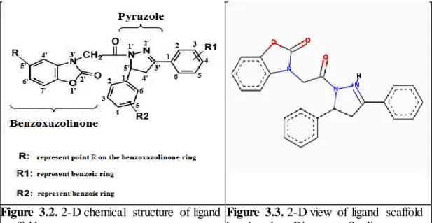
Figure 3.2. 2-D chemical structure of ligand scaffold.
Figure 3.3. 2-D view of ligand scaffold by Accelrys Discovery Studio 3.1.
Figure 3.4. Preparing code names of ligands.
Figure 4.1. 3-D view of ligand 1d3r and active side of MAO-A at the lowest energy conformation.
Figure 4.2. 3-D view of ligand 1d6r and active side of MAO-A at the lowest energy conformation.
Figure 4.3. 3-D view of ligand 1a3r and active side of MAO-A at the lowest energy conformation.
Figure 4.4. 3-D view of ligand 1d1r and active side of MAO-A at the lowest energy conformation.
Figure 4.5. 3-D view of ligand 1a6r and active side of MAO-A at the lowest energy conformation.
Figure 4.6. 3-D view of ligand 1c3r and active side of MAO-A at the lowest energy conformation.
Figure 4.7. 3-D view of ligand 1c6r and active side of MAO-A at the lowest energy conformation.
Figure 4.8. 3-D view of ligand 3e6r and active side of MAO-A at the lowest energy conformation.
Figure 4.9. 3-D view of ligand 2c7s and active side of MAO-A at the lowest energy conformation.
Figure 4.10. 3-D view of ligand 1d7s and active side of MAO-A at the lowest energy conformation.
Figure 4.11. 3-D view of ligand 2d1r and active side of MAO-B at the lowest energy conformation.
Figure 4.12. 3-D view of ligand 1d4s and active side of MAO-B at the lowest energy conformation.
Figure 4.13. 3-D view of ligand 1d5s and active side of MAO-B at the lowest energy conformation.
Figure 4.14. 3-D view of ligand 1d6s and active side of MAO-B at the lowest energy conformation.
Figure 4.15. 3-D view of ligand 1d3s and active side of MAO-B at the lowest energy conformation.
Figure 4.16. 3-D view of ligand 1a3s and active side of MAO-B at the lowest energy conformation.
Figure 4.17. 3-D view of ligand 1a1s and active side of MAO-B at the lowest energy conformation.
Figure 4.18. 3-D view of ligand 3b4r and active side of MAO-B at the lowest energy conformation.
Figure 4.19. 3-D view of ligand 1c6s and active side of MAO-B at the lowest energy conformation.
Figure 4.20. 3-D view of ligand 3d4s and active side of MAO-B at the lowest energy conformation.
Figure 5.1. Detailed docking results and chemical structures of MAO-A and MAO-B inhibitor candidates.
List of Symbols
Ǻ: Angstrom α: Alpa angle ß: Beta angle γ: Gama angle
∆S: Entropy lost upon binding R: Point of benzoic ring R1: Name of a benzoic ring R2: Name of a benzoic ring
g: Gram L: Ligand P: Protein E: Enzyme W: Constant V: Volume of atoms S: Solvation patameter
Dij: Maximal well depth for hydrogen bonds, oxygen and nitrogen Cij: Maximal well depth for hydrogen bonds, oxygen and nitrogen -R: Right isomer
-S: Left isomer pM: Picomolar nM: Nanomolar μM: Micromolar
List of Abbreviations
MAO: Monoamine oxidase
MAO-A: Monoamine oxidase – A MAO-B: Monoamine oxidase – B FAD: Flavine di nucleotide
π-interaciton: A type of non-covalent interaction X-RAY: A form of electromagnetic radiation
3-D: Three dimension
SET: Single electron transfer
GA: Genetic algorithm
LGA: Lamarcian genetic algorithm SASA: Solvent accessible solvent area GPF: Grid parameter file
DPF: Docking parameter file
VDW: Van der waals
NMR: Nuclear magnetic resonans SSRI: Selective serotonin inhibitors
Hbond: Hydrogen bond
Tor: Torsion Sol: Desolvation Elec: Electrostatic Ala: Alanine Arg: Arginine Asn: Asparagine
Asp: Aspartic acid
Cys: Cysteine
Glu: Glutamic acid
Gln: Glutamine
Gly: Glycine
His: Histidine
Hyp: Hydroxyproline
Leu: Leucine Lys: Lysine Met: Methionine Phe: Phenylalanine Pro: Proline Glp: Pyroglutamatic Ser: Serine Thr: Threonine Trp: Tryptophan Tyr: Tyrosine Val: Valine
Chapter 1
1. Introduction
Monoamine oxidase enzymes are classified as flavoproteins because they include flavine adenine dinucleotide (FAD). In most cell type, MAO is observed at the mitochondrial outer membrane. MAO includes two different isoforms such as MAO-A isozyme and MAO-B isozyme. Both isozymes found in brain but also reside in different kinds of cell. MAO-A is generally observed in catecholaminergic
neurons whilst MAO-B is found in serotonergic neurons also both of these isozymes accessible in the astroglial cell [1].
MAO plays so crucial role in the mammalian body system cause of catalyze oxidation of monoamines. Even amino acid sequences of MAO isozymes %70 similar, they are encoded from separate genes and they have been identified by their substrate selectivity and their inhibitor sensitivity [2]. General view, MAO-A selectivity inhibitor is clorgline and selectivity substrate serotonin while MAO-B selectivity inhibitor is R-deprenyle and selectivity substrate is dopamine [3]. Both isozymes play active role for deamination of dopamine, tyramine and tryptamine. According to Edmonson [4] MAO-A preferable deaminates aromatic monoamines for instance the neurotransmitters serotonine, adrenaline. On the other hand MAO -B generally oxidazes β-phenylethylamines and benzylamines.
Present, close relation between neurotransmitter amines and such diseases is evidenced by chemical, biological computations, pharmaceutical studies and clinical researches. Especially, after the discovery of X-RAY crystallography enabled proteins virtual visualization so vast data of compound which related structure of proteins are accessible for novel drug design and developing of medicinal industry. According to Yelekçi [5] after the specification of the 3-D crystal structure of MAO-B pave the way for development of computer-assisted more selective and reversible inhibitor design.
In the major depression diseases, deficit of monoamines is observed basically neuroephinephrine and serotonine where critical synapses in the central nervous
system [6].In the Parkinson diseases, concentration of dopamine is decreased. Consideration of MAO deamination function of monoamines, these results shows that MAO inhibitors have critical role in the therapy of severe neurological and psychological disorders. Combinational chemistry offers vast data about new molecule compounds so clearly known that experimental st udies of novel drug design lead to time-consuming and loss of money.
In this thesis, chapter 2 provides detailed information of MAO isozymes, their function mechanisms and amines which are catalyzed by MAO. Also comparison of MAO-A and MAO-B isozymes is presented. In the chapter 3, two main target
enzymes are prepared that are MAO-A, MAO-B isozymes and 210 many designed inhibitor (which are includes both of –R and –S stereoisomers) candidates are created by considered to previous MAO inhibition study literature. Also information of molecular docking tools is given and their parameters are set up. Finally results of docking studies, detailed analysis of ligand candidates and discussion are given in chapter 4.
Aim of in this thesis studies is designing of inhibitor candidates and finding the best MAO-A and MAO-B inhibitors by using the automated docking tools. Results of this thesis can help for the next studies for treatment of Parkinson, Alzheimer and depression diseases.
Chapter 2
2. Background of Monoamine Oxidase
2.1. Introduction of Monoamine Oxidase
MAO is a flavo protein situated at the mithocondria's outer membrane of neural, glial and other cells, specially appertain in the brain and liver. MAO enzyme has an important role in some psychiatric and neurological disorders, including depression and Parkinson’s disease because inhibition of MAO increases the concentration of neurotransmitters in the central nervous system, searching for the effective inhibitors represent one significant approach to thriving novel drugs to treat such diseases [7]. Enzyme of MAO was discovered in liver as tyramine oxidase by Mary Bernheim in 1928.
After the 1950's, MAO inhibitors were thought for clinical treatments but using with other type drugs and nutrients that including tyramine was caused a significant risk of rapid increased blood pressure (hypertansive crise) and other different side effects. Thus, developments of therapeutic monoamine oxidase inhibitors are regressed. With the discovery of human monoamine oxidase's two isoforms crystal structure which called monoamine oxidase A (Mao A) and monoamine oxidase B (Mao B), Protein – ligand selectivity interactions and
catalytic mechanisms better understood.
Today, vast number compounds are examining by the development of
computational chemistry and computer-aid bioinformatics. After the determination of three dimensional crystralized structures of MAO isozymes by X-RAY
crystallography and NMR, in last decade found in the literature has increased a great deal of space. Also over the therapeutic potency of rational selectivity enzymes, lot of research has been done.
2.1.1. Comparing of Monoamine Oxidase Isozymes
MAO-A and MAO-B have own specific substrates in addition they have own specific inhibitors too. MAO-A is mostly located at the catecholaminergic neurons, and MAO-B is located at serotonergic neurons and glia [8].
Simply MAO-A oxidizes serotine and noradrenaline while MAO-B oxidizes dopamine and phenilethilamine. Monoamine oxidases isozymes (A and MAO-B) have about % 70 amino acid sequence similarity [9]. Human MAO-A has 59.700 molecular weight and human MAO-B has 58.800 molecular weight.
a. Monomeric crystal structure of MAO-A
b. Dimeric crystal structure of MAO-A. Balls represent FAD
Figure 2.1. 3-D crystal structures of human MAO-A in complex with harmine. on
the figure a. and on the figure b.
a. Monomeric crystal structure of MAO-B
b. Dimeric crystal structure of MAO-B. Balls represent FAD
Figure 2.2. 3-D crystal structure of MAO-B in complex with the selective inhibitor
safinamide
MAO-A and MAO-B isozymes are compared with the software programs Blast2 and Fatcat. Blast2 software program displays the similarity of sequence alignments and as a result calculating of pairwise sequence alignments display that specially % 68 identities, % 81 positives sings for MAO-A and MAO-B isozymes.
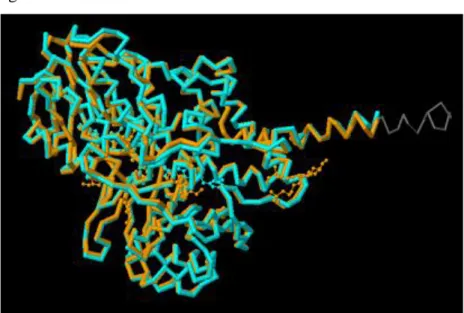
On the other hand comparing of MAO-A and MAO-B isozymes by the Fatcat program displays that their 3-D structures have high degree similarity as figure 2.3
Figure2.3. Comparing 3-D structures of MAO-A isozyme & Mao-B isozyme.
Orange color represent the Mao-A and turquoise color represent the Mao-B
2.1.2. Active site of Monoamine Oxidase
In the enzymes, the area that includes catalytic residues, makes binding the substrate, in this way actualizes the reaction is known as the active site. Monoamine oxidases contain the covalent bound cofactor flavine adenine dinucleotide (FAD) in the active site. The FAD is bound to the protein in an extended conformation with the most of the bonds to the protein detected as hydrogen bonds with amino acid side chains, amide bonds, and water molecules. Since those amino acids interacting with the FAD are conserved in A, it is proposed that the FAD binding site in MAO-A is quite similar to that in MMAO-AO-B. MAO-According to Edmonson et al [10] comparing active side of MAO-A (human) and MAO-B (human) is illustrated as Figure 2.4.
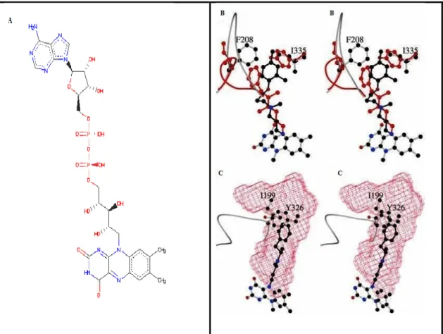
Figure 2.4. (A) Chemical structure of FAD that is located at the active side of
MAO. B and C figures are taken from national center for biotechnology information. On the figure 2.4(B) indicate that active site comparing of human MAO-A and human MAO-B with the critical Phe-208 and Ile-335 residues of MAO-A superimposed to the correspondent residues of MAO-B such as Ile-199 and Tyr-326. Atoms of inhibitor and protein molecules of MAO-B are shown in the red cage. According to A, the pattern has been rotated by ≈90° around the vertical axis in the plane of the model. On the figure 2.4(C) represent that the active site cavern (red surface) of MAO-B in complex with deprenyl (black) is shown.
Flavin di nucleotide that is depended to a cysteine amino acid residue of Mao's 8α position with covalent bind as figure 2.5.
Figure 2.5. 2-D chemical structure of FAD in active side of MAO-A & MAO-B
2.1.3. Amines of Monoamine Oxidase Catalyze
2.1.3.1 Dopamine
Dopamine is a type of the catecholamine neurotransmitters that accessible in the brain. It is derived from tyrosine and is the precursor to neurephinephrine and epinephrine. Dopamine has important role in the many cerebrum processes that movement of control, emotional and mental response, and ability to experience delight and ache. In the respect of chemical, molecular formula is C8H11NO2 and molecular weight is 153.17844 [11].
Regulation of dopamine has critical role in human intellectual and physical health. Neurons which including the neurotransmitter dopamine are clustered in the midbrain in an area called the substantia nigra, especially in disease of Parkinson, the dopamine- transmitting neurons die in this area. [12]. Eventual, brains of patients with disease of Parkinson do not include almost any dopamine.
Two major degradation pathways for dopamine exist. In most areas of the brain, including the striatum and basal ganglia, dopamine is inactivated by reuptake via the dopamine transporter, then enzymatic breakdown by monoamine oxidase into 3,4-dihydroxyphenylacetic acid [13].
Figure 2.6. 2-D chemical structure of dopamine
2.1.3.2. Seratonin
Seratonine (5-hydroxtryptamine 5HT) is firstly observed in the gastrointestinal tract, platels and nervous system of human [14]. Seratonine’s molecular formula is known as C10H12N2O and molar mass is measured 176.215 g/mol and slightly soluble in the water. The functions of serotonin are in large quantities and appear to involve control of regulating appetite, dexterity of learning and memory, temperature regulation, state of mind, behavior (including sexual and hallucinogenic behavior), cardiovascular function, muscle contraction, endocrine regulation, and depression.
Lately, selective serotonin inhibitors (SSRIs) are prefered and used for treatment of supress depression diseas.These drugs process by altering the function of neurons that release serotonin by blocking the reuptake of serotonin back into the cell. Thus the concentration level of serotonin efficiency is increased in any where of the nervous system that make uses this neurotransmitter as a chemical signal between cell transmission mechanizm. The SSRIs are improved into the drug of preference because they present lesser side effects due to their limited action to reuptake serotonin alone and no other neurotransmitters [15].
Serotonin synthesis occurs in a several process first step of that begins with the amino acid tryptophan. The second step in serotonin production includes an enzyme named tryptophan hydroxylase. Tryptophan hydroxylase appends a hydroxl group to tryptophan, generating 5-hydroxytryptophan. Another enzyme, amino acid decarboxylase, eliminations a carboxyl group from hydroxytryptophan, leaving 5-hydroxytryptamine, or serotonin. Then the serotonin is inactivated by a MAO. MAO degrades serotonin by cleaving the amine group off, which is the deamination
reaction. The two products are ammonium ion and 5-hydroxyindole-3-acetaldehyde, this essentially leaves the neurotransmitter deactivated [16].
Figure 2.7. 2-D chemical structure of seratonin
2.1.3.3. Ephinephrine
Ephinephrine (adrenaline) plays role as a hormone and plays role as a neurotransmitter. It responsible for increasing heart rate, constricts blood vessels, dilates air passages and participates in the fight-or-flight response of the sympathetic nervous system [17]. The significant physiological triggers of ephinephrine release center upon stresses, such as physical threat, enthusiasm, loudness, bright lights, and high environment temperature [18]. Chemically, epinephrine is a catecholamine and further only the adrenal glands from the amino acids tyrosine and phenylalanine produce as a monoamine.
Figure 2.8. 2-D chemical structure of ephinephrine (adrenaline)
2.1.3.4. Norepinephrine
Norepinephrine consists of a catecholamine and a phenethylamine. The inherent stereoisomer is L- (-) - (R) - norepinephrine. This monoamine has an
important role of brain's notice and response stimulations. Noradrenaline’s molecular formula is C8H11NO3 and molecular weight is 169.18 g mol−1 [19].
A major important function of norepinephrine is its role as the
neurotransmitter released from the sympathetic neurons affecting the heart. Enhance in norepinephrine from the sympathetic nervous system leads to increasing the rate of contractions [20].
Figure 2.9. 2-D chemical structure of noradrenaline or (R)-(–)-norepinephrine
2.2. Mechanism of Monoamine Oxidases Amine Catalyze
Monoamine oxidase is a flavin-dependent enzyme that catalyzes the oxidative deamination of a variety of amine neurotransmitters and toxic amines, to the
corresponding imines which are nonenzymatically hydrolyzed to aldehydes [21]. The cofactor FAD plays active role in the reaction. This could schematized in three steps which are same for both enzymes as figure 2.10.
Figure 2.10. Steps of the oxidative deamination of amines catalyzed by MAO.(a)
As a result of enzymatic reaction, aldehyde derives from imine by water. Reduced FAD is oxidized by aid of molecular oxygen and returns to form of before reaction as catalyzer.
Various mechanisms are proposed for the oxidation of the biological amines by monoamine oxidases. According to Yelekci these mechanisms are handled in four section such as Two Electron Mechanism, Carbanion Followed by One-Electron Transfers, One-Electron Mechanism Hydride Mechanism [22].
According to Hydride Mechanism, amine α-H is transported to Flavine as Hydride. Thence two electrons and one proton are transferred to the flavine so reaction is completed but hydride transfer requires high energy and this causes some questions about Hydride Mechanism [23].
Figure 2.11. 2-D chemical structure of hydride mechanism.
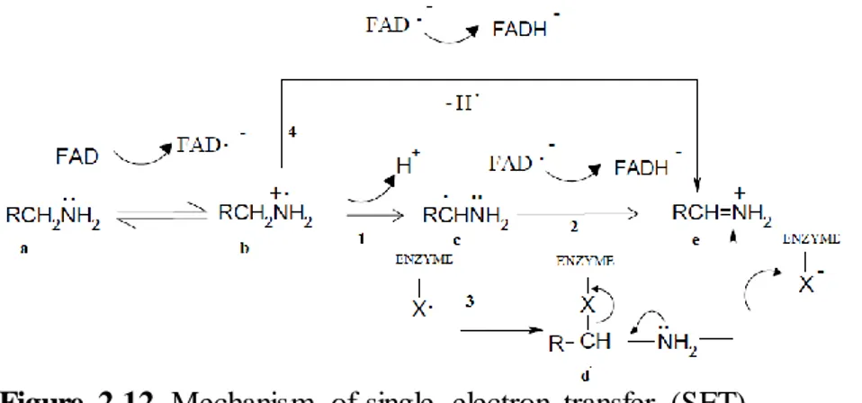
According to suppose of Silverman, one electron flavin mechanism is
possible for both monoamine oxidase isozymes, α-carbon oxidation of amines begins by the one electron transfer. One electron of the unmatched electron pairs where located nitrogen atom of the monoamine (a) is transferred to the flavine thus radikal amine cation (b) and FADH occur. After the leaving hydrogen of α-Carbon where located radical amine cation, by the path of ‘’1’’ compound transforms to α-radical amine (c). In this situation reaction can follow two different path as ‘’2’’ or ‘’3’’. If reaction continues according to path ‘’2’’ second electron could transfers to FADH, can formation of iminium ion (e). On the other hand if reaction proceed pursuant to path ‘’3’’ could radical combine with a radical that located other regions of enzyme so does covalent bounding by X and transforms to compound (d). If this consisting intermediate product can be stable enough that can make the enzyme inactive and then it transforms iminium ion (e) and free enzyme [24].
Figure 2.12. Mechanism of single electron transfer (SET)
In the disease of depression consantre of amines neurotransmitters such as serotonine, dopamine decreases. Important involved enzyme of these amines that monoamine oxidase is considered for target enzyme for depression disease
2.3. Background of Known MAO Inhibitors
A great number of studies for inhibition of MAO are inspired from old inhibitiors such as Moclobemide, Iproniazid.
First potent MAO inhibitor is Iproniazid that was introduced in therapy of depression diaese, in 1957. Its side effects with other chemicals and particular nutrients, the therapeutic implementation of Iproniazid have been reduced [25]. In 1961 Iproniazid was withdrawn owing to the inadmissible ratio of hepatitis and was replaced by less hepatotoxic drugs such as isocarboxazid , phenelzine, and
tranylcypromine [26].
Figure 2.13. 2-D chemical structure of Iproniazid
Moclobemide was the first nonhydrazine, reversible MAO-A selective inhibitor approved for depression treatment. That was a potent inhibitor in vivo but
on the other hand it was a feeble inhibitor in vitro. It has been described as a 'slow binding inhibitor', whereby conformational changes to either Moclobemide or the enzyme to MAO-A slowly form a more tightly bound complex, resulting in the non-competitive MAO inhibition by Moclobemide [27].Moclobemide is a benzamide derivative of morpholine (O(CH2CH2)2NH.) which acts pharmacologically as a selective, reversible inhibitor of MAO-A, a type of increases levels of especially serotonin, dopamine and norepinephrine [28].
Chapter 3
3. Materials and Methods
3.1.1. General Properties of Molecular Docking
Docking is finding the binding geometry of two interacting molecules with known structures. Molecular docking is the crucial tool in computational biology and structure based drug design. After the determination of three-dimensional structure of proteins by X-ray crystallography and NMR importance of these 3-D structures has increased in last decade because of function of a protein depends primarily on its structure.
In the broadest sense, aim of the molecular docking is predicting molecular recognition and spatial conformation, to detection of potential binding positions withal optimal energy between two structures. Generally molecular docking is performed for a small molecule and a target macromolecule. Regard of the computer-aided drug design, molecular docking is computer simulation process to estimate of possible conformations of a binary complex that mostly between protein and ligand. Each docking software program uses own specific algorithms. Almost all docking software programs handle the rigid body protein, as an exception, Autodock 4.2. Software program allows side chains in the macromolecule to be flexible. Moreover molecular docking algorithms can be classified by two different forms. This classify is defined according to ligands structure selection such as flexible ligand or rigid body ligand.
Figure 3.1. Workflow of docking studies
Rigid body docking algorithm is based on the geometric, volumetric, the atomic coordinates of the structure. Thus rigid body docking relates more the formal structure, but that causes some disadvantages as rigid body molecule cannot be a model for all molecules conformations. Owing to the fact that the computational costs increase exponentially with the degrees of freedom of molecules . As well as the rigid body ligand dockings occurs quickly than flexible ligand docking. Flexible docking algorithm is much harder than performing rigid-body docking. These flexible docking approaches consider free to rotate around its rotatable bounds of ligand molecule, that provides more accural docking results of ligand and receptor cause of ligand is could alter of spatial conformations and that prevent such only one sample template for docking process. But disadvantage of this method causes too much computational costs and waste of the time.
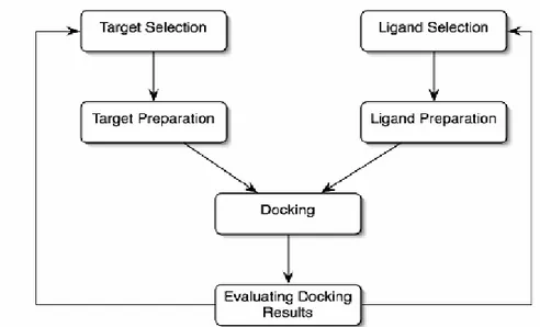
The first step in docking studies is to identify regions on the protein structure that upon binding a ligand would interfere with normal function, such as an enzyme active site, the binding site of a receptor, or an allosteric site [29].
3.1.2. General Properties of AUTODOCK
Autodock (automated docking) is a most common docking software program and a major advantage is that open source. The main reason for arising Autodock is
bioactive compounds design and especially for computer aided drug design. It is conceived to estimate how small molecules, such as drug candidates, or substrates bind to a receptor of known three dimensional structure [30]. 3-D X-Ray
crystallography structures that particularly proteins are stunning targets for understanding the principle biological activity and drug candidates. Herein,
autodock is a crucial calculation tool for drug candidates because of measuring and predicting to molecular interactions and affinity.
Autodock offers a several of search algorithms to explore a given docking problem process. These search algorithms are Monte Carlo Simulated Annealing ( SA ), a Genetic Algorithm ( GA ) and a hybrid local search genetic algorithm which is known the Lamarcian Genetic Algorithm ( LGA ). Genetic algorithm is a search heuristic and mimics the natural evoulation process. Genetic algorithms are inspired by natural evolution for generating solutions to optimization problems and that use similar methods such as mutation, crossover, selection and inheritance. These algorithms offer a set of solution instead only one solution. In a genetic algorithm a population of string which is also represent chromosomes or genotype of genome encode candidate solutions that includes creatures or phenotypes to an optimization problem and evolves toward better solutions.
Creators of Autodock are clearly specify that, the most efficient method is Lamarcian Genetic Algorithm that includes both of global search and local search methods.
Autodock 4.2 uses a semi empirical free energy force field for assessment of conformation in the course of docking simulation process. Force field could be consider in two parts for Autodock 4.2 model. Macromolecule or protein and the ligand launch in unbound conformation. Firtsly, the intramolecular energetics are estimated for the modulation from these unbound states to the conformation of the ligand and protein in the bound state, this is considerable for estimate of the conformational entropy lost upon binding. Secondly, intermolecular energetics of protein and the ligand combination is evaulated in their bound conformation. Force field includes six pairwise evaluations (V) and an estimate of the conformational entropy lost upon binding also includes the weighting constants (W) for optimize to calibre the empirical free energy in the binding equation that based on a several of
experimentally-determined binding constant and basis of molecular mechanics terms and entropic terms which are Van der Waals forces, Hydrogen Bonding, Coulombing potential, desolvation and torsional parameters are use these weighting constants. Free energy of binding equation represent as below equations.
In this equation L represent ligand compound and P represent protein for procces of protein-ligand docking.
For the purpose of calculation for free binding energy has a several process. The final intermolecular energy is calculated from total of van der Waals, hydrogen bond, desolvation, electrostatic energies and final internal energy, torsional energy of the ligand and the total energy of the unbound system.
3.1.3. General Properties of Accelrys Discovery Studio
Accelrys Discovery Studio is a software program that provides molecular design solutions for computational chemists and computational biologist especially in the areas of drug discovery and materials science. Accelrys Disovery Studio includes modern methods for docking. In this thesis Libdock is used by Accelrys Discovery Studio 3.1. During the docking process enzymes are handled as rigid and ligands are prepared flexibly so rotatable bonds are considered. [31]
Libdock based on work of David J. Diller [32]. Workflow of Libdock starts by conformational search such as Fast, Best, Caesar. Second step is hotspot
generation that related protein aggregation. Next step is triplet match between hotspot and molecule which knowledge-based docking. The last step is final optimization.
Libdock provides four different conformation methods such as high quality, fast search, fast search for SASA (solvent accessible solvent area) and user specified but according to producers fast search is most appropriate for docking a very large
data set so in this study conformation method is setup fast search. Details of Libdock docking parameters are shown in Chapter 3.3.2
3.2. Preparing Compounds
3.2.1. Preparing Monoamine Oxidase Isozymes
Monoamine oxidase isozymes are classified in two forms which are
monoamine oxidase A (MAO-A) and monoamine oxidase B (MAO-B). In this thesis known crystallized 3-D structures of both MAO isoenzmes are utilized from Protenin Data Bank. (PDB) Crystal structure of Human Monoamine Oxidase A with Harmine
[33] is used in this studies and experimental details of X-Ray diffraction in table 13.
Table 3.1
Method X- Ray Diffraction Resolution [Ǻ] 2.20 R-Value 0.204 (obs.) R-Free 0.255 Unitcell Length [Ǻ] Angles [ ◦ ] a = 135.26 ɑ = 90.00 b = 218.71 ß = 90.00 c = 54.37 ϒ = 90.00
Other known crystallized 3D structure of human Mao B in complex with safinamide [34] (2v5z) and two coumarin derivates all sharing a common bezylox substituent, were defined by X-RAY crystallography. Experimental details of X-Ray diffraction in table 14.
Table 3.2
Method X- Ray Diffraction Resolution [Ǻ] 1.60 R-Value 0.208 (obs.) R-Free 0.227 Unitcell Length [Ǻ] Angles [ ◦ ] a = 132.55 ɑ = 90.00 b = 223.60 ß = 90.00 c = 86.59 ϒ = 90.00
Both of subtypes of Monoamine oxidase are obtained as the pdb format from protein data bank cause of the docking simulation studies are constituted for macromolecule based drug design.
3.2.2. Preparing Ligands of Monoamine Oxidase Isozymes
In this thesis, preparing of ligands are designed by based on the two different compound which are benzoxazolinone ring and pyrazole.
Figure 3.2. 2-D chemical structure of ligand
scaffold
Figure 3.3. 2-D view of ligand scaffold
by Accelrys Discovery Studio This scafold is basis of the all ligands for MAO-A and MAO-B isozymes. On the other hand this scafold was thought the lead compound for this thesis studies. Process of creating this scaffold is inspired by the preceding experimental and computational researches.
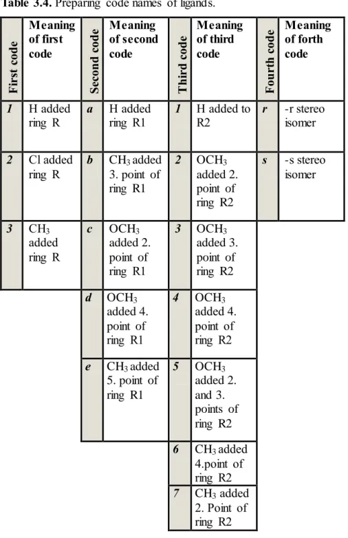
Table 3.4. Preparing code names of ligands. F ir st c od e Meaning of first code S ec on d c od e Meaning of second code T h ir d c od e Meaning of third code F ou rt h c od e Meaning of forth code 1 H added
ring R a H added ring R1 1 H added to R2 r
-r stereo isomer 2 Cl added ring R b CH3 added 3. point of ring R1 2 OCH3 added 2. point of ring R2 s -s stereo isomer 3 CH3 added ring R c OCH3 added 2. point of ring R1 3 OCH3 added 3. point of ring R2 d OCH3 added 4. point of ring R1 4 OCH3 added 4. point of ring R2 e CH3 added 5. point of ring R1 5 OCH3 added 2. and 3. points of ring R2 6 CH3 added 4.point of ring R2 7 CH3 added 2. Point of ring R2
In the figure 3.2, ‘’R’’ represent fifth point of benzoxazolinone ring, ‘’R1’’ represent ring R1 and ‘’R2’’ represent ring R2.
Ligands name are generated by 4 part codes. These codes contain 2 special numbers and 2 special letters. Some ligands code names are such as 1a2r, 2c4s, 3e7r.
All ligands names start with a number that first part of code names are between 1 and 3.
Meaning of these codes;
1 → hydrogen (H) is added to R point of benzoxazolinone ring 2 → chloride (Cl) is added to R point of benzoxazolinone ring 3 → methyl (CH3) is added to R point of benzoxazolinone ring.
For example, code ‘1’ determinates that point R of scaffold has only hydrogen ( H ) atom. Code ‘’ 2 ‘’ determinates that point R of scaffold has only chloride (Cl) atom. Code ‘’ 3 ‘’ displays that point R of scaffold has only methyl (CH3) compound.
Second code is related with another benzoike* ring that is indicated with R1. This second code is consist of letters as a, b, c, d and e. That letters display that spesific binding points and special compounds that are binding ring R1. Ring R1 has got 6 corners. Each corner of R1 has a number. These numbers are determinate for binding point of hydrogen (H), methoxy (OCH3) and methyl (CH3) .
Meaning of these codes:
a→ only hydrogen atoms are added to R1 ring
b→ methyl (CH3) is added to number 3 point of R1 ring. c→ methoxy (OCH3) is added to number 2 point of R1 ring. d→ methoxy (OCH3) is added to number 4 point of R1 ring. e→ methyl (CH3) is added to number 5 point of R1 ring.
Third code is that has a number between 1 and 7, each number is related with binding points and adding compounds of ring R2. R2 ring has 6 corners as figure 3.2.
Meaning of third codes:
1→ only hydrogen element is added to ring R2
2→ methoxy (OCH3) is attached to point number 2 of the ring R2 3→ methoxy (OCH3) is attached to point number 3 of ring R2 4→ methoxy (OCH3) is attached to 4.point of ring R2
5→ two methoxy (OCH3) are attached to 2. point and 3. point of ring R2 6→ methyl (CH3) is attached to 4.point of ring R2
Fourth and last code is consist of only two letters as ‘’r and s’’. These codes are related chiral center of ligands stereoisomerism. 105 ligands are created as –s stereoisomer and other 105 ligands are created as –r stereoisomer.
3.3. Molecular Docking
3.3.1. Docking Setup of AUTODOCK 4.2
AUTODOCK 4.2 requires 4 different input parameter files for properly running. These input files for each docking are, PDBQ file, GPF file and DPF file on the other hand AUTODOCK Tools (ADT) is a primary auxiliary for creating these files.
Macromolecule files are prepared by first adding polar hydrogen atoms. Ligands are prepared by first nonpolar hyrogen atoms are merged and Gasteiger charges are added. Bonds with torsional freedom are marked obtained to PDBQ file.
Grid files constitute GPF files. During the producing GPF, points of grid size, grid center points, spacing between grid points and quantity of grid files which based on the atom types are defined as parameters. In this thesis two set of grid file is used. The first grid set is designed for MAO-A isozyme and second set is designed for MAO-B isozyme. Both of settings are designed for relatively fine docking. Map parameters are defined as 80x80x80 grid points for three dimension and spacing is kept default 0.375 Ǻ. These parameters circle whole active side of the proteins. Grid center points of MAO-A isozyme are defined x: 33.625 y: 30.159 z: -18.303 on the xyz coordinate and grid center points of MAO-B isozyme are defined x: 53.506 y: 147.816 z: 24.376on the xyz coordinate.
DPF contains docking algorithm and essential variable parameters for Autodock docking. Population size, number of energy evaulation, number of generation, number of runs, rate of crossover, rate of mutations are defined in
docking parameter file. In this thesis these parameters are improved from default units for fine docking. Population size: 150, number of energy evaulation: 5000000, number of generation 27000, number of run: 50-100, crossover rate: 0.8, mutation rate: 0.02.
3.3.2 Enzyme-Ligand Molecule Docking by AUTODOCK 4.2
In this thesis Autodock is performed for MAO-A and MAO-B isozymes. For each isozymes of MAO is prepared in the AUTODOCK 4.2 using the parameter set up.
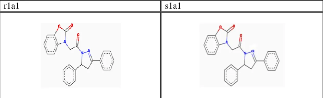
MAO A and MAO B enzymes were handled separately in this software program. Firstly MAO A enzyme is docked with 210 many ligands specially half of these ligands have r stereoisomerism and other half of these ligands have s
stereoisomerism. Goal of the using that two different stereoisomerism is detecting of the best prediction lowest energy value during the docking of ligand and enzyme. This stereoisomerism is generated for all ligands thus all ligands are obtained in pairs.
r1a1 s1a1
Figure 3.4. Comparing –r and –s stereoisomers of ligands by Accelerys Discovery
Studio
MAO A enzyme is performed respectively 105-r stereoisomer ligands and 105-s stereoisomer ligands by the using docking set up parameters as mentioned in chapter 3.3.1. Docking results are in the supplementary.
MAO-B enzyme is performed with the same ligands as MAO-A docking process. 105-r stereoisomer ligands and 105 s isomer ligands are docked one by one with MAO B isozyme. Docking set up parameters created as mentioned in chapter
3.3.1. The only difference between MAO A is grid box parameters cause of active side of MAO isozymes have different coordinates.
3.3.3 Docking Setup of LIBDOCK
Accelrys Discovery Studio 3.1 requires two main input parameters for properly docking process. These parameters are enzyme file and ligand file. Enzyme file format is converted to dsv format from format pdb for each MAO-A and MAO-B by Accelrys likewise ligand file is converted to sd format from format pdb by
Accelrys. During prepare of ligands, according to results of Autodock the best 10 ligands for MAO-A and the best 10 ligands for MAO-B are prepared for Libdock process.
Firstly enzyme and ligand parameters are inserted to Accelrys, secondly enzyme and ligand are defined. Before the defined of enzyme cavity, hydrogen atoms are added for the best work of cavity method. Before the docking process, special parameters of Libdock are defined. In the course of MAO-A Libdock
perform, input enzyme sapphire xyz coordinates are defined as x: 25.717, y: 37.50, z: -15.287 and radius: 14.169 other parameters set up as number of hotspots: 100, docking tolerance: 0.25, docking preferences: high quality, conformation method: fast, minimized algorithm: do not minimize. On the other side, MAO-B enzyme sapphire xyz coordinates defined as x: 46.238, y: 162.147, z: 36.200 and radius: 19.60 other parameters set up as number of number of hotspots: 100, docking tolerance: 0.25, docking preferences: high quality, conformation method: fast, minimized algorithm: do not minimize.
Chapter 4
4. Result and Discussion
4.1. Results of Molecular Docking Studies by Autodock 4.2
The data set of 210 ligand compounds described in Chapter 3 are docked to monoamine oxidase A isozyme by runing AUTODOCK 4.2 software program that is calculated results using the Lamarcian genetic algorithm.
The best ligand molecules and their scoring functions are categorized which are top ten ranked in classify of r and s stereoisomerism used to rank them are listed in table 4.1. These molecules are chosen from 210 different ligands and classified for drug candidates for MAO-A and MAO-B isozymes.
Docking studies by MAO-A show that ligands of first code name include ‘1’ have high score than other ligands such as code names starting with ‘2’ and ‘3’. Thus, when only hydrogen is attached R point of scaffold bind to MAO-A enzyme more effective than other ligands which have chloride and methyl on R point of benzoxazolinone. The best docking result for MAO-A is obtained by ligand 1d3r. Free binding energy of 1d3r is -13.09 kcal/mol and estimated inhibition constant is 252.50 pM. This result indicates at the lowest docking energy conformation, methoxy that is on 3 position of ring R1 makes hydrogen bonding by Lys305 and methoxy which is on the 4 position of ring R2 makes another hydrogen bonding by Tyr197. Second good docking result is obtained by 1d6r docking that has -12.77 kcal/mol free binding energy and estimated inhibition constant is 435.24 pM. Other high score ligands after the Autodock studies are ordinarily 1a3r, 1d1r, 1a6r, 1c3r, 1c6r, 3e6r, 2c7s and 1d7s. Detailed result values are in the table Y.
Considerable that r-stereoisomerism much effective than s-stereoisomerism for MAO-A isozyme. According to results, only two s-isomers such as 2c7s and 1d7s are observed in the top ten results of docking MAO-A studies.
On the other hand, MAO-B docking results show that ligand 2d1r consist the best docking with MAO-B. Free binding energy is -13.35 kcal/mol and estimated inhibition constant is 165.01 pM. Methoxy (OCH3) on the ring R1 makes sigma - π interaction with FAD. Also Tyr435 and FAD make sigma – π interactions with ring R1, Tyr188 and Tyr398 makes π - π interactions with ring R1. Second good docking result is obtained by 1d4s. Free binding energy is -11.69 kcal/mol and estimated binding constant is 2.69 nM. Hydrogen atom that determinate s-stereo isomer makes close interaction with Tyr 326. Other good docking results are obtained by ligands 1d5s, 1d6s, 1d3s, 1a3s, 1a1s, 3b4r, 1c6s and 3d4s.
According to these results, s-stereo isomers give high scores for docking MAO-B isozyme on the contrary MAO-A isozyme. Although the best result is an r-isomer (2d1r), eight docking results are obtained by s-r-isomers in the best 10 results for MAO-B docking.
On the other hand, according to docking results estimated inhibition constants (Ki) of MAO-A docking are better than MAO-B docking results.
Table: 4.1. Top 20 Autodock docking results of MAO-A and MAO-B isozymes
MAO-A MAO-B
Ligands Average binding free energy (kcal/mol) Estimated Inhibition Constant (Ki) Ligands 1d3r -13.09 252.50 pM 2d1r 1d6r -12.77 435.24 pM 1d4s 1a3r -12.63 50.71 pM 1d5s 1d1r -12.61 570.20 pM 1d6s 1a6r -12.59 589.50 pM 1d3s 1c3r -12.56 621.24 pM 1a3s 1c6r -12.53 654.87 pM 1a1s 3e6r -12.38 848.42 pM 3b4r 2c7s -12.24 1.16 nM 1c6s 1d7s -12.09 1.38 nM 3d4s
4.2. Results of Molecular Docking Studies by LIBDOCK
After the Autodock 4.2 studies the best docking results are analyzed by Libdock tool of Accelrys Discovery Studio 3.1. These results include 20 ligands for MAO-A and MAO-B. These ligands are prepared for the Libdock as chapter 3.3.2. …
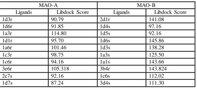
Libdock docking based on poses of ligand and enzyme. Each docking process come true by these poses. Every one of ligands makes their specific conformation at the active side of enzyme. Thus produced poses are key role for the docking. MAO-A docking studies shows that less poses produced and as a result scores of ligands could not effective for MAO-A ligands. In respect of Libdock, score of ligand 1a3r is 114.80 and score of ligand 1a6r is 101.46 thus these ligands are better candidates for MAO-A inhibitor.
On the other hand, MAO-B docking process produced a greater number poses than MAO-A docking process, thus scores of MAO-B docking are observed much more effective. Specially ligand 2d1r, 1d6s, 3b4r, 1a1s, 1d3s are strong candidate. Score of ligand 2d1r is 141.08, score of ligand 1d6s is 145.86, score of ligand 3b4r is 143.824, score of ligand 1a1s is 143.66 and score of ligand 1d3s is 138.28.These results indicate that ligands are much effective for MAO-B than MAO-A. Detailed libdock scores of both MAO-A and MAO-B are given in the table x.
Table 4.2. Results of Libdock docking for MAO-A and MAO-B isozymes
MAO-A MAO-B
Ligands Libdock Score Ligands Libdock Score
1d3r 90.79 2d1r 141.08 1d6r 91.85 1d4s 97.16 1a3r 114.80 1d5s 92.16 1d1r 95.70 1d6s 145.86 1a6r 101.46 1d3s 138.28 1c3r 98.75 1a3s 125.50 1c6r 94.16 1a1s 143.66 3e6r 105.318 3b4r 143.824 2c7s 92.16 1c6s 112.02 1d7s 87.24 3d4s 111.30
4.3. Analysis of Monoamine Oxidase A – Ligand Docking
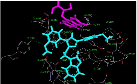
Ligand 1d3r
Pyrazole, benzoxazolinone and R2 parts of ligand 1d3r are positioned vertical towards FAD and R1 ring of ligand is positioned horizontal. Also inhibitor is
surrounded by hydrophobic Leu337, Phe352, Tyr407 and Tyr197 residues at the lowest energy conformation moreover Ile335, Ile180, Asn181, Ile207, Asp338, Phe208 other residues that surround ligand 1d3r. In addition residues Tyr69, Ala68 and Gly67 are located close to FAD.
Tyr407 makes hydrogen bound (1.8Ǻ) with the oxygen atom of benzoxazolinone ring and shows π- π interaction (3.6 Ǻ) by R1. Another π- π interaction (4.4 Ǻ) occurs between Phe352 and benzoxazolinone. Tyr197 located close to aromatic cage and positioned horizontally to R2 make one hydrogen bond (2.4 Ǻ) by the oxygen atom of methoxy on the ring R2. Other hydrogen bounds are observed between the Lys305 and oxygen atom of methoxy on the ring R1.
Distances of these hydrogen bounds are 2.3Ǻ and 2.6Ǻ.
Figure 4.1. 3-D view of ligand 1d3r and active side of MAO-A at the lowest energy
conformation. Pink color represent FAD compound, cyan color represent inhibitor, green dashed line represent hydrogen bonding.
Ligand 1d6r
Ligand 1d6r has hydrogen at the R point, mehthoxy at the fourth point of ring R1, methyl at the fourth point of ring R2. Benzoxazolinone ring get involved among of two amino acid side chains range where are Tyr444, Gln215, Ile180 and Tyr407, Leu337, Phe352, Tyr69. Especially residues Tyr444 and Tyr407 are located so close to aromatic cage. Other residues of surrounding ligand are Tyr197, Ile207, Phe208, Met 350, Ile335 and hydrophilic Asp338 at the lowest energy conformation of docking MAO-A isozyme.
There is 2 hydrogen bounds are obtained. First one occurs between Tyr407 and oxygen of benzoxazolinone (1.9 Ǻ). Second hydrogen bond is observed between Lys305 and oxygen of methoxy group of R1 ring. Otherwise FAD does a sigma – π interaction with R2 ring (2.4 Ǻ). Tyr 407 makes π - π interactions with ring R2 (3.3 Ǻ), Tyr444 makes another π - π interactions with benzoxazolinone.
Figure 4.2. 3-D view of ligand 1d6r and active side of MAO-A at the lowest energy
conformation. Pink color represent FAD compound, cyan color represent inhibitor, green dashed line represent hydrogen bonding.
Ligand 1a3r
Chemical structure of ligand 1a3r that scaffold has hydrogen at the point R, only hydrogen atoms are at the ring R1 and methoxy is at the third point of ring R2. Ligand 1a3r is located to the active side of MAO-A by a good clean geometry. Benzoxazolinone ring of the ligand is located to between two hydrophobic pockets that are Tyr407, Phe352, Leu337 and Ile335, Gln215, Tyr444. Pyrazole is positioned
between the Tyr69, Lys218 amino acid side chains and FAD. Also Ile207, Asn181, Ile207 and Met350 are other residues which surrounding of the ligand.
FAD and Ring R2 have sigma – π interaction (2.4 Ǻ). Oxygen of
benzoxazolinone ring makes hydrogen bound by hydrophobic Tyr407 (2.2 Ǻ). Also Tyr407 does π - π interaction with the ring R1 (3.4 Ǻ). Other π - π interaction is obtained between the Phe352 and benzoxazolinone ring (4.4 Ǻ).
Figure 4.3. 3-D view of ligand 1a3r and active side of MAO-A at the lowest energy
conformation. Pink color represent FAD compound, cyan color represent inhibitor, green dashed line represent hydrogen bonding.
Ligand 1d1r
Benzoxazolinone, pyrazole and ring R2 positioned horizontally to each other and positioned vertically to ring R1 and FAD at the active side of MAO-A isozyme. Ligand conformation is located between amino acid side chains of hydrophobic Tyr444, Ile180, Ile207 and Leu337, Phe352, Tyr407 thence makes close contact with FAD. Also hydrophilic Asp339, Lys305, Lys218, Asp64 residues surrounder ring R1 within 4.5 Ǻ at the lowest energy conformations.
Tyr407 makes a hydrogen bond of good geometry with the oxygen of benzoxazolinone (2.0 Ǻ). Another hydrogen bond that length is 2.4 Ǻ occurs between oxygen of ring R1 and hydrophilic Lys305. π – π interaction consist of Tyr407 and ring R2 (3.5 Ǻ). Another π – π interaction that distance is 4.3 Ǻ occurs between the oxygen of benzoxazolinone ring and residue Leu337.
Figure 4.4 3-D view of ligand 1d1r and active side of MAO-A at the lowest energy
conformation. Pink color represent FAD compound, cyan color represent inhibitor, green dashed line represent hydrogen bonding.
Ligand 1a6r
Conformation of ligand 1a6r is similar with ligand 1d3r at the lowest energy conformation of docking MAO-A. Aromatic cage is surrounded by Tyr407, Tyr444, Gln215, Ile180 and Leu337 residues. Also residues Phe208, Asn181, Gln215, Phe352 are close to benzoxazolinone ring. Other residues that surround of ligand 1a6r are Ala68, Gly67, Lys305, Tyr197.
Residue Tyr407 and oxygen atom of benzoxazolinone makes hydrogen bonding (2.1Ǻ) and same residue where located to aromatic cage generates π-π interaction (3.3Ǻ) with ring R2. Hydrophobic Phe352 makes π-π interaction (4.4Ǻ) with benzoxazolinone. In addition significant sigma-π interaction (2.3Ǻ) occurs between FAD and ring R2.
Figure 4.5. 3-D view of ligand 1a6r and active side of MAO-A at the lowest energy
conformation. Pink color represent FAD compound, cyan color represent inhibitor, green dashed line represent hydrogen bonding.
Ligand 1c3r
Conformation of inhibitor 1c3r is located quite close to FAD. Aromatic cage is clearly shown with a good geometry. When ring R2 and benzoxazolinore are positioned horizontally for each other FAD and ring R1 are positioned vertically at the lowest energy conformation. Analysis of 3D structure of inhibitor position shows that aromatic cage remains among to hydrophobic residues Tyr407, Leu337, Phe352 and Tyr444. Also residues such as Tyr197, Tyr444, Lys305, Leu317, Ile180, Phe208 and Ile207 surround ligand 1c3r.
Important residue Tyr407 is associated with oxygen atom of benzoxazolinone by hydrogen bond (2.2Ǻ) and this residue makes π-π interaction (3.4Ǻ) with ring R2. Hydrophobic residue Phe352 makes another π-π interaction (4.4Ǻ) by five carbon ring of benoxazolinone.
Figure 4.6. 3-D view of ligand 1c3r and active side of MAO-A at the lowest energy
conformation. Pink color represent FAD compound, cyan color represent inhibitor, green dashed line represent hydrogen bonding.
Ligand 1c6r
This inhibitor shows high degree similarity with inhibitor 1c3r about position at the lowest energy conformation. Resiudes Tyr407, Tyr69 and Tyr444 are located close to aromatic cage. Other considerable residues Leu337, Tyr197, Ser442, Gln215, Ile180 and Lys305 surround ligand 1c6r.
In the ligand 1c6r docking process small number interactions are obtained. Tyr407 and benzoxazolinone has hydrogen bond (2.4Ǻ) and Tyr407 makes π-π interaction with R2 (3.0Ǻ). Other π-π interaction occurs between Phe352 and benzoxazolinone.
Figure 4.7 3-D view of ligand 1c6r and active side of MAO-A at the lowest energy
conformation. Pink color represent FAD compound, cyan color represent inhibitor, green dashed line represent hydrogen bonding.
Ligand 3e6r
Hydrophobic residues Tyr407, Phe352, Leu337 are located close to aromatic cage. When Ile207 and Ile180 positioned near to benzoxazolinone, residues Tyr444, Gln215, Tyr197 are positioned around ring R2. Other residues Gly443, Ser442, Tyr69 Lys305 and Asn181 surround the ligand 3e6r.
In this docking analysis, three interactions are obtained. Residue Tyr407 and ring R2 makes π-π interaction (3.4Ǻ). Tyr444 and benzoxazolinone ring does hydrogen bounding (1.7Ǻ). Last interaction for ligand 3e6r is sigma-π that occurs between the benzoxazolinone and ring R2.
Figure 4.8. 3-D view of ligand 3e6r and active side of MAO-A at the lowest energy
conformation. Pink color represent FAD compound, cyan color represent inhibitor, green dashed line represent hydrogen bonding.
Ligand 2c7s
Residue Tyr444 is located almost central of aromatic cage. Benzoxazolinone ring is positioned between residues Ile335, Asn181, Ile180, Tyr197 and hydrophobic pocket residues such as Tyr407, Ile207, Leu337. Other residues Tyr69, Ala68, Gly67, Phe208, Phe352 and Val182 surround ligand.
Tyr407 has important role for 2c7s docking results. Ring R2 and Tyr407 makes cation-π interaction (4.2Ǻ). Also hydrogen bounding occurs between the Tyr407 and benzoxazolinone (2.1 Ǻ). Another interaction is observed as π-π (5.5Ǻ). This interaction occurs between the Tyr444 and ring R1.
Figure 4.9. 3-D view of ligand 2c7s and active side of MAO-A at the lowest energy
conformation. Pink color represent FAD compound, cyan color represent inhibitor, green dashed line represent hydrogen bonding.
Ligand 1d7s
Residues Leu337, Gly67, Gln215 are positioned at the aromatic cage. Pyrazole is located between the hydrophobic packet residues Ile335, Phe177, Ile180, Phe208 and other hydrophobic residues Tyr407 and Phe352. Also Tyr444, Tyr197, Ala68, Tyr69 and Gly443 are other residues that surround the ligand 1d7s.
Benzoxasazolinone and Lys305 make cation-π interaction (4.0Ǻ). Another cation-π interaction occurs between the Phe352 and nitrogen atom of pyrazole (5.4Ǻ) and also Phe352 makes π-π interaction with ring R1 (5.6Ǻ). Other π-π interaction occurs between the Tyr407 and ring R2 (3.3Ǻ). On the other hand, FAD makes a cation-π interaction with nitrogen atom of pyrazole (6.0Ǻ). Moreover FAD makes hydrogen bonding with oxygen atom of benzoxazolinone.
Figure 4.10. 3-D view of ligand 1d7s and active side of MAO-A at the lowest energy
conformation. Pink color represent FAD compound, cyan color represent inhibitor, green dashed line represent hydrogen bonding.
4.4. Analyses of Monoamine Oxidase B - Ligand Docking
Ligand 2d1r
Ligand 2d1r is located active side of MAO-B isozyme and ring R1 of ligand is surrounded by hydrophobic pockets such as Tyr435, Tyr188 Tyr398. Also ligand has close interaction by FAD. Oxygen of pyrazole makes hydrogen bounding with Gln206 (2.3 Ǻ). Besides hydrogen bonding very significant interactions are watched around the ring R1. π – π interaction is shown between Try435 and R1 ring (3.9Ǻ). Also FAD performs 2 many sigma – π interactions. (2.9 Ǻ, 2.6 Ǻ)
Figure 4.11. 3-D view of ligand 2d1r and active side of MAO-A at the lowest energy
conformation. Pink color represent FAD compound, cyan color represent inhibitor, green dashed line represent hydrogen bonding.