the relationship between the
structural characteristics of
lactobacilli-eps and its ability to
induce apoptosis in colon cancer
cells in vitro
Ummugulsum tukenmez
1, Busra Aktas
2, Belma Aslim
1& serkan Yavuz
3Colon cancer is one of the most common cancer around the world. exopolysaccharides (epss) produced by lactobacilli as potential prebiotics have been found to have an anti-tumor effect. In this study, lyophilized epss of four Lactobacillus spp. for their impact on apoptosis in colon cancer cells (HT-29) was evaluated using flow cytometry. The relationship between capability of a lactobacilli-EPS to induce apoptosis and their monosaccharide composition, molecular weight (MW), and linkage type was investigated by HpLC, seC, and NMR, respectively. Changes in apoptotic-markers were examined by qpCR and Western Blotting. epss were capable of inhibiting proliferation in a time-dependent manner and induced apoptosis via increasing the expression of Bax, Caspase 3 and 9 while decreasing Bcl-2 and Survivin. All EPSs contained mannose, glucose, and N-acetylglucosamine with different relative proportions. Some contained arabinose or fructose. MW ranged from 102–104Da with two or three
fractions. eps of L. delbrueckii ssp. bulgaricus B3 having the highest amount of mannose and the lowest amount of glucose, showed the highest apoptosis induction. In conclusion, lactobacilli-EPSs inhibit cell proliferation in HT-29 via apoptosis. Results suggest that a relationship exists between the ability of eps to induce apoptosis and its mannose and glucose composition.
Today, cancer is one of the most important health problem leading to death. Colorectal cancer is the second most common cause of cancer deaths in the world1,2. Patients with colorectal cancer have been treated with surgery,
radiotherapy, or chemotherapy with toxic drugs such as 5-fluorouracil and oxaliplatin. Most of the anti-cancer drugs used in chemotherapy cause immunotoxicity and slow down the healing process3. Identification of
anti-tumor therapy with low side effects is essential in cancer studies. In preventing colon cancer, therefore, diet style rich in fibers, fermented foods such as probiotics have been suggested3.
Gut microbiota has found to be related to the risk of colorectal cancer4. A well-balanced bidirectional
rela-tionship between gut microbiota and host immune system exists5. Disruption of this balance with dysbiosis, the
perturbation of the healthy/normal gut microbiota, can result in a wide range of disorders or diseases such as metabolic disorders and inflammatory bowel diseases6,7. A study on patients with colorectal cancer showed that
the abundance of Fusobacteria was enriched in the colon tissue with tumor8. Zackular et al. reported that
abnor-mal microbiota in mice is associated with inflammation and tumor in colon9. Another study showed that patients
with colon cancer had lower counts of total Bifidobacterium in the colon microbiota4. It was assumed that targeted
alterations in the gut microbiota could be used to prevent or treat colorectal cancer in the future4,10.
Probiotics are one of the strategies that could be used to alter the microbial composition of the gut. A diverse set of health benefits have been ascribed to probiotics including: immunomodulation, improved ability to tolerate lactose; reduction in gastrointestinal pathogens; and reduction in colorectal cancer11–13. Probiotics come from a
variety of genera, including Lactobacillus, Bifidobacterium, Propionibacterium, Escherichia, and Saccharomyces; 1faculty of Science, Department of Biology, Gazi University, Ankara, turkey. 2faculty of Arts and Science, Department of Molecular Biology and Genetics, Burdur Mehmet Akif ersoy University, Burdur, turkey. 3faculty of Science, Department of chemistry, Gazi University, Ankara, turkey. correspondence and requests for materials should be addressed to B.A. (email: [email protected])
Received: 29 November 2018 Accepted: 28 February 2019 Published: xx xx xxxx
however, Lactobacillus are the most common genera used as probiotics14. Studies have demonstrated that
bacte-rial secondary products can also alter the gut environment and affect cancer development15,16. In a study
evaluat-ing the impact of cell fractions and exopolysaccharides (EPSs) from Lactobacillus on colon cancer cells, Liu et al. reported that EPS has the highest cytotoxic effect among the tested fractions and reduced proliferation of colon cancer cells17. Another study investigating the effect of lactobacilli EPS on cervical tumor cells demonstrated that
lactobacilli EPS induced apoptosis in tumor cells and showed an anti-proliferative effect15. Microbial EPSs are
primary or secondary metabolites produced by microorganisms and they have been used as prebiotics, which defined as the specific fermented ingredient resulting in changes in the gastrointestinal microbiota and provide health benefit18,19. There is a great diversity among EPS produced by lactic acid bacteria (LAB)20. LAB EPSs can be
divided into homopolysaccharides (HoPSs) consisting one type of monosaccharide, and heteropolysaccharides (HePSs) consisting a backbone of repeating units that are composed of two or more types of monosaccharides21,22.
LAB mostly produces HePSs which consist of different sugars such as pentose (D-arabinose, D-ribose, D-xylose), hexose (D-glucose, D-galactose, D-mannose), or uronic acids (D- glucuronic acid, D-galacturonic acid). They mostly consist different types of linkages and branches such as α-(1,2) or α-(1,6) linkages which are rigid, β-(1,4) or β-(1,3) which are less rigid23,24. Studies reported that the composition and the structure of EPSs tend to be
strain dependent25,26. Moreover, it has been shown that structural and compositional diversity among EPSs might
be responsible for the variation in their health benefits20,27. Li et al. purified three fractions of EPS isolated from L.
helveticus MB2-1 and evaluated their structure and antioxidant activities in vitro28. They reported that although
the molecular weights of EPSs were similar, their sugar compositions and antioxidant effect were different as well as their anti-cancer impact on colon cancer cells28,29. Additionally, anti-cancer activity of polysaccharides can be
affected by other physicochemical properties, such as presence of β-type glycosidic linkages, uronic acid, sulfate groups, and glucose increasing anti-cancer activity30–33. Therefore, the determination of chemical composition
and structure of EPS must be taken into consideration when predicting potential applications of EPSs30.
Although lactobacilli EPSs have been reported to have cytotoxic effect on various cancer cell lines, the mech-anism of action and the impact of its structure on cytotoxic effect have not been understood yet. In this study, we investigated the EPS produced by various Lactobacillus spp. and their impact on proliferation and apoptosis in colon cancer cells. We performed a chemical and structural characterization of lactobacilli EPSs including molecular weight, monosaccharide composition, and linkage type. Additionally, we evaluated their structural, characteristic effects on apoptosis.
Materials and Methods
Bacterial strains.
A total of four previously described Lactobacillus spp. isolated from healthy infant feces (L. plantarum GD2, L. rhamnosus E9, L. brevis LB63) and yogurt (L. delbrueckii ssp. bulgaricus B3) were used in this study34,35. Bacterial species were confirmed by 16S rRNA sequence analysis using universal primers (Uni27F,5′ AGAGTTTGATCCTGGCTCAG 3′ and Uni1492R, 5′ GGTTACCTTGTTACGACTT 3′). Lactobacilli stock cultures were maintained at −30 °C in MRS broth (Oxoid, Istanbul, Turkey) with 10% (v/v) glycerol. Working cultures were prepared from frozen stocks by two sequential transfers in MRS broth and incubations were con-ducted aerobically at 37 °C for 18 h.
Isolation and lyophilization of exopolysaccharide.
The method of Frengova et al. was followed to isolate EPS36. The growth culture with an optical density of 0.6 at 600 nm (~8.5 log CFU/ml) was heated at 100 °Cfor 15 min. After cooling, the cell suspension was treated with 17% (v/v) of 85% trichloracetic acid solution and centrifuged at 15, 493 × g for 20 min to remove cells and proteins. The exopolysaccharide was precipitated using two volume of cold absolute ethanol followed by centrifugation at 15, 493 × g for 15 min. The resulting pellet con-taining EPS was suspended in deionized water. Total carbohydrate was measured at 490 nm by phenol-sulfuric acid method37 using glucose as standard. The EPSs isolated were stored at −80 °C until being lyophilized in Christ
Alpha 2–4 freeze dryer (Marin Christ Co. FL, USA). The freeze-dried EPS powder was stored at 4 °C38.
physico-chemical characterization of the eps produced by Lactobacillus strains.
Monosaccharide composition. The method of Ledezma et al. was followed to hydrolyze EPSs isolated from Lactobacillus spp.39.Briefly, EPSs (10 mg/ml) were incubated with 1 M H2SO4 for 3 hours at 90 °C and then neutralized with 1 M NaOH to pH 7. After the complete hydrolysis, monosaccharide composition of EPS isolated from Lactobacillus spp. was quantified by high pressure liquid chromatography (HPLC) using an AGILENT 1260 system equipped with a refractive index detector at the Middle East Technical University, Central Laboratory. The separation (25 µl volume of injection) was carried out in the Metacarb 67 C columns (300 mm × 6.5 mm) maintained at 90 °C. For N-acetylglucosamine composition, the separation was carried out in the Metacarb 87 H (300 mm × 7.8 mm) column.
The mobile phase was water with a fixed flow rate of 0.5 ml/min and the separation was utilized for 30 min. Runs were performed at least in triplicate and the data was presented as mean ± SEM.
Molecular weight. This analysis was performed at the Bilkent University, National Nanotechnology Research Center (UNAM) following the protocol of Boymirzaev et al.40. Molecular weight of EPSs was determined by Size
Exclusion Chromatography (SEC) with an Agilent 1200 series system equipped with a PL aquagel-OH MIXED-H column and a refractive index detector. 10 µl of EPS at 0,05–0,2% (w/V) was injected and was eluted with 0.2– 0.8 M NaNO3 at a flow rate of 0.6 ml/min. Polysaccharide (pullulan) was used as standard.
Structural analysis. Nuclear Magnetic Resonance Spectroscopy Analysis (NMR) was performed at Çankırı Karatekin University Research Center. NMR spectrum of the EPS isolated from Lactobacillus spp. (30 mg/500 µl) was recorded with 99.96% D2O as the solvent at 600 MHz (Agilent, 600 MHz, 14.1 Tesla Premium Compact
NMR). Two-dimensional (2D) 1H–1H correlated spectroscopy (COSY), and nuclear overhauser effect spec-troscopy (NOESY) measurements were used to assign signals and to determine the sequence of sugar residues. Spectra was referenced to internal trimethylsilylpropanoic acid41.
Cell culture.
HT-29, Human Colorectal Adenocarcinoma Cell Line, (ATCC®
HTB-38™
) was kindly provided by Prof. Hakan Akbulut (Ankara University, Medical Oncology). The cells were grown in Dulbecco’s modified Eagle’s medium (ThermoFisher Scientific) containing high glucose (4.5 g/l), sodium pyruvate (1 mM), and sup-plemented with 10% of heat-inactivated fetal bovine serum (ThermoFisher Scientific), penicillin/streptomycin (100 units/ml of penicillin and 100 μg/ml of streptomycin) (ThermoFisher Scientific), and L-glutamine (2 mM) (ThermoFisher Scientific). HT-29 cell culturing was carried out in 25 cm2 or 75 cm2 cell culture flasks at 37 °C in a humidified incubator with 5% CO2 atmosphere. The cell culture medium was changed every 48 h from the sec-ond day after seeding, and cells were harvested by 0.05% trypsin/EDTA (ThermoFisher Scientific) after reached 80–90% confluence.Anti-proliferation activity.
Impact of EPSs from lactobacilli on HT-29 cell proliferation was evaluated using a WST-1 cell proliferation assay kit (Cayman Chemical Company, Ann Arbor, Michigan, USA). The lyophilized EPSs were dissolved in distilled water and filtered using a 0.2 µm syringe filter prior to analyses. HT-29 cells were seeded into a 96-well plate at a density of 1 × 10⁴ cells/well and treated with EPSs at a final concentra-tion of 400 µg/ml followed by 24 h or 48 h incubaconcentra-tion at 37 °C with 95% air and 5% CO2. After incubation, 10 μl of the WST-1 mixture was added to each well and the plates were incubated for 2 h at 37 °C with 95% air and 5% CO2. Formation of formazan was measured at 450 nm by a microplate reader (Epoch, Biotek, Winooski, VT, USA) and the absorbance was correlated with the cell number. The anti-proliferative effect was evaluated by com-paring to viability of the treated samples with the untreated control (ultrapure water and DMEM mix without test sample for EPS). The percentage of viability was calculated as follows:= ×
%Viability (Absorbance Sample/Absorbance Control) 100
Cell distribution by flow cytometry.
Briefly, treated and untreated cells (control cells) were washed twice with PBS and harvested by scraping from 6 well plate using a cell scraper in PBS and collected by centrifugation (367 × g, 4 min). Flow cytometry analysis were performed according to kit manufacturer’s directions42,43. The cellpellets were resuspended into 1 ml of DMEM. Following that, 4 ml of Annexin V binding buffer was added (Cat. No: BB10X-50ml, Immunostep, Spain) and centrifuged (500 × g, 5 min). After centrifugation, the supernatant was aspirated and the cells were resuspended in 200 µl of 1X BB (BB10X diluted 1X with distilled ultrapure water). 100 µl of the cell suspension was incubated for 30 minutes at room temperature and in the dark by adding 5 µl of Annexin V-FITC (FITC Annexin V, Immunostep, Spain) and propidium iodide (PI) (as in the final concentration of 40 µg/ml, Immunostep, Spain). 1X BB (100 µl) was added into each cell tube and the cells were analyzed using ACEA NovoCyte 3000 Flow cytometer. Data analysis was performed using ACEA NovoExpress software.
RNA isolation and gene expression analysis by Rt-pCR.
Total RNA was isolated from HT-29 cells using GeneJET RNA Purification Kit (Thermo Scientific, Cat No: K0731). Concentration and purity of RNA samples were determined using a Take3 Micro-Volume Plate (Epoch, Biotek, Winooski, VT, USA). First strand cDNA was synthesized from 1 μg of RNA using RevertAid First Strand cDNA Synthesis Kit (Fermentas, K1 621). PCR conditions were as follows; 3 min at 94 °C for initial denaturation, 30 s at 94 °C 35 cycles of denaturation, 30 s at 58 °C for annealing, and 45 s at 72 °C for extension. cDNA samples were stored at −20 °C until gene expression analysis. Real-time PCR was performed using ABI 7500 Fast Real Time-PCR and QuantiFast SYBR Green PCR Mix (Qiagen). The primers are shown in Table S1. PCR conditions were as follows; 5 min at 95 °C for initial activa-tion, 10 s at 95 °C for 40 cycles of denaturation and 30 s at 60 °C for combined annealing–extension. All reactions were performed in triplicate and repeated at least 2 times. Cyclophilin A (PPIA) gene was used as an internal control to normalize the target transcripts by the 2−ΔΔCT method44.Western blot analysis.
Total protein extracts from untreated cells or cells treated with EPS at different time intervals (24 h or 48 h) were subjected to Western blot analysis as described by Huang et al.45. HT-29 cellsat a density of 1 × 10⁶ cells were treated with EPSs at a final concentration of 400 µg/ml and incubated for 24 and 48 h. After treatment, the medium was aspirated and the cell culture plate placed on ice washed twice with ice-cold phosphate buffer saline (PBS). After PBS was drained, the cells were lysed by 250 µl of lysis buffer (NP-40 buffer-150 mM NaCl, 50 mM Tris, pH 8.0, and 1% NP-40) containing protein inhibitor cocktail. The cells scraped from the plate gently transferred into the pre-cooled centrifuge tubes. The tubes were kept on ice for 30 min with constant agitation. The lysates were centrifuged at 13,201 × g for 20 minutes at 4 °C and the supernatant was stored at 4 °C until use46,47. The total protein was determined using the Bradford assay (Sigma-Aldrich). 40 μg of
protein lysates denatured in loading buffer at 95°C for 10 min was separated by 4–12% Acrylamide-Bisacrylamide gel and then transferred onto PVDF membranes using iBlot
®
(ThermoFisher). The membranes were then blocked in blocking solution (Western Breeze, ThermoFisher) at room temperature before incubating with antibodies for one hour. The expression patterns of Bax, Bcl-2, Caspase 3, Caspase 9, Survivin were detected using specific antibodies and β-actin was used as loading control48. After washes in Antibody Wash, the membrane wasincu-bated in secondary antibody (anti-rabbit IgG) for 30 min. After second washes, the membrane was incuincu-bated in Chromogenic Substrate until the bands develop on the membrane. The molecular weight of the protein bands was determined using BIORAD ImageLab 5.21 program compared to the protein marker (Thermo Scientific PageRuler Prestained Protein Ladder 26616).
statistical analysis.
All experiments were carried out with three replicates and values were reported as means ± standard deviation (SD), unless otherwise indicated. Statistical analysis was performed using SPSS 16.0 (SPSS Inc., Chicago, IL, USA). Statistical difference was assessed with one-way analysis of variance (ANOVA) followed by Tukey test. For Western blot analysis, t-Test (Excel 2007) was performed. Additionally, post hoc Dunnett’s test for pair-wise comparison was ran to analyze flow cytometer data. Statistical difference was deter-mined at a P value of 0.05 or less. Monosaccharide composition of EPSs produced by Lactobacillus spp. were used to generate a dendrogram by the Ward method of hierarchical clustering (JMP version 12, SAS Institute Inc., Cary, NC).Results and Discussion
production of eps by Lactobacillus spp. in culture medium and their sugar composition
anal-ysis.
The bidirectional interaction between host and probiotic bacteria results in health benefits to the host49.Several factors in lactobacilli have been shown to impact human health in vitro and in vivo, including cell surface components and metabolites49. EPSs produced by LAB are one of the important component that have a key role
in probiotic activity including anti-proliferative effect, immunomodulation, and adhesion13,17,50. Here we
inves-tigated the production of EPSs by four Lactobacillus strains (L. plantarum GD2, L. rhamnosus E9, L. brevis LB63, and L. delbrueckii ssp. bulgaricus B3) and their compositional analysis. EPSs were freeze-dried and lyophilized. EPS production by various lactobacilli strains differed significantly from each other (p < 0.05). L. plantarum GD2, L. rhamnosus E9, L. brevis LB63, and L. delbrueckii ssp. bulgaricus B3 yielded 397 ± 4, 298 ± 5, 347 ± 4, and 449 ± 4 mg/l, respectively. L. delbrueckii ssp. bulgaricus B3 produced the highest amount of EPS among other lactobacilli. L. delbrueckii ssp. bulgaricus B3 was isolated from yogurt while the others were isolated from feces samples. Mozzi et al. reported that most of the HePS are produced by Lactobacillus from food origin as seen in our results51. Studies on EPS production by Lactobacillus to date reported varying amount of EPS. Sungur et al. stated
that two of the L. gasseri strains produced 242 ± 3 mg/l and 255 ± 4 mg/l of lyophilized EPS15. In another study,
EPS production by L. crispatus in different carbon sources ranged from 200 to 400 mg/l52. Van Geel-Schutten et al.
examined the production of EPSs by a total of 182 Lactobacillus strains in MRS medium with relatively high sugar concentration53. Only 60 of them produced EPS and from those strains only 10% produced EPS more than
100 mg/l which is referred to as the large amount of production. The lactobacilli strains studied here produced in a decent amount of EPS relative to the lactobacilli EPSs studied in the literature15,52,53.
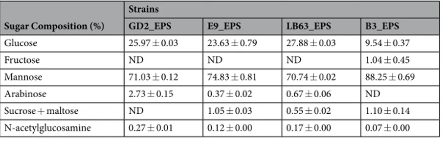
EPS produced by LAB exhibit a large variation in their sugar composition54. Here, we examined
monosaccha-ride composition of EPSs produced by various Lactobacillus spp., using HPLC. Different types of sugar unite were found in the lactobacilli EPSs studied. The EPSs were mainly composed of mannose and glucose ranging between 71–88% and 10–28%, respectively. Additionally, fructose, arabinose, sucrose + maltose, and N-acetylglucosamine were detected at different ratios (Table 1). Detection of maltose and sucrose, disaccharide molecules, might be due to incomplete hydrolysation of EPS. A review on functional properties of EPSs derived from yeast reported that EPSs consisting mannose with more than 50% are classified as biologically active55. Anti-cancer activity of EPSs
has thought to be related with high amount of mannose in sugar composition56–58. Shao et al. showed that
poly-saccharides consisting of glucose and mannose can interact with Toll-like receptors and activate host immunity56.
Vidhyalakshmi and Vallinachiyar reported that macrophages carry mannose and glucose specific receptors which are important in triggering anti-cancer activities and suppressing cell proliferation in tumor57. Similarly, Jin et al.
studied anti-tumor activity of EPS and concluded that anti-tumor activity of polysaccharides tends to correlate with mannose presence as major monomer in the sugar composition59.
The presence of different monomers among the EPSs suggest that EPSs of lactobacilli strains studied here are heteropolysacharides as identified mostly in other LAB54. Additionally, cluster analysis of sugar composition in
EPSs of Lactobacilli spp. revealed that the L. delbrueckii ssp. bulgaricus B3 clustered by itself separated from the other strains (Table 1). Suggesting that this strain tends to have different biological functions compared to the other strains. Based on the variations in the ratio of monomers among various Lactobacillus, there is a composi-tional diversity of EPSs and this diversity likely contributes strain to strain variation in their biological functions including proliferation inhibition and apoptosis induction.
structural analysis of epss produced by Lactobacillus spp.
It has been shown that an impressive differences exist in molecular weights and structure of EPSs among LAB50,60. To understand better the diversityamong Lactobacillus spp. and compare the structure of EPSs produced we also performed a molecular weight
Sugar Composition (%)
Strains
GD2_EPS E9_EPS LB63_EPS B3_EPS
Glucose 25.97 ± 0.03 23.63 ± 0.79 27.88 ± 0.03 9.54 ± 0.37 Fructose ND ND ND 1.04 ± 0.45 Mannose 71.03 ± 0.12 74.83 ± 0.81 70.74 ± 0.02 88.25 ± 0.69 Arabinose 2.73 ± 0.15 0.37 ± 0.02 0.67 ± 0.06 ND Sucrose + maltose ND 1.05 ± 0.03 0.55 ± 0.02 1.10 ± 0.14 N-acetylglucosamine 0.27 ± 0.01 0.12 ± 0.00 0.17 ± 0.00 0.07 ± 0.00
Table 1. Monomer composition of the EPSs produced by Lactobacillus spp.*. ND; Not determined. *Hierarchical clustering of four Lactobacillus spp. strains based on monomer ratio in their EPS sugar composition. The results presented in average.
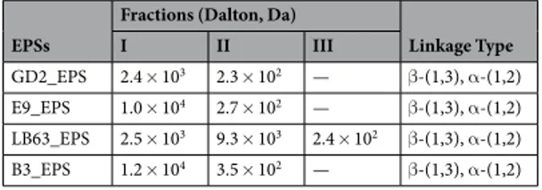
analysis by SEC and a structural analysis by NMR. SEC results showed that molecular weight of the EPSs ranged from 102 to 104 Da consisting of two fractions except LB63_EPS (Table 2). Interestingly, LB63_EPS has three frac-tions. Similarly, Tallon et al. isolated EPS from L. plantarum EP56 that has two fractions with molecular mass of 8.5 × 10⁵ and 4 × 10⁴ Da61. However, a study on structure analysis of EPS isolated from another strain of L.
plan-tarum (YW32) reported that L. planplan-tarum EPS has only one fraction with molecular weight of 1.03 × 10⁵ Da62.
Hamet et al. examined the EPSs of 28 different Lactobacillus and showed that the fraction number ranged from one to three with molecular weight distribution being strain dependent63. Biofunctinality of EPS has been shown
to be affected by molecular weight. Xu et al. reported that EPS from Bifidobacterium animalis RH has a stronger atioxidant activity due to its low molecular weight64. Based on literature, EPS ≤104 Da is considered as low molec-ular weight64–66. It has been reported that low molecular weight polysaccharides can easily pass through the host
cell membrane barriers and exhibit biological activity better30. On the contrary, in other studies investigating
chemical composition and anti-tumor activity of polysaccharides, high molecular weight polysaccharides tend to have more anti-tumor impact than those of low molecular weight67,68.
Furthermore, NMR chemical shifts were determined as described in literature41,69 by performing 1H-NMR, COSY and NOESY NMR analysis. Generally, the 1H NMR spectrum of a polysaccharide can be divided into three main regions: the anomeric region (δ 4.5–5.5), the ring proton region (δ 3.1–4.5) and the alkyl region (δ 1.2–2.3)70.
In the present study, the chemical shift of the anomeric H1 protons of the EPS was observed at δ 4.8, δ 4.9 and δ 5.2 ppm. 1H chemical shifts of the EPS from Lactobacillus spp. is shown in Table 3.
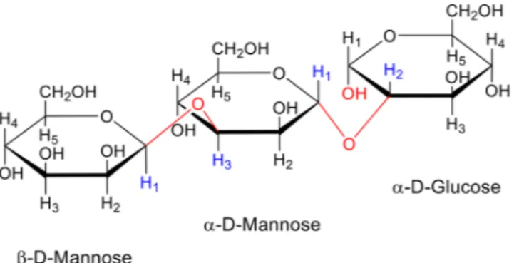
Similar H–H interactions was observed in all NOESY spectra. The released peaks were observed as a result of interaction between H1 proton of α-D-mannose (5.2 ppm) and H2 proton of α-D-glucose (4.0 ppm pro-tons). Furthermore, the H3 proton of α-D-mannose and H1 proton of β-D-mannose were found to be in interaction. These results helped us to understand the sequencing and binding stereochemistry of mon-osaccharide units of EPSs of Lactobacillus spp. The results showed that all of the lactobacilli studied here produced EPS with the same type of sugar linkage which are β-H1-H3 (β-D-Mannose-α-D-Mannose) /α-H1-H2 (α-D-Mannose-α-D-Glucose), designated as β-1,3 (β-D-Mannose-α-D-Mannose) and α-1,2 (α-D-Mannose-α-D-Glucose), respectively. The primary structure of EPSs is shown in Fig. 1.
Two types of linkage were found among Lactobacillus spp. Main sugar linkage types present in the EPS frac-tions isolated from Lactobacillus spp. are β-(1,3) and α-(1,2) (Table 2). Compared to β-linkages, α-linkages result in more flexible polymers22. It has been shown that molecular properties including type of linkages of the
poly-saccharides strongly impact the interactions with proteins71. Biological activities such as anti-tumor and
apop-tosis inducing activity by polysaccharides are strongly associated with their structures. It has been reported that anti-tumor polysaccharides mainly show β-1,3-linkages and polysaccharides containing mainly β-1,6- linkages have less anti-tumor activities71,72. Another study mainly focused on the structure of β-glucan demonstrated
that (1,3)- β-glucan with the (1,6)- β-glucan branches increased immuno competent cell activity and have an important role in anti-tumor activity of the polysaccharides67. However, anti-tumor activity of EPS with different
linkage have also been reported such as EPS from L.plantarum with β-D-(1–4), β-D-(1–6)-linked glucose resi-dues73. As a result, similar linkage structure was determined in all EPSs used in this study. Variations in the ratio
of the monomers and molecular weight among Lactobacillus spp. strains suggest that the compositional diversity of EPSs isolated from Lactobacillus spp. likely contributes strain to strain variation in their ability to inhibit pro-liferation and induce apoptosis. To better understand the contribution of this variation, we further analyzed the impact of EPSs of Lactobacillus spp. on colon cancer cells, HT-29.
EPSs
Fractions (Dalton, Da)
Linkage Type I II III GD2_EPS 2.4 × 10³ 2.3 × 10² — β-(1,3), α-(1,2) E9_EPS 1.0 × 10⁴ 2.7 × 10² — β-(1,3), α-(1,2) LB63_EPS 2.5 × 10³ 9.3 × 10³ 2.4 × 10² β-(1,3), α-(1,2) B3_EPS 1.2 × 10⁴ 3.5 × 10² — β-(1,3), α-(1,2)
Table 2. Molecular weight and linkage type of EPSs produced by Lactobacillus spp.
Proton
Chemical shift (δ) in residue
β-D-Mannose α-D-Mannose α-D-Glucose
H-1 4.9 5.2 4.8 H-2 4.4 4.1 4.0 H-3 3.9 3.9 3.8 H-4 — 3.8 3.6 H-5 — 3.7 3.4 H-6 — 3.5 3.2
Anti-proliferative impact of epss produced from Lactobacillus spp. strains on HT 29 cells.
Studies have suggested that LAB products including EPS have anti-tumor activity21,41,74. Researches on the impactof EPS on therapeutic functions including anti-tumor activity and immunomodulation brings new expectations to biomedical fields18,73,75. Here, we examined the cytotoxic effect of EPSs from Lactobacillus spp. on HT-29 cells
at two time points, 24 h and 48 h by WST-1 assay. EPSs showed an anti-proliferative effect on HT-29 cells in a time dependent manner and differed significantly (p < 0.05) from the control (Fig. 2). The cell death in the cells exposed to GD2_EPS, E9_EPS, LB63_EPS, and B3_EPS was higher at 48 h time point than at 24 h time point, with 80.7 ± 1.8%, 71 ± 1.6%, 78.7 ± 1.9%, and 75.3 ± 1.7% cell viability, respectively. Wang et al. tested the inhib-itory effect of EPS from L. plantarum strain against HT-29 cells for two time intervals, 24 h and 72 h. While they barely saw an impact at 24 h time point, the strongest anti-proliferative impact was seen after 72 h62. However, it
is not known that if the cell death happened due to necrosis or apoptosis. As mentioned previously, type of the linkage in lactobacilli EPS may correlate with the anti-tumor activity. β-1,3-linkage has been shown to have a better anti-tumor activity71,72. All of the strains we studied here contain β-(1,3), α-(1,2) as the main linkage. The
capability of the Lactobacillus to inhibit proliferation in HT-29 cells are likely to be related with their structure. In addition to the chain linkage, anti-proliferative effect of polysaccharides has been demonstrated to be related to their chemical characteristics including molecular weight and molecular composition28. Here in this study we
showed that Lactobacillus varied in molecular weight and sugar composition of their EPSs. This variation might have impact on their different anti-proliferative effect. Overall, these results suggest that all of the EPSs from the lactobacilli strains studied here are capable of inhibiting proliferation of HT-29 cells in a time dependent manner.
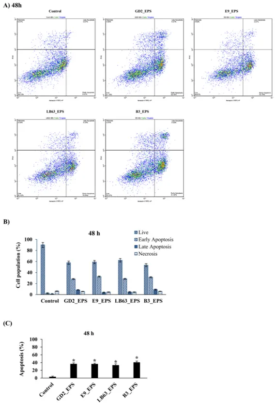
Impact of EPS produced from Lactobacillus spp. strains on apoptosis in HT-29 cells.
Apoptosis is a programmed cell death and is crucial in development and tissue homeostasis76. Induction of apoptosis couldbe used in control of proliferation of cancer cells. Most anti-cancer drugs in use affect via the induction of apopto-sis77,78. To further analyze the anti-proliferative effect by EPSs of Lactobacillus spp., we treated the HT-29 cells with
EPSs at two incubation time points, 24 h or 48 h, and performed a flow cytometric analysis. EPSs of Lactobacillus
Figure 1. Repeating unit structure of the exopolysaccharides produced by Lactobacillus spp. NMR
chemical shifts were determined by performing 1H-NMR, COSY and NOESY NMR analysis for the binding stereochemistry of monosaccharide units of EPSs of Lactobacillus spp. (L. plantarum GD2, L. rhamnosus E9, L. brevis LB63, and L. delbrueckii ssp. bulgaricus B3). All of them produced EPS with the same type of sugar linkage which are β-H1-H3 (β-D-Mannose-α-D-Mannose)/α- H1-H2 (α-D-Mannose-α-D-Glucose), designated as β-1,3 (β-D-Mannose-α-D-Mannose) and α-1,2 (α-D-Mannose-α-D-Glucose), respectively.
Figure 2. Anti-proliferative effect of the EPSs produced by Lactobacillus spp. against HT-29 colon cancer cells
at two time points. HT-29 cells seeded at a density of 1 × 10⁴ cells/well were treated with EPSs of Lactobacillus spp. (L. plantarum GD2, L. rhamnosus E9, L. brevis LB63, and L. delbrueckii ssp. bulgaricus B3) at a final concentration of 400 µg/ml for 24 h or 48 h and the anti-cytotoxicity effect of the EPSs was evaluated by WST-1 assay. *p < 0.05, significant difference from the control (n:3 for each bar).
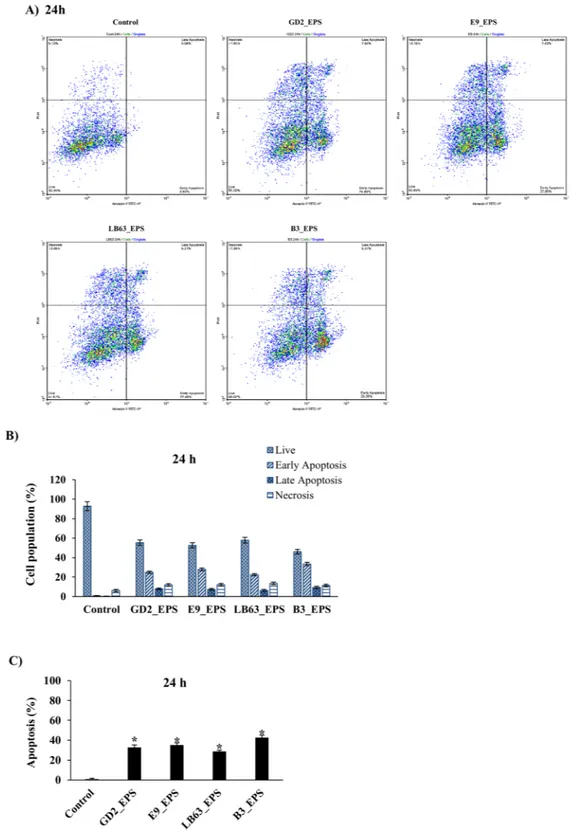
spp. induced apoptosis in HT-29 cells at both time points with an apoptosis percentage ranging from 28 to 43 at 24 h and 33 to 41 at 48 h. (Figs 3B, 4B). EPSs from L. delbrueckii ssp. bulgaricus B3 showed the highest apoptosis percentage (42.9 ± 2.4% and 40.6 ± 3.3, respectively) at both time points. Distribution of viable, early apoptotic, late apoptotic and necrotic cells showed that EPSs of Lactobacillus spp. induced both early and late apoptosis in HT-29 cells with the early apoptosis being higher (Figs 3C, 4C). Our results suggest that anti-proliferative impact of Lactobacillus studied here could be due to their capability to induce apoptosis (Figs 2–4). This is important in developing a new anti-cancer drug with good efficacy leading the cancer cells to apoptosis.
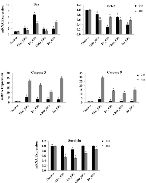
Genes and proteins involved in apoptotic pathway induced by epss of Lactobacillus spp.
To further understand the underlying mechanism of EPS-induced apoptosis, we next investigated changes in gene expression of apoptosis markers in HT-29 cells treated with EPSs of Lactobacillus spp. We targeted five genes associated with the apoptotic pathway, which are Bax, Bcl-2, Caspase 3, Caspase 9, and Survivin and measured both relative gene expression and protein expression.Bcl-2 is a large family of proteins that regulate cell death and cell survival. Bcl-2 family proteins take an impor-tant role in mitochondria-mediated apoptotic pathway and control the integrity of the mitochondrial outer membrane79. In this study, two of Bcl-2 family proteins were examined, Bcl-2 which is an anti-apoptotic protein
and Bax which is a pro-apoptotic protein. Effect of EPSs from Lactobacillus spp. on both anti-apoptotic and pro-apoptotic Bcl-2 family proteins were analyzed. The gene expression results showed that Lactobacillus spp. significantly increased the Bax gene expression at two time points, relative to the control, non-treated HT-29 cells (Fig. 5). However; a significant decrease in expression of Bcl-2 gene was observed in HT-29 cells treated with EPSs of Lactobacillus spp. for 24 h or 48 h, relative to the control (Fig. 5). The induction of Bax gene was higher at 24 h time point compared to 48 h time point. The highest increase of Bax gene expression at 24 h time point was observed in the HT-29 cells treated with EPS of L. rhamnosus E9, which was 6.79 ± 0.12 -fold change relative to the control (Fig. 5). The highest increase of Bax gene expression at 48 h time point was observed in the HT-29 cells treated with EPS of L. delbrueckii ssp. bulgaricus B3, which was 4.3 ± 0.01-fold change relative to the control (Fig. 5). Once apoptosis is induced by any agent, Bax proteins reach the mitochondrial outer membrane from cytoplasm. At the same time, anti-apoptotic Bcl-2 family proteins such as Bcl-2 binds Bax to prevent their transfer to the mitochondrial membrane. Therefore, in a cell underwent apoptosis, an increase in Bax gene and a decrease in Bcl-2 gene is expected. Bax/Bcl-2 ratio, hence, could be used to determine the fate of the cells in the apoptotic system80. Protein expression data confirmed these results (Figs 6 and S1).
Most of the molecules taking role in cell death are controlled by caspases. Apoptotic caspases can be grouped in two categories; caspases that initiate apoptosis are Caspase 2, 8, 9, and 10; and effector caspases with Caspase 3, 6, and 781. We examine the Caspase 3 and 9 gene expression in HT-29 cells to evaluate the impact of EPSs of
Lactobacillus spp. on expression of genes encoding caspases. Of the caspases examined, all of the Lactobacillus spp. EPSs resulted in a significant increase in gene expression at both 24 h (Fig. 5) and 48 h time point (Fig. 5). Levels of Caspase 3 and 9 gene expression at 24 h time point were the highest in the HT-29 cells treated with EPS of L. plantarum GD2, which were 5.68 ± 0.07 and 3.77 ± 0.09 -fold change relative to the control, respectively. At 48 h time point, both L. plantarum GD2 and L. delbrueckii ssp. bulgaricus B3 EPSs resulted in a high increase in Caspase 3 with 22 ± 0.26 and 24.5 ± 0.69 - fold change, respectively. The highest Caspase 9 gene expression was in HT-29 cells treated with EPS of L. plantarum GD2 with 29.5 ± 0.05 -fold change relative to the control (Fig. 5). Protein expression results also confirmed the gene expression data (Figs 6 and S1).
Studies have shown that polysaccharides induce apoptosis in cancer cells in a time dependent manner32,45. In
this study, administration of lactobacilli EPSs to HT-29 cells resulted in a time dependent induction in expression of caspases with a higher impact at 48 h compared to the impact at 24 h. The results indicate that EPSs induce apoptosis by caspase activation. Mitochondrial depolarization was dependent on caspase activation, suggesting a positive amplification loop for mitochondrial dysfunction. Loss of mitochondrial membrane potential would lead to release of cytochrome C and activation of Bax and Caspase 3/982.
Survivin is a member of apoptosis inhibitor family76 and inhibits Caspase 3 and 9. The expression of Survivin
gene was suppressed by the EPS of L. rhamnosus E9 (only 0.14 ± 0.01 fold) at 24 h (Fig. 5). However, all of the EPSs resulted in a significant suppression (p < 0.05) in the gene expression of Survivin at 48 h (Fig. 5). This sug-gest that suppression of Survivin just started at 24 h and reached a high suppression level at 48 h. However, the protein suppression of Survivin has started earlier than the suppression of the gene expression did (Figs 6 and S1). Similarly, Stolfi et al. showed that reduction of Survivin protein was seen as early as 8 h following the application of an apoptosis inducing agent whereas the inhibition of Survivin gene expression occurred at later time points (i.e., 32 hours)83 suggesting that a posttranscriptional control of Survivin could be involved in this process. EPS
from Aphanothece halaphytica has been shown to induce apoptosis in cancer cells by modulating p53-survivin pathway and target unfolded Protein Response Regulator Grp7884. The EPS of A. halaphytica induce the
expres-sion of CHOP and suppress the expresexpres-sion of Survivin, which leads p53-survivin pathway and cause apoptosis by activating Caspase 3.
The gene and protein expression results demonstrated that the ability to induce apoptosis by EPSs of Lactobacillus spp. associated with an upregulation of Bax, Caspase 3, and 9 and a downregulation of Bcl-2 and Survivin. In vitro studies have suggested that colon cancer could be inhibited by activation of Caspase 3 and 9 and inhibition of Bcl-285–87. In another study, EPS isolated from A. halophytica induce apoptosis similar to our study.
They reported that endoplasmic reticulum (ER) signaling pathway leads apoptosis. Based on their explanation for the molecular mechanism, ER targets Grp78 regulating cell respond UPR and suppresses Survivin and Bcl-2 expression while induces Caspase 3 expression, as a results, leads the cells to apoptosis84. Our results suggest that
EPSs released by Lactobacillus spp. studied here inhibit proliferation via apoptosis in HT-29 cells. It has been reported that biofunctional activities of polysaccharides including anti-tumor and apoptosis inducing activity are strongly associated with their molecular weight, monomer composition, structure, and linkage type67,72,73,88.
Figure 3. Flow cytometric analysis of the impact of Lactobacillus spp. EPSs on apoptosis in HT-29 cells at 24 h
time point. HT-29 cells seeded at a density of 1 × 10⁴ cells/well were treated with EPSs of Lactobacillus spp. (L. plantarum GD2, L. rhamnosus E9, L. brevis LB63, and L. delbrueckii ssp. bulgaricus B3) at a final concentration of 400 µg/ml for 24 h and then stained with Annexin V-FITC and PI. Fluorescence intensities were detected by flow cytometry to determine the effect of the EPSs on earlier apoptosis, late apoptosis and necrosis. (A) Distribution of viable, early apoptotic, late apoptotic and necrotic cells analyzed by flow cytometry. (B) Percentage of cells in viable, early apoptotic, late apoptotic and necrotic stages. (C) Percentage of apoptosis in HT-29 cells exposed to the lactobacilli EPSs for 24 h. *p < 0.05, significant difference from the control.
Figure 4. Flow cytometric analysis of the impact of Lactobacillus spp. EPSs on apoptosis in HT-29 cells at 48 h
time point. HT-29 cells seeded at a density of 1 × 10⁴ cells/well were treated with EPSs of Lactobacillus spp. (L. plantarum GD2, L. rhamnosus E9, L. brevis LB63, and L. delbrueckii ssp. bulgaricus B3) at a final concentration of 400 µg/ml for 48 h and then stained with Annexin V-FITC and PI. Fluorescence intensities were detected by flow cytometry to determine the effect of the EPSs on earlier apoptosis, late apoptosis and necrosis. (A) Distribution of viable, early apoptotic, late apoptotic and necrotic cells analyzed by flow cytometry. (B) Percentage of cells in viable, early apoptotic, late apoptotic and necrotic stages. (C) Percentage of apoptosis in HT-29 cells exposed to the lactobacilli EPSs for 48 h. *p < 0.05, significant difference from the control.
The capability of EPSs from Lactobacillus spp. to induce apoptosis at a high level, relative to the EPSs inducing apoptosis in the literature, might be associated with the main monomer in their structure, mannose, and their linkage type which are β-(1,3), α-(1,2)75,88–90. Glucose and mannose are known to have highly specific receptors
on macrophages, which is important in tumor immunology. The ability of all EPSs studied here to induce apop-tosis in colon cancer cells could be related to their sugar composition having mannose and glucose as major com-ponent. B3_EPS, which has the highest amount of mannose in sugar composition, showed the highest apoptosis induction on HT-29 cells. Suggesting that there might be a relationship exists between the ability of an EPS to induce apoptosis and its mannose composition. Additionally, B3_EPS has the lowest amount of glucose relative to the other strains. Having low amount of glucose with a high mannose content might have a role in the ability of B3_EPS to induce apoptosis strongly.
Figure 5. Fold change in mRNA expression of target genes of the HT-29 cells in the control group and the
group treated with EPSs from Lactobacillus spp. (L. plantarum GD2, L. rhamnosus E9, L. brevis LB63, and L. delbrueckii ssp. bulgaricus B3) at a final concentration of 400 µg/ml for 24 h (black bar) or 48 h (gray bar). *p < 0.05, significant difference from the control (n: 3).
Conclusion
EPSs produced by LAB are one of the important component that have a key role in probiotic activity including anti-tumor effect, immunomodulation, and adhesion13,17,50. In this study we evaluated EPSs of four previously
described Lactobacillus spp. isolated from healthy infant feces (GD2_EPS, E9_EPS, and LB63_EPS) and yogurt (B3_EPS) for their health effect on colon cancer cells (HT-29) and for their physicochemical properties34,35. We
demonstrated that a compositional and structural diversity exists within Lactobacillus spp., and this diversity
Figure 6. Protein expression of target genes of the HT-29 cells in the control group and the group treated
with EPSs from Lactobacillus spp. (L. plantarum GD2, L. rhamnosus E9, L. brevis LB63, and L. delbrueckii ssp. bulgaricus B3) at a final concentration of 400 µg/ml for 24 h. *p < 0.05, significant difference from the control (n:3).
likely contributes to variation in the ability to inhibit proliferation and induce apoptosis. Relative proportions of the individual sugars among Lactobacillus spp. are different and mannose of which portion size impact biological activities55, is the major sugar component in EPSs. All of the EPSs contain β-1,3-linkage which has been found in
anti-tumor polysaccharides71,72. The results showed that EPSs of Lactobacillus spp. inhibit proliferation in colon
cancer cells via apoptosis. The level of their capability to induce apoptosis was time dependent. While B3_EPS and GD2_EPS have better impact on inducing Caspases, E9_EPS and B3_EPS showed better impact on Bax and Survivin modulation. B3_EPS showed the highest apoptosis induction on HT-29 cells and has the highest amount of mannose in sugar composition with a quite low amount of glucose. There might be a relationship exists between the ability of an EPS to induce apoptosis and its high mannose and low glucose composition. Collectively these findings are important for further evaluating Lactobacillus spp. EPSs for cancer therapy depending on EPS structure and monomer composition. Particularly mannose ratio in EPS composition should be taken into con-sideration when designing anti-cancer agents. Further research is required to elucidate the mechanism of action of mannose and glucose on anti-cancer functionality.
Data Availability
All data generated or analysed during this study are included in this published article (and its Supplementary Information files).
References
1. The International Agency for Research on Cancer (IARC). Latest Global Cancer Data, 2018. World Health Organization (2018). 2. Bray, F. et al. Global Cancer Statistics 2018: GLOBOCAN Estimates of Incidence and Mortality Worldwide for 36 Cancers in 185
Countries. CA Cancer J. Clin 68, 394–424 (2018).
3. Hill, C. et al. The International Scientific Association for Probiotics and Prebiotics consensus statement on the scope and appropriate use of the term probiotic. Nat. Rev. Gastroenterol. Hepatol 11, 506–514 (2014).
4. Gueimonde, M., Ouwehand, A., Huhtinen, H., Salminen, E. & Salminen, S. Qualitative and quantitative analyses of the bifidobacterial microbiota in the colonic mucosa of patients with colorectal cancer, diverticulitis and inflammatory bowel disease. World J Gastroenterol 13, 3985–3989 (2007).
5. Kamada, N. & Núñez, G. Regulation of the immune system by the resident intestinal bacteria. Gastroenterology 146, 1477–88 (2014). 6. Ohno, H. Impact of commensal microbiota on the host pathophysiology: focusing on immunity and inflammation. Semin
Immunopathol 37, 1–3 (2015).
7. Wang, Q. et al. Alteration of gut microbiota in association with cholesterol gallstone formation in mice. BMC Gastroenterol. 17, 1–9 (2017).
8. Pedamallu, C. S. et al. Genomic analysis identifies association of Fusobacterium with colorectal carcinoma. Genome Res. 22, 292–298 (2012).
9. Zackular, J. P. et al. The Gut Microbiome Modulates Colon Tumorigenesis. Am Soc Microbiol 4, e00692–13 (2013).
10. Schrezenmeir, J. & de Vrese, M. Probiotics, prebiotics, and synbiotics-approaching a definition. Am J Clin Nutr 73, 361S–364S (2001).
11. Leyer, G. J., Li, S., Mubasher, M. E., Reifer, C. & Ouwehand, A. C. Probiotic effects on cold and influenza-like symptom incidence and duration in children. Pediatrics 124, e172–e179 (2009).
12. Hörmannsperger, G. et al. Post-translational inhibition of IP-10 secretion in IEC by probiotic bacteria: impact on chronic inflammation. PLoS One 4, e4365 (2009).
13. Lebeer, S., Claes, I. J. J., Verhoeven, T. L. A., Vanderleyden, J. & De Keersmaecker, S. C. J. Exopolysaccharides of Lactobacillus rhamnosus GG form a protective shield against innate immune factors in the intestine. Microb. Biotechnol. 4, 368–374 (2011). 14. Kearney, N. et al. Handbook of fermented functional foods. (CRC Press, 2008).
15. Sungur, T., Aslim, B., Karaaslan, C. & Aktas, B. Impact of Exopolysaccharides (EPSs) of Lactobacillus gasseri strains isolated from human vagina on cervical tumor cells (HeLa). Anaerobe 47, 137–144 (2017).
16. Zakostelska, Z. et al. Lysate of probiotic Lactobacillus casei DN-114 001 ameliorates colitis by strengthening the gut barrier function and changing the gut microenvironment. PLoS One 6, e27961 (2011).
17. Liu, C.-T., Chu, F.-J., Chou, C.-C. & Yu, R.-C. Antiproliferative and anticytotoxic effects of cell fractions and exopolysaccharides from Lactobacillus casei 01. Mutat. Res. 721, 157–62 (2011).
18. Caggianiello, G., Kleerebezem, M. & Spano, G. Exopolysaccharides produced by lactic acid bacteria: from health-promoting benefits to stress tolerance mechanisms. Appl. Microbiol. Biotechnol. 100, 3877–3886 (2016).
19. Hong, J.-H. & Hee, K. J. Antioxidant and Antitumor Activities of β-glucan- rich Exopolysaccharides with Different Molecular Weight from Paenibacillus polymyxa JB115. J Korean Soc Appl Biol Chem 57, 105–112 (2014).
20. Ciszek-Lenda, M. et al. Strain specific immunostimulatory potential of lactobacilli-derived exopolysaccharides. Centr Eur J Immunol
36, 121–129 (2011).
21. Ruas-Madiedo, P., Hugenholtz, J. & Zoon, P. An overview of the functionality of exopolysaccharides produced by lactic acid bacteria. Int. Dairy J. 12, 163–171 (2002).
22. Mende, S., Rohm, H. & Jaros, D. Influence of exopolysaccharides on the structure, texture, stability and sensory properties of yoghurt and related products. Int. Dairy J. 52, 57–71 (2016).
23. Gorret, N., Maubois, J. L., Engasser, J. M. & Ghoul, M. Study of the effects of temperature, pH and yeast extract on growth and exopolysaccharides production by Propionibacterium acidi-propionici on milk microfiltrate using a response surface methodology. J. Appl. Microbiol. 788–796 (2001).
24. Rinker, K. D. & Kelly, R. M. Effect of carbon and nitrogen sources on growth dynamics and exopolysaccharide production for the hyperthermophilic archaeon Thermococcus litoralis and bacterium Thermotoga maritima. Biotechnol. Bioeng. 69, 537–547 (2000). 25. Li, C. et al. Microbiological, physicochemical and rheological properties of fermented soymilk produced with exopolysaccharide
(EPS) producing lactic acid bacteria strains. LWT - Food Sci. Technol. 57, 477–485 (2014).
26. Zhang, L. et al. Antioxidant activity of an exopolysaccharide isolated from Lactobacillus plantarum C88. Int. J. Biol. Macromol. 54, 270–275 (2013).
27. Xu, R., Ye, H., Sun, Y., Tu, Y. & Zeng, X. Preparation, preliminary characterization, antioxidant, hepatoprotective and antitumor activities of polysaccharides from the flower of tea plant (Camellia sinensis). Food Chem. Toxicol. 50, 2473–2480 (2012).
28. Li, W. et al. Structural elucidation and antioxidant activities of exopolysaccharides from Lactobacillus helveticus MB2-1. Carbohydr. Polym. 102, 351–359 (2014).
29. Li, W. et al. Characterization of a novel polysaccharide with anti-colon cancer activity from Lactobacillus helveticus MB2-1. Carbohydr. Res. 411, 6–14 (2015).
30. Li, S. et al. Molecular Modification of Polysaccharides and Resulting Bioactivities. ComprehensiveReviewsinFoodScienceandFoodSafety
15, 237–250 (2016).
32. Ma, L. et al. Preparation, preliminary characterization and inhibitory effect on human colon cancer HT-29 cells of an acidic polysaccharide fraction from Stachys floridana Schuttl. ex Benth. Food Chem. Toxicol. 60, 269–276 (2013).
33. Wang, K. et al. Characterization of a novel exopolysaccharide with antitumor activity from Lactobacillus plantarum 70810. Int. J. Biol. Macromol. 63, 133–139 (2014).
34. Gürsoy, A., Durlu-Özkaya, F., Yıldız, F. & Aslım, B. Ekzopolisakkarit Üretimi Yüksek Yerli Streptococcus thermophilus (W22) ve Lactobacillus delbrueckii ssp. bulgaricus (B3). Kafkas Univ. Vet. Fak. Derg. 16, 81–86 (2009).
35. Yildiz, G. G., Öztürk, M. & Aslim, B. Identification of Lactobacillus strains from breast-fed infant and investigation of their cholesterol-reducing effects. World J. Microbiol. Biotechnol. 27, 2397–2406 (2011).
36. Frengova, G. I., Simova, E. D., Beshkova, D. M. & Simov, Z. I. Production and monomer composition of exopolysaccharides by yogurt starter cultures. Can. J. Microbiol. 46, 1123–1127 (2000).
37. Dubois, M., Gilles, K. A., Hamilton, J. K., Rebers, P. A. & Smith, F. Colorimetric Method for Determination of Sugars and Related Substances. Anal. Chem. 28, 350–356 (1956).
38. Tsuda, H., Hara, K. & Miyamoto, T. Binding of mutagens to exopolysaccharide produced by Lactobacillus plantarum mutant strain 301102S. J. Dairy Sci. 91, 2960–2966 (2008).
39. Ledezma, O. E. V. et al. Characterization of extracellular polymeric substances (EPS) produced by marine Micromonospora sp. J. Chem. Pharm. Res. 8, 442–451 (2016).
40. Boymirzaev, A. S., Shomurotov, S. & Turaev, A. S. Secondary Effects in Aqueous Size-Exclusion Chromatography of Polysaccharides. Chem. Plant Mater. 2, 51–55 (2013).
41. Ismail, B. & Madhavan Nampoothiri, K. Exposition of antitumour activity of a chemically characterized exopolysaccharide from a probiotic Lactobacillus plantarum MTCC 9510*. Biol. Sect. Cell. Mol. Biol. 68, 1041–1047 (2013).
42. Monsalve, D. M. et al. Human VRK2 modulates apoptosis by interaction with Bcl-xL and regulation of BAX gene expression. Cell Death Dis. 4, 1–10 (2013).
43. Moro, L. et al. Placental microparticles and MicroRNAs in pregnant women with Plasmodium falciparum or HIV infection. PLoS One 11, 1–17 (2016).
44. VanGuilder, H. D., Vrana, K. E. & Freeman, W. M. Twenty-five years of quantitative PCR for gene expression analysis. Biotechniques
44, 619–626 (2008).
45. Huang, F. et al. Sepia ink oligopeptide induces apoptosis in prostate cancer cell lines via caspase-3 activation and elevation of Bax/ Bcl-2 ratio. Mar. Drugs 10, 2153–2165 (2012).
46. Ramadoss, D. P. & Sivalingam, N. Vanillin Extracted From Proso Millet and Barnyard Millet Induce Apoptosis in Ht-29 and Mcf-7 Cell Line Through Mitochondria Mediated Pathway. Asian J. Pharm. Clin. Res. 10, 226 (2017).
47. Harlow, E. D., Lane, D. A laboratory manual. (Cold Spring Harbor Laboratory, 1988).
48. Baselga, J. & Arteaga, C. L. Critical update and emerging trends in epidermal growth factor receptor targeting in cancer. J. Clin. Oncol. 23, 2445–2459 (2005).
49. Lebeer, S., Vanderleyden, J. & De Keersmaecker, S. C. J. Host interactions of probiotic bacterial surface molecules: comparison with commensals and pathogens. Nat. Rev. Microbiol. 8, 171–84 (2010).
50. Burns, P. et al. Technological characterization and survival of the exopolysaccharide-producing strain Lactobacillus delbrueckii subsp. lactis 193 and its bile-resistant derivative 193 + in simulated gastric and intestinal juices. J. Dairy Res. 78, 357–364 (2011). 51. Mozzi, F. et al. Diversity of Heteropolysaccharide-Producing Lactic Acid Bacterium Strains and Their Biopolymers. Appl. Environ.
Microbiol. 72, 4431–4435 (2006).
52. Donnarumma, G. et al. Lactobacillus crispatus L1: high cell density cultivation and exopolysaccharide structure characterization to highlight potentially beneficial effects against vaginal pathogens. BMC Microbiol. 14, 137 (2014).
53. Van Geel-Schutten, G. H., Flesch, F., Ten Brink, B., Smith, M. R. & Dijkhuizen, L. Screening and characterization of Lactobacillus strains producing large amounts of exopolysaccharides. Appl. Microbiol. Biotechnol. 50, 697–703 (1998).
54. Broadbent, J. R., McMahon, D. J., Welker, D. L., Oberg, C. J. & Moineau, S. Biochemistry, genetics, and applications of exopolysaccharide production in Streptococcus thermophilus: a review. J. Dairy Sci. 86, 407–23 (2003).
55. Gientka, I., Błażejak, S., Stasiak-Różańska, L. & Chlebowska-Śmigiel, A. Exopolysaccharides from yeast: insight into optimal conditions for biosynthesis, chemical composition and functional properties – review. Acta Sci. Pol. Technol. Aliment 14, 283–292 (2015).
56. Shao, B. M., Dai, H., Xu, W., Lin, Z. B. & Gao, X. M. Immune receptors for polysaccharides from Ganoderma lucidum. Biochem. Biophys. Res. Commun. 323, 133–141 (2004).
57. Vidhyalakshmi, R. & Vallinachiyar, C. Apoptosis of Human Breast Cancer Cells (MCF-7) Induced by Polysacccharides Produced by Bacteria. J. Cancer Sci. Ther. 5, 031–034 (2013).
58. Ghada, S., Mahmoud, G., Asker, M. S. & Ghazy, A. Production and Biological Evaluation of Exopolysaccharide From Isolated Rhodotorula glutinins. Aust. J. Basic Appl. Sci. 6, 401–408 (2012).
59. Jin, Y. et al. Antitumor activities of heteropolysaccharides of Poria cocos mycelia from different strains and culture media. Carbohydr. Res. 338, 1517–1521 (2003).
60. Cerning, J. Production of exopolysaccharides by lactic acid bacteria and dairy propionibacteria. Lait 75, 463–472 (1995). 61. Tallon, R., Bressollier, P. & Urdaci, M. C. Isolation and characterization of two exopolysaccharides produced by Lactobacillus
plantarum EP56. Res. Microbiol. 154, 705–712 (2003).
62. Wang, J., Zhao, X., Yang, Y., Zhao, A. & Yang, Z. Characterization and bioactivities of an exopolysaccharide produced by Lactobacillus plantarum YW32. Int. J. Biol. Macromol. 74, 119–126 (2015).
63. Hamet, M. F., Piermaria, J. A. & Abraham, A. G. Selection of EPS-producing Lactobacillus strains isolated from kefir grains and rheological characterization of the fermented milks. LWT - Food Sci. Technol. 63, 129–135 (2015).
64. Xu, R., Qian, S., Ding, X., Wengeng, G. & Li, P. Chemical characterization and antioxidant activity of an exopolysaccharide fraction isolated from Bifidobacterium animalis RH. Eur Food Res Technol 232, 231–240 (2011).
65. Zarour, K. et al. Rheology and bioactivity of high molecular weight dextrans synthesised by lactic acid bacteria. Carbohydr. Polym.
174, 646–657 (2017).
66. Nwodo, U. U., Green, E. & Okoh, A. I. Bacterial exopolysaccharides: Functionality and prospects. Int. J. Mol. Sci. 13, 14002–14015 (2012).
67. Wasser, S. Medicinal mushrooms as a source of antitumor and immunomodulating polysaccharides. Appl. Microbiol. Biotechnol. 60, 258–274 (2002).
68. Ooi, V. E. C. & Liu, F. Immunomodulation and Anti-Cancer Activity of Polysaccharide- Protein Complexes. Curr. Med. Chem. 7, 715–729 (2000).
69. Ferreira, J. A. et al. Identification of cell-surface mannans in a virulent Helicobacter pylori strain. Carbohydr. Res. 345, 830–838 (2010).
70. Nordmark, E.-L. Structural and Interaction Studies of Bacterial Polysaccharides by NMR Spectroscopy. (Stockholm University, 2004). 71. Diemer, S. K. et al. Binding Interactions Between α-glucans from Lactobacillus reuteri and Milk Proteins Characterised by Surface
Plasmon Resonance. Food Biophys. 7, 220–226 (2012).
72. Wang, Y.-Y. et al. Studies on the Immuno-Modulating and Antitumor Activities of Ganoderma lucidum (Reishi) Polysaccharides: Functional and Proteomic Analyses of a Fucose-Containing Glycoprotein Fraction Responsible for the Activities. Bioorg. Med. Chem. 10, 1057–1062 (2002).
73. Haroun, B. M., Refaat, B. M., Menoufy, H. A. E.-, Amin, H. A. & Amr, A. Structure Analysis and Antitumor Activity of the Exopolysaccharide from Probiotic. J. Appl. Sci. Res. 9, 425–434 (2013).
74. Oleksy, M., Klewicka, E. & Zbieta Klewicka, E. Critical Reviews in Food Science and Nutrition Exopolysaccharides produced by Lactobacillus sp.: Biosynthesis and applications. Crit. Rev. Food Sci. Nutr. 58, 450–462 (2018).
75. Cao, W. et al. A novel polysaccharide, isolated from Angelica sinensis (Oliv.) Diels induces the apoptosis of cervical cancer HeLa cells through an intrinsic apoptotic pathway. Phytomedicine 17, 598–605 (2010).
76. Riedl, S. J. & Shi, Y. Molecular mechanisms of caspase regulation during apoptosis. Nat. Rev. Mol. Cell Biol. 5, 897–907 (2004). 77. Bergamo, A., Dyson, P. J. & Sava, G. The mechanism of tumour cell death by metal-based anticancer drugs is not only a matter of
DNA interactions. Coord. Chem. Rev. J. 360, 17–33 (2018).
78. Reed, J. C. Bcl-2 on the brink of breakthroughs in cancer treatment. Nat. Publ. Gr. 25, 3–6 (2018).
79. Adams, J. M. & Cory, S. The BCL-2 arbiters of apoptosis and their growing role as cancer targets. Nat. Publ. Gr. 25, 27–36 (2018). 80. Fan, T. J., Han, L. H., Cong, R. S. & Liang, J. Caspase family proteases and apoptosis. Acta Biochim. Biophys. Sin. (Shanghai). 37,
719–727 (2005).
81. Wu, H. et al. Caspases: A molecular switch node in the crosstalk between autophagy and apoptosis. Int. J. Biol. Sci. 10, 1072–1083 (2014).
82. Wu, Y. J., Wong, B. S., Yea, S. H., Lu, C. I. & Weng, S. H. Sinularin induces apoptosis through mitochondria dysfunction and inactivation of the pI3K/Akt/mTOR pathway in gastric carcinoma cells. Mar. Drugs 14 (2016).
83. Stolfi, C. et al. 2-Methoxy-5-Amino-N-Hydroxybenzamide Sensitizes Colon Cancer Cells to TRAIL-Induced Apoptosis by Regulating Death Receptor 5 and Survivin Expression. Mol Cancer Ther 10, 1969–81 (2011).
84. Ou, Y., Xu, S., Zhu, D. & Yang, X. Molecular mechanisms of exopolysaccharide from Aphanothece halaphytica (EPSAH) induced apoptosis in HeLa cells. PLoS One 9, e87223 (2014).
85. Altonsy, M. O., Andrews, S. C. & Tuohy, K. M. Differential induction of apoptosis in human colonic carcinoma cells (Caco-2) by Atopobium, and commensal, probiotic and enteropathogenic bacteria: Mediation by the mitochondrial pathway. Int. J. Food Microbiol. 137, 190–203 (2010).
86. Wan, Y. et al. Fermentation supernatants of lactobacillus delbrueckii inhibit growth of human colon cancer cells and induce apoptosis through a caspase 3-dependent pathway. Oncol. Lett. 7, 1738–1742 (2014).
87. Chen, Z.-Y., Hsieh, Y.-M., Huang, C.-C. & Tsai, C.-C. Inhibitory Effects of Probiotic Lactobacillus on the Growth of Human Colonic Carcinoma Cell Line HT-29. Molecules 22, 107 (2017).
88. Zhao, K., Jin, M., Chen, Q. & Zheng, P. S. Polysaccharides produced by enterobacter cloacae induce apoptosis in cervical cancer cells. Int. J. Biol. Macromol. 72, 960–964 (2015).
89. Liu, G., Kuang, S., Wu, S., Jin, W. & Sun, C. A novel polysaccharide from Sargassum integerrimum induces apoptosis in A549 cells and prevents angiogensis in vitro and in vivo. Sci. Rep. 6, 1–12 (2016).
90. Di, W. et al. Exopolysaccharides produced by Lactobacillus strains suppress HT-29 cell growth via induction of G0/G1 cell cycle arrest and apoptosis. Oncol. Lett. 16, 3577–3586 (2018).
Acknowledgements
This work supported by the TUBITAK [grant number 115R282].
Author Contributions
Ideas, designing the study: Belma Aslim. Performed the research: Ummugulsum Tukenmez, Serkan Yavuz, Belma Aslim. Analyzed data: Ummugulsum Tukenmez, Belma Aslim, Serkan Yavuz, Busra Aktas. Wrote the paper: Busra Aktas, Belma Aslim, Ummugulsum Tukenmez, Serkan Yavuz.
Additional Information
Supplementary information accompanies this paper at https://doi.org/10.1038/s41598-019-44753-8.
Competing Interests: The authors declare no competing interests.
Publisher’s note: Springer Nature remains neutral with regard to jurisdictional claims in published maps and
institutional affiliations.
Open Access This article is licensed under a Creative Commons Attribution 4.0 International
License, which permits use, sharing, adaptation, distribution and reproduction in any medium or format, as long as you give appropriate credit to the original author(s) and the source, provide a link to the Cre-ative Commons license, and indicate if changes were made. The images or other third party material in this article are included in the article’s Creative Commons license, unless indicated otherwise in a credit line to the material. If material is not included in the article’s Creative Commons license and your intended use is not per-mitted by statutory regulation or exceeds the perper-mitted use, you will need to obtain permission directly from the copyright holder. To view a copy of this license, visit http://creativecommons.org/licenses/by/4.0/.