Summary
The ratio of gray and white matter is an important clinical parameter in the diagnosis of diffuse and compressive diseases of the spinal cord. Although histological methods are used to determine this parameter, there are some difficulties encountered in histological studies related to tissue size. The aim of this study was to evaluate possible modifications to overcome these difficulties. In the study, nine tissue samples taken from the C6 segment of a female Shetland pony and selected by systematic random sampling were used. The dehydration process of the spinal cord of the horse was supported by applying a vacuum. Paraffin blocks were prepared and cut into 10 µm sections to be stained separately with the different staining methods. Six different staining methods, including Modified May - Grunwald - Giemsa (MMGG), were compared and used to image entire slides. The stains, Hematoxylin & eosin (H&E), May-Grunwald-Giemsa (MGG), Masson’s trichrome (MT), AgNORs, Kluver Barrera (KB) and MMGG, were evaluated macroscopically and microscopically by participants who were unaware of which staining methods had been used. The staining methods were scored from worst (1) to best (5) using a Likert scale. Vacuum application was found to reduce the difficulties related to inadequate tissue dehydration. MMGG was selected as the best staining method in differentiating gray and white matter in the spinal cord of the horse.
Keywords: Gray matter ratio, Horse, Imaging, Spinal cord, Stain comparison
At Omuriliğinin Gri ve Ak Maddesinin Seçici Boyanması
Özet
Medulla spinalis’te yer alan gri ve ak madde oranları diffuz ve kompresif omurilik hastalıklarının teşhisinde önemli klinik para-metrelerdendir. Bu parametrelerin tespitinde histolojik metotlar kullanılmasına rağmen, histolojik çalışmalarda doku büyüklüğüne bağlı bazı güçlüklüklerle karşılaşılmaktadır. Bu çalışmanın amacı, bahsedilen zorlukları aşmaya yönelik olası modifikasyonları değerlendirmektir. Çalışmada, Shetland pony ırkına ait C6 segmentinden sistematik rastgele örnekleme kuralına sadık kalınarak elde edilen 9 doku kesiti kullanıldı. Medulla spinalis’in dehidrasyon işlemi sırasında vakum uygulaması yapıldı. Dokuların parafin blokları hazırlandı, dokular 10 µm kalınlığında kesilerek farklı histolojik boyalar ile boyandı. Modifiye May - Grunwald - Giemsa (MMGG)’nın yer aldığı 6 farklı boyama metodunun boyama performansları karşılaştırıldı. Hematoxylin & eosin (H&E), May-Grunwald-Giemsa (MGG), Masson’s trichrome (MT), AgNORs, Kluver Barrera (KB) ve MMGG ile boyanan kesitler tek kör grup tarafından makroskopik ve mikroskopik olarak değerlendirildi. Boyaların performansları Likert skalası kullanılarak en kötü (1) en iyi (5) olmak üzere değerlendirildi. Vakum uygulamasının yetersiz doku dehidrasyonundan kaynaklanan problemleri ortadan kaldırdığı görüldü. MMGG, at medulla spinalis’inde gri ve ak madde ayrımında en başarılı boya olarak tespit edildi.
Anahtar sözcükler: Gri madde oranı, At, Görüntüleme, Medulla spinalis, Boya karşılaştırması
Selective Gray and White Matter Staining of the Horse Spinal Cord
Durmus BOLAT *
Sadullah BAHAR **
Emrah SUR ***
Muhammet L SELCUK **
Sadettin TIPIRDAMAZ **
* ** ***
Kirikkale University, Faculty of Veterinary Medicine, Department of Anatomy, Campus, TR-71451Yahsihan, Kirikkale - TURKEY Selcuk University, Faculty of Veterinary Medicine, Department of Anatomy, Campus, TR-42000 Selcuklu, Konya - TURKEY Selcuk University, Faculty of Veterinary Medicine, Department of Histology and Embryology, Campus, TR-42000 Selcuklu, Konya - TURKEY
Makale Kodu (Article Code): KVFD-2011-5371
The part of the central nervous system situated within the vertebral canal, namely, the spinal cord, is composed of 42-43 segments in horses, 8 of which are cervical, 18 thoracic, 6 lumbar, 5 sacral and 5-6 caudal. The spinal cord, enveloped by the meninges, extends from the foramen
magnum to the level in-between the first and second sacral vertebrae. In general, the spinal cord is dorso-ventrally compressed and elliptic with two enlargements at the cervical intumescence and the lumbar intumescence, and terminates forming the medullary cone. In histological
INTRODUCTION
İletişim (Correspondence)
+90 318 3574242sections, the gray matter, resembling the letter H in shape and composed of nerve and glia cells and blood vessels, is observed to be situated centripetally, whilst the white matter, composed mostly of myelinated axons and blood vessels, is located peripherally. The central canal is located in the center of histological sections. Of all domestic animals, horses have the largest spinal cord, which measures 180-200 cm in length and 250-300 g in weight 1.
The segmental anatomical features of the spinal cord have been demonstrated in the cat 2, monkey 3, dog 4, sheep 5, goat 6, impala 7, horse and cattle 8. Furthermore, histological and some histomorphometric features of the spinal cord have been ascertained in research conducted in humans 9-11, rats 12, sheep 13, donkeys 14 and horses 15. However, during the conduct of research on sections pertaining to spinal cord segments of humans, goats, sheep, donkeys and horses, the necessity for full images arose in order to obtain histomorphometric data. For this purpose, several methods were applied, including direct drawings using millimetric paper and a calliper 6,13,14, indirect drawings 15, photograph combining 9, projection 10 and digital tablets 11. Literature review revealed that different fixation, processing and staining methods were applied for histological tissue processing in the researches referred to above, and yet limited information was provided by the researchers on the methods applied.
The gray and white matter in the central nervous system have different anatomical and cellular properties. Investigation of chronic diseases that affect the central nervous system, such as multiple sclerosis and schizophrenia, using the ratio of gray and white matter is of great importance 16-18. Imaging techniques, for instance Magnetic Resonance Imaging (MRI) and Diffusion Tensor Imaging (DTI) 19-21, and histological methods are used routinely to determine this parameter 12,22. Although obtaining data using imaging methods is very rapid and practical, some difficulties (in tissue processing, staining and imaging) are encountered during application of the histological process, owing to the size of tissue samples.
A search of the literature found that a limited number of morphometric and histological studies have been conducted on the spinal cord of the horse. The aim of this study was to overcome difficulties related to tissue processing, to decide which stain is best for the clear differentiation of gray and white matter of the spinal cord of horse, to acquire tissue images as a whole, and to compare the staining performance of various stains macroscopically and microscopically.
MATERIAL and METHODS
Material
A 15-year-old female Shetland pony (230 kg) that was
suffering from various orthopedic disorders was sent to the Department of Anatomy from the Equestrian Facility of the Faculty of Veterinary Medicine, Selcuk University for use in this study. The research was approved by the ethical committee of Faculty of Veterinary Medicine, Selcuk University (2011/16). The animal was anesthetized by administration of 10% chloral hydrate (80 mg/kg, IV) 23 and killed under general anesthesia by draining blood from the common carotid artery. Ten percent neutral formaldehyde was administered to the animal through a cannula in the common carotid artery after death. The spinal cord was dissected out from the vertebral column by laminectomy after the cadaver had been kept in a container of 10% neutral formaldehyde for 10 days. Segmentation was performed over the spinal cord following removal of the dura mater and pia mater. The C6 segment was used in this study; it was 68.8 mm in length and weighed 12.8 g.
Methods
Sampling and Tissue Processing
The C6 segment was divided into 18 pieces using a tissue slicer prior to histological processing. Each tissue sample was 3.8 mm in length. Systematic random sampling was performed on the separated pieces 24. A sampling ratio of 1/2 was used, and nine tissue samples were obtained for routine histological processing. The tissue samples were placed on trays, taking into account their cranial-caudal orientation, and dehydrated in an ethanol series. A vacuum was applied during the second application of 96% ethanol (200 mmHg, 30 min), the third application of 100% ethanol (200 mmHg, 1 h), the third application of xylene (200 mmHg, 30 min), and in paraffin (56-58ºC, 300 mmHg, the last 2 h), and the samples were blocked in paraffin wax. Samples in paraffin blocks were sectioned consecutively on a rotary microtome at a thickness of 10 μm, and six sections were obtained from each block. The sections were kept in an incubator (37ºC) for 24 h, stained according to the order given in Table 1, and mounted with Entellan under a glass coverslip.
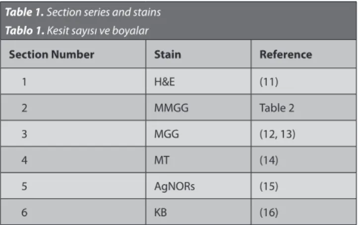
Table 1. Section series and stains Tablo 1. Kesit sayısı ve boyalar
Section Number Stain Reference
1 H&E (11) 2 MMGG Table 2 3 MGG (12, 13) 4 MT (14) 5 AgNORs (15) 6 KB (16)
H&E (Hematoxylin & eosin), MGG (May-Grunwald-Giemsa), MT (Masson’s trichrome), KB (Kluver Barrera), MMGG (modified May-Grunwald-Giemsa)
Preparation of Giemsa Solution
Giemsa stock solution (Merck KGaA, Darmstadt, Germany) was added at 1 drop per ml to 250 mL distilled water using a Pasteur pipette (The solution prepared in this way is not recommended to be used more than two times).
Acquiring Images from Slides Using an Office Scanner Images of gross biological structures (brain or cerebellum) that are difficult to view under the
micro-scope can be obtained with the help of a standard office scanner 31. In this study, all slides were scanned as positive image using a standard office flatbed scanner (Hp Scanjet G4010) at 300 dpi for macroscopic evaluation.
Area Calculation and Evaluation of Variations among Stains with Point Counting Frame
The point counting method has been used frequently to assess morphological parameters such as the area, volume, and area or volume ratio 32-34. This method was used to predict possible variations among stains that could affect measurement of the areas of gray matter and gross section. For this purpose, the grid function of the image analysis software ImageJ was applied to the scanned images. The area per point value was set at 0.4 mm2 (0.2 × 0.2 mm) and 4 mm2 (2 × 2 mm) to acquire an optimum CE value 24 for gray matter and the gross section respectively (Fig. 1). The areas of gray and gross section
on the scanned images were calculated three times by each of six different operators using the random function of the software. The results were analyzed using ANOVA (Table 3).
Evaluation of the Ability of White and Gray Matter Staining with A Survey
This part of the study was designed as a single blinded experiment. The survey group was composed of 20 healthy and non-colorblind students who were selected randomly
Table 3. The mean cross-sectional areas of gray matter and gross section and responses of survey participants (median) Tablo 3. Transversal kesitlerde ortalama gri ve ak madde oranları (mean±SE) ve anket sonuçları (median)
Stain Gray Matter (mm2) Spinal Cord (mm2) Median Score
H&E 11.97±0.641 138.20±0.577 1d MMGG 11.91±0.255 139.01±0.376 5a MGG 11.72±0.513 138.52±0.565 3.5b MT 12.00±0.391 138.05±0.425 2c AgNORs 11.64±0.444 138.15±0.465 2cd KB 11.99±0.320 138.52±0.565 4a
There was no statistical difference among the areas of the structures with different staining methods (P>0.05, ANOVA)
a,b,c,d Different letters in the same column are significantly different (P<0.05, Mann-Whitney U test)
Table 2. Modified May-Grunwald-Giemsa staining procedure Tablo 2. Modifiye edilmiş May-Grunwald-Giemsa boya prosedürü
Staining Stages
1 Xylene, 5 min 8 Rinse in distilled water 15 96% alcohol dip once 2 Xylene, 5 min 9 Tap water for 5 min. 16 96% alcohol dip once 3 100% alcohol, 3 min 10 Rinse in distilled water 17 100% alcohol dip once 4 100% alcohol, 3 min 11 Place in 250 ml May-Grunwald stock solution at room temperature for 10 min 18 100% alcohol dip once 5 96% alcohol, 3 min 12 Rinse in distilled water 19 Xylene 2 min 6 80% alcohol, 3 min 13 Place in Giemsa solution (250 ml) in 56°C incubator for 45 min 20 Xylene 2 min 7 70% alcohol, 3 min 14 Cool at room temperature 21 Xylene 2 min
Fig 1. Superimposed point counting frame on cross-section of spinal cord using ImageJ (area per point = 4 mm2,bar=5 mm)
Şekil 1. Medulla spinalis kesitleri üzerine ImageJ kullanılarak uygulanan noktalı alan ölçüm cetveli (bir noktanın alanı = 4 mm2, bar=5 mm)
from the senior students of the Faculty of Veterinary Medicine. The students were given a figure (Fig. 2) that showed six different stains and were asked to evaluate these stains in terms of the differentiation of gray and white matter according to a Likert scale (1: worst, 5: best). The results were analyzed using the Mann-Whitney U test (Table 3).
Light Microscopic Evaluation of Sections
The ability of the six dyes to stain neurons, glia, ependymal cells, endothelium, axon and dendrites were investigated using a Likert scale and the results are given in Table 4.
RESULTS
Collapsed areas were detected particularly in the gray matter region in paraffin blocks of tissues to which vacuum had not been applied during dehydration. Ruptures occurred during the cutting of these blocks with the rotary microtome, and low quality staining was observed especially in that area. This problem was solved by the application of vacuum. As a result of calculations made using the point counting frame superimposed on each slide stained with the different staining methods, the mean cross-sectional surface area of C6 was found to be 138±0.197 mm2 and the mean area of gray matter
Table 4. Microscopic assessment using Likert scale
Tablo 4. Likert skalası kullanılarak mikroskopik değerlendirme
Stains
White Matter Gray Matter
Ep endymal c ells D iff er an tia tıon of gr ay and whit e ma tt er D iff er an tia tıon of ax on and dendrit e A pp ear anc e of N issl b odies A xo n G
lia Endothel Neur
on G lia Endothel N C N C N C N C N C N C H&E 3 4 - 4 - 4 4 4 - 4 - 4 4 1 3 4 MMGG 4 3 - 2 - 2 2 3 - 2 - 1 1 5 2 2 MGG 2 3 - 3 - 3 3 3 - 3 - 2 2 2 3 3 MT 3 4 - 5 - 4 4 4 - 5 - 5 5 2 4 4 AgNORs 3 4 - 5 - 3 2 4 - 5 - 5 3 3 1 1 KB 4 4 - 3 - 5 5 4 - 3 - 4 4 5 5 5 N: Nucleus, C: Cytoplasm
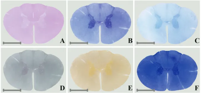
Fig 2. The image used by the survey group to evaluate the performance of stains using a Likert scale (bar= 5mm) (A: H&E, B: MMGG, C: MGG, D: MT, E: AgNORs, F: KB)
Şekil 2. Anket grubu tarafından Likert skalası kullanarak boya performanslarının değerlendirilmesinde kullanılan resim dosyası (bar= 5mm) (A: H&E, B: MMGG, C: MGG, D: MT, E: AgNORs, F: KB)
was estimated to be 11.87±0.170 mm2. The ratio of gray matter to gross section was determined to be 8.58%. A statistical difference was not detected (P>0.05, Table 3) among the areas of gray and white matter calculated from sections made using the six different stains.
The calculated coefficient of error value (CE) for the gray matter was 0.053 and for the spinal cord was 0.048. Statistical analysis of the responses given by the survey group showed that MMGG and KB were thought to be the best staining methods for differentiation of gray and white matter (Table 3, Fig. 2).
DISCUSSION
The application of a vacuum during the dehydration process is not recommended because of the fragile character of central nervous system tissue 25. In the present study, collapse caused by inadequate dehydration of the surface of paraffin blocks were seen, especially in the gray matter region, as a result of the routinely applied dehydration process without vacuum application. These problems were solved with vacuum application, as described in the section describing sampling and tissue processing. Different staining techniques have been utilized to differentiate gray and white matter macro-scopically 35,36 and microscopically 9,37 because of the poor contrast between these structures. Although KB is the staining method used most commonly for the central nervous system, MMGG (Table 2) is a preferable staining method to differentiate gray and white matter in the spinal cord of the horse because of its selective quality for this area and rapid reliable results (Fig. 3, Table 4).
The ratio of gray and white matter is used as an important parameter in the diagnosis of several diseases (schizophrenia, multiple sclerosis) in the central nervous system. Although imaging methods such as MRI are used most commonly to obtain this information 16,17,21, its use is limited in large domestic animals. Histological methods are frequently preferred when investigating the spinal cord of these animals 13,15. However, the entire structure must be displayed to estimate the ratio from histological sections. For this reason, a photographic camera 9,10, Edingerschen Apparatus 15, slide scanner 38 and office scanner 31 have been used to view the entire sections taken from large biological structures. In the current study, stained slides were scanned as JPEG files using a standard flatbed office scanner with a positive image scan option at 300 dpi. Measurement of the scanned images with a point counting frame estimated the ratio of gray matter to be 8.58% in the C6 segment (Table 3). The ratios of gray matter in C6 segments were reported as 11.86% of donkey 14, 18.3% of human 10, and 35-40% of rat 12 respectively. Braun 15 reported that the ratio of gray matter was 12.7% in the C6 segment of horse. It is thought that the dissimilarity between the two results could have resulted from differences in methodology. However, use of an office scanner to obtain images of large structures such as the brain and cerebellum was found to be a rapid method to view the spinal cord of the horse. Under microscopic examination, H&E, AgNORs, MT and KB for glia, MT and AgNORs for endothelial cells, KB for differentiation of axons and dendrites and also Nissl bodies, MT and AgNORs, especially for ependymal cells, were found to be good staining options (Fig. 3, Table 4).
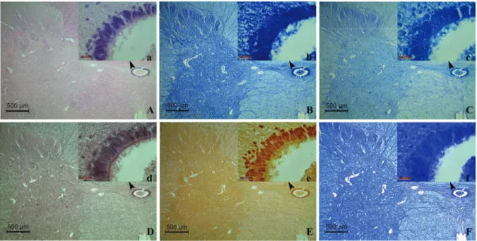
Fig. 3. Cross-sectional images obtained by 4× and 100× objectives under a light microscope (A: H&E, B: MMGG, C: MGG, D: MT, E: AgNORs, F: KB; Upper-case letter: 4×, Lower-case letter: 100×)
Şekil 3. Işık mikroskopu ile 4× ve 100× objektif kullanılarak elde edilen kesit görüntüleri (A: H&E, B: MMGG, C: MGG, D: MT, E: AgNORs, F: KB; Büyük harf: 4×, Küçük harf: 100×)
The current study showed that a vacuum can be applied during dehydration of large structures such as the spinal cord of the horse in order to acquire acceptable results. MMGG is a useful staining method for the differentiation of gray and white matter, and it is advised to be used when the spinal cord is examined both macroscopically and microscopically. Use of an office scanner is a cheap and practical method to view and scan large biological tissues. These methods are suggested as tools for use in morphometric studies related to the central nervous system.
A
cknowledgementsThe Authors are grateful to senior students of Faculty of Veterinary Medicine, Selcuk University for their kindly assistance.
REFERENCES
1. Nickel R, Seiferle E. Eingeweide: Lehrbuch der Anatomie der Haustiere. Band 4 Nervensystem, Sinnesorgane, Endokrine Drüsen. 3th ed., pp. 34-39, Stuttgart, Parey, 2004.
2. Thomas CE, Combs CM: Spinal cord segments. A. Gross structure in the adult cat. Am J Anat, 110 (1): 37-47, 1962.
3. Thomas CE, Combs CM. Spinal cord segments. B. Gross structure in the adult monkey. Am J Anat, 116 (1): 205-216, 1965.
4. Fletcher T, Kitchell R: Anatomical studies on the spinal cord segments of the dog. Am J Vet Res, 27 (121): 1759-1767, 1966.
5. Rao GS: Anatomical studies on the ovine spinal cord. Ann Anat, 171, 261-264, 1990.
6. Kahvecioğlu K, Özcan S, Çakır M: Anatomic studies on the medulla spinalis of the Angora goat (excluding the coccygeal segmentis). YYÜ Vet
Fak Derg, 6 (1-2): 76-80, 1995.
7. Rao G, Kalt D, Koch M, Majok A: Anatomical studies on the spinal cord segments of the impala. Anat Histol Embryol, 22 (3): 273-278, 1993. 8. Habel R.The topography of the equine and bovine spinal cord. J Am
Vet Med Assoc, 118, 379-382, 1951.
9. Makino M, Mimatsu K, Saito H, Konishi N, Hashizume Y: Morphometric study of myelinated fibers in human cervical spinal cord white matter. Spine, 21, 1010-1016, 1996.
10. Kameyama T, Hashizume Y, Sobue G: Morphologic features of the normal human cadaveric spinal cord. Spine, 21, 1285-1290, 1996. 11. Ko H, Park JH, Shin YB, Beak SY: Gross quantitative measurements of spinal cord segments in human. Spinal Cord, 42, 35-40, 2004.
12. Portiansky EL, Barbeito CG, Goya RG, Gimeno EJ, Zuccolilli GO: Morphometry of cervical segments grey matter in the male rat spinal cord. J Neurosci Meth, 139, 217-229, 2004.
13. Done J, Woolley J, Barnard V, Upcott D, Hebert C, Terlecki S: Border disease of sheep: Spinal cord morphometry. J Comp Pathol, 95, 325-333, 1985.
14. Ocal M, Hazıroglu RM: Comparative morphological studies on the spinal cord of the donkey. I. The cross-sectional areas of the spinal cord segments. Ankara Üniv Vet Fak Derg, 35, 55-68, 1988.
15. Braun A: Der segmentale feinbau des rückenmarks des pferdes. Cells
Tissues Organs, 10, 5-75, 1950.
16. Sastre-Garriga J, Ingle G, Chard D, Cercignani M, Ramio-Torrenta L, Miller D, Thompson A: Grey and white matter volume changes in early primary progressive multiple sclerosis: A longitudinal study. Brain, 128, 1454-1460, 2005.
17. Fornito A, Yücel M, Patti J, Wood S, Pantelis C: Mapping grey matter reductions in schizophrenia: an anatomical likelihood estimation analysis
of voxel-based morphometry studies. Schizophr Res, 108, 104-113, 2009. 18. Gilmore C, Geurts J, Evangelou N, Bot J, Van Schijndel R, Pouwels P, Barkhof F, Bö L: Spinal cord grey matter lesions in multiple sclerosis detected by post-mortem high field MR imaging. Mult Scler, 15, 180-188, 2009.
19. Tench CR, Morgan PS, Jaspan T, Auer DP, Constantinescu CS: Spinal cord imaging in multiple sclerosis. J Neuroimaging, 15, 94S-102S, 2005. 20. Ellingson BM, Ulmer JL, Schmit BD: Morphology and morphometry of human chronic spinal cord injury using diffusion tensor imaging and fuzzy logic. Ann Biomed Eng, 36, 224-236, 2008.
21. Hassen WB, Bégou M, Traore A, Moussa AB, Boehm N, Ghandour MS, Renou JP, Boespflug-Tanguy O, Bonny JM: Characterisation of spinal cord in a mouse model of spastic paraplegia related to abnormal axono-myelin interactions by in vivo quantitative MRI. Neuroimage, 46, 1-9, 2009.
22. Bjugn R, Gundersen HJG: Estimate of the total number of neurons and glial and endothelial cells in the rat spinal cord by means of the optical disector. J Comp Neurol, 328, 406-414, 1993.
23. Taylor P: Effects of surgery on endocrine and metabolic responses to anaesthesia in horses and ponies. Res Vet Sci, 64, 133-140, 1998.
24. Gundersen HJ, Jensen EB, Kieu K, Nielsen J: The efficiency of systematic sampling in stereology--reconsidered. J Microsc, 193, 199-211, 1999.
25. Culling CFA, Allison R, Barr W: Cellular pathology technique: Butterworth-Heinemann, 1985.
26. Serviere J, Dubayle D, Menetrey D: Increase of rat medial habenular mast cell numbers by systemic administration of cyclophosphamide.
Toxicol Lett, 145, 143-152, 2003.
27. Beghdadi W, Porcherie A, Schneider BS, Dubayle D, Peronet R, Huerre M, Watanabe T, Ohtsu H, Louis J, Mécheri S: Inhibition of histamine-mediated signaling confers significant protection against severe malaria in mouse models of disease. JEM, 205, 395-408, 2008. 28. Clark G: Staining procedures: 3rd. ed., Baltimore, Williams & Wilkins Co, 1973.
29. Plate K, Ruschoff J, Mennel H: Cell proliferation in intracranial tumours: selective silver staining of nucleolar organizer regions (AgNORs). Application to surgical and experimental neuro-oncology. Neuropath
Appl Neuro, 17, 121-132, 1991.
30. Sandoz P, Meier E: A differential stain for neuronal nucleoli in unfixed cryostat sections. Biotech Histochem, 53, 195-197, 1978.
31. Bush EC, Allman JM: The scaling of white matter to gray matter in cerebellum and neocortex. Brain Behav Evol, 61, 1-5, 2003.
32. Zacha ová G, Kubínová L: Stereological methods based on point counting and unbiased counting frames for two-dimensional measurements in muscles: Comparison with manual and image analysis methods. J Muscle Res Cell M, 16, 295-302, 1995.
33. Acer N, Sahin B, Usanmaz M, Tatoglu H, Irmak Z: Comparison of point counting and planimetry methods for the assessment of cerebellar volume in human using magnetic resonance imaging: A stereological study. Surg Radiol Anat, 30, 335-339, 2008.
34. Bas O, Acer N, Mas N, Karabekir HS, Kusbeci OYI, Sahin B: Stereological evaluation of the volume and volume fraction of intracranial structures in magnetic resonance images of patients with Alzheimer’s disease. Ann Anat, 191, 186-195, 2009.
35. Heller MW, Stoddard SL: Procedure for staining fixed human brain slices. Biotech Histochem. 61, 71-73, 1986.
36. Loftspring M, Smanik J, Gardner C, Pixley S: Selective gray matter staining of human brain slices: Optimized use of cadaver materials.
Biotech Histochem, 83, 173-177, 2008.
37. Deb C, Francis C: Modified romanowsky staining of the spinal cord and the cerebellum. Proc Nat Inst Sci, 26, 189-191, 1960.
38. Huisman A, Looijen A, van den Brink SM, van Diest PJ: Creation of a fully digital pathology slide archive by high-volume tissue slide scanning.