ORIGINAL ARTICLE
1Department of Pediatrics, Başkent University Faculty of Medicine, Dr Turgut Noyan Teaching and Medical Research Center, Adana, Turkey 2Department of Pediatrics, Division of Neurology, Başkent University Faculty of Medicine, Dr Turgut Noyan Teaching and Medical Research Center, Adana, Turkey Submitted 12.03.2018 Accepted 09.04.2018 Correspondence Yasemin Özkale, Department of Pediatrics, Başkent University Faculty of Medicine, Dr Turgut Noyan Teaching and Medical Research Center, Adana, Turkey Phone: 0322 458 68 68
e.mail: [email protected]
©Copyright 2018 by Erciyes University Faculty of Medicine - Available online at www.
erciyesmedj.com
Serum Magnesium and Calcium Levels in Children
With Breath-holding Spells
Yasemin Özkale
1, İlknur Erol
2, Murat Özkale
1ABSTRACT
Objective: Although breath-holding spells (BHS) are the most common form of non epileptic paroxysmal events in infancy, the pathophysiology of these events remain unknown. Several studies have indicated that multiple factors can be involved in the pathogenesis of BHS. The aim of this study was to assess the associations between BHS and serum magnesium and calcium levels.
Materials and Methods: This prospective, case-control study enrolled 79 consecutive children with BHS and 114 healthy children, who were included as controls, without any illness between October 2012 and January 2014. Mean hemoglobin (Hb), mean corpuscular volume, serum iron, serum iron binding protein, magnesium(Mg), calcium(Ca), phosphorus (P), and alkaline phosphatase levels and Ca/Mg ratiowere compared between the two groups.
Results: Overall, the Hb, Ca, and P levels were in the normal range in both groups; however, the mean Hb, Ca, and P levels were significantly lower in the BHS group than in the control group. Furthermore, there was no significant difference in the mean serum Mg level or Ca/Mg ratio between the groups.
Conclusion: Therefore, low Hb, Ca, and P levels may decrease the threshold of BHS and thus constitute a risk factor for the development of BHS.
Keywords: Breath-holding spells, magnesium, calcium, phosphorus
INTRODUCTION
Breath-holding spells (BHS) are the most common form of non epileptic paroxysmal events in infancy,yet to date, their precise etiopathogenesis remains unknown. Genetic and environmental factors, such as micronutri-ent deficiency and immunologic reactions, are thought to be involved inthe pathophysiology of BHS. BHS most commonly begins during the first 18 months of life, and most children outgrow BHS by the age of 6 years (1). Although BHS is benign and the prognosis is excellent, the phenomena can be rather frightening for parents. Serious complications of BHS are rare, but cases of sudden death, prolonged asystole, and status epilepticus have been reported. A detailed history and examination are important to successfully diagnose BHS and help distinguish it from epileptic seizures and other causes of syncope (2).
The pathophysiology of BHS is not well understood. Some investigations have revealed significantly lower serum iron (SI) levels in patients with BHS than in healthy children (2, 3). However, Saad et al. (1) showed significantly decreased seleniumlevels in children withBHScompared to healthy controlsHowever, among the majority ofstudies conducted regardingBHS, autonomic dysregulation has consistently been postulated to have an important role in its pathophysiology (4-6).
Autonomic dysregulation leads to alterations in cardiac function resulting in brain hypoperfusion, seizure, and loss of consciousness. Heart rate variability (HRV), which controls cardiac function via efferent fibers to the vasculature of the heart as well as the sinoatrial node and myocardium, is an important marker for autonomic dysregulation (4, 6, 7). Serum electrolyte levels, including sodium(Na), potassium (K), calcium (Ca), and magnesium (Mg), can affect HRV directly by influencing cardiac muscle excitability and heart rate and indirectly by modifying blood pres-sure (8,9). In addition, several authors have suggested that both Ca and Mg levels are involved in cardiovascular diseases, including sudden cardiac death (10, 11).
Mg, a natural antagonist of Ca, is an essential element in the regulation of circulation and plays a role in numerous enzymatic processes in the cardiovascular system. Therefore, Mg affects pivotal cardiac functions, including cardiac contraction, beating rhythm, vasomotor control, and proliferation of smooth muscle cells in vessels. However, Cite this article as:
Özkale Y, Erol İ, Özkale M. Serum Magnesium and Calcium Levels in Children With Breath-holding Spells. Erciyes Med J 2018; 40(2): 75-80
studies elucidating the influence of Mg and Ca on the autonomic nervous system are relatively rare. Murasato et al. (12) suggested that Mg deficiency induces sympathetic excitation and enhances the sensitivity of the sinus node to autonomic regulation. Further-more, Devisetty et al. (13) found that decreased serum Ca levels causes disturbances in the autonomic nervous system.Although BHS are closely related to autonomic dysfunction and Mg and Ca are suspected to have effects on autonomic functions, there areno studies reporting the relationship between serum Mg and Ca levels in BHS. Therefore, the aim of the present study was to assess the relationship between BHS and serum Mg, Ca, and P levels as well as Ca/Mg ratio in patients in a single institution in Adana, Turkey.
MATERIALS and METHODS
All consecutive children admitted to Baskent University, Dr. Turgut Noyan Teaching and Medical Research Center Adana Hospital, Adana, Turkey, between October 2012 and January 2014 with or without BHS were eligible to participate. Patients with sepsis and signs of central nervous system infection or any confirmed neu-rological illness; cardiac, hepatic, renal, or endocrine impairment or pulmonary disease; and developmental delay and regular blood transfusion were excluded. Informed consent was obtained from the parents of the participants.
Based on these criteria, 193 patients were enrolled. The BHS group comprised 79 children with BHS (43 boys and 36 girls; mean age, 2.0±1.3 years; range, 0.5–6.0 years) and the control group comprised 114 healthy children (52 boys and 62 girls; mean age, 2.0±1.2 years; range, 1–5.3 years) with a similar age distribu-tion.
All patients in the BHS group underwent a comprehensive clinical evaluation. Each participant underwent a diagnostic examination with clinical and laboratory techniques. Electroencephalography (EEG) was performed when clinically indicated. In children with BHS, the frequency of episodes, type of BHS at presentation, history of BHS in their family, onset time of BHS, and findings regarding any underlying illness, and frequency of BHS episodes were recorded.
The control group participants had normal clinical and laboratory findings.
All participants had a single fasting blood sample drawn from the antecubital vein within 24 h after admission. Fasting time ranged from 4 h to overnight. Laboratory testing included complete blood count; serum iron, serum iron binding protein (SIBP), Mg, Ca, P, and alkaline phosphatase (ALP) levels; and Ca/Mg ratio. Serum testing was performed using chemiluminescent microp article im-munoassay using a commercial kit (Abbott Laboratories, Abbott Park, IL, USA) and an Abbott Architect I2000 system (Abbott Laboratories).
Anemia was defined as hemoglobin (Hb) level below the normal range for age (<11 g/dL for age range 6 months–4 years; <11.5 g/dL for age range 5−7 years). The age-based normal ranges for mean corpuscular volume (MCV) is70−76 f/L for age range6 months–2 years, 73–75 f/L for age range2−4 years, and 75–95 f/L for age range5–7 years (13).
The normal ranges for biochemical parameters were as follows: the age-based normal range for serum Mg is 1.6–2.6 mg/Dl for 7 days–2 years and 1.5–2.3 mg/dL for 2–14 years and the age-based normal range for serum P was 3.8–6.5 mg/dL for 1–3 years and 3.7–5.6 mg/dL for 4–11 years. The normal range for serum Ca is 8.8–10.8 mg/dL (13).
The mean results for Hb levels; hematocrit (htc) levels; MCV; white blood cell (WBC) count; red cell distribution width (RDW); platelet count; SI; serum ıron bonding protein (SIBP); Mg, Ca, P,ALP lev-els; and Ca/Mg ratio were compared between the groups.
Statistical Analysis
Data were analyzed using Statistical Package for the Social Sci-ences (SPSS version 17.0. IBM, USA). Measurements were sum-marized as mean±standard deviation (and, where necessary, me-dian and minimum–maximum). Distributions were checked when comparing continuous measurements between groups. Student’s t tests were used when parametric test assumptions were provided, and Mann–Whitney U test was used when assumptions were not provided. In the study, the area under the ROC curve was calcu-lated using sensitivity and specificity values to determine threshold values for serum Mg and Calevels. The statistical significance was set at p<0.05.
RESULTS
The ages and genders of the participants in the BHS group and the control group were similar (p>0.05) (Table 1).
Of the 79 children with BHS, 71 (90%) were diagnosed with cya-notic type and (10%) with pallid type; 6 (8%) had a family history of BHS; and 55 (70.0%) had recurrent attacks, who were evaluated using EEG: One patienthad an EEG abnormality consistent with epileptiform discharges and the EEG studies were normal in the remaining 54.
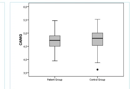
The mean Hb levels; MCV; SI; SIBP; serum Mg, Ca, P levels; and Ca/Mg ratioin the BHS and control groups are presented in Table 1. When these findings were compared, the only significant differences between the two groups were in Hb, Ca, and P levels. Although Hb levels were in the normal range in both groups, the mean Hb levels were significantly lower in the BHS group than in the control group (Table,p=0.03). However, no significant differ-ences were found in the mean SI and MCV levels between the two groups (p>0.05). Furthermore, the number of anemic patients was also similar in the BHS and control groups (20 patients (25.3%) in the BHS group and 24 patients (32.0%) in the control group). When serum Ca and P levels were compared, both levels were in the normal range in both groups, but mean serum Ca and P lev-els were significantly lower in the BHS group than in the control group (Table 1, p=0.001, p=0.007, respectively; figures 1 and 2).There were no significant differences in either mean serum Mg levels or Ca/Mg ratio between the BHS and control groups (Table 1, p=0.189, p=0.403, respectively; figures 3 and 4).
DISCUSSION
The main finding of the present study was that children with BHS had significantly lower mean serum Ca and P levels than those
without BHS. Furthermore, no differences were observed with re-spect to Ca/Mg ratio or serum Mg and SI levels, although the mean serum Hb levels differed significantly between the groups. BHS is a common non epileptic paroxysmal disorder in infancy and most commonly begins during the first 18 months of life, with an incidence of 4.6%–4.7% in the total population and a male to female ratio of 3:1 (14, 15).Nearly all children out grow BHS by the age of 6 years, and the persistence of BHS beyond 6 years is extremely rare. Consistent with previous reports, we found that the mean age of occurrence of BHS was 2.0±1.3 years with no significant difference between girls and boys (15, 16). In a previous study, a family history of BHS was found in 20%–30% of children with BHS (17, 18). Di Mario et al. (18) hypothesized that a consid-erable BHS cases that had a family history of these events are pos-sibly because of autosomaldominant inheritance with incomplete penetrance. In contrast to the study Di Mario et al. (18), only 8% of patients in the present study had a family history of BHS in a firstdegree relative.
BHS occurs when a child has had a minor accident or a frightening experience after which the child cries and holds his or her breath. Based on the child’s coloration during the spell, BHS is classified as “pallid type” if there is a powerful cardiac inhibition with a slight respiratory effect and as “cyanotic type” if there is an intense re-spiratory inhibition with a slight cardiac effect (18, 19). However, occasionally children experience a mixed type, indicating that the pathogenesis of both types is similar. Furthermore, it has been found that cyanotic BHS are more common than pallid BHS. Cya-notic BHS has been reported in 54%–62% of cases, pallid BHS has been reported in 19%–22% of cases, and mixed or unclassifi-able BHS has been reported in 19%–24% of cases (18, 20).In the present study, 90% of children had cyanotic spells, which was a higher proportion than previously reported.
The mechanism of BHS is not fully understood and is most likely multifactorial. Several studies have examined the potential roles of iron, anemia, and changes in trace element contents in biologi-cal fluids in the pathogenesis of BHS (1-4). Several studies found significantly higher rates of iron deficiency anemia in children with BHS than in healthy children. Indeed, several studies suggest an association between BHS and anemia in children (2, 3, 21). Mo-can et al. (21) followed 91 children aged 6–40 months and found that 69% of patients had iron deficiency anemia. Yilmaz et al. (16) revealed that approximately half of the 165 children with BHS also had iron deficiency anemia Interestingly, results from several studies found that iron supplementation in anemic or iron-deficient patients appeared to reduce the frequency of BHS, with one clini-cal trial indicating complete remission of spells in 32%–52% of patients (2, 3). In contrast, Azab et al. (22) found that patients with BHS had no significant statistical differenceregarding anemia and had low SI compared with controls. They suggested that because no other types of anemia were associated with BHS, these spells were related to iron and not anemia. However, iron deficiency is not the only factor responsible for BHS, because not all children with BHS were iron deficient at baseline in the present study. In addition, it remains to be determined why children with BHS who were not iron deficient responded to iron treatment (21). Colina et al. (23) suggested that BHS occurs in children without iron
de-ficiency andthat the other causes of anemia appear to exert an effect on BHS.We found that the mean Hb level was significantly lower in the BHS group than in the control group, but there was no statistically significant correlation in SI and MCV levels between two groups. Children with low Hb levels have been found to have decreased cerebral oxygen transport capacity, making them more susceptible to the chain of events leading to loss of consciousness during BHS, which supports the hypothesis that low serum Hb lev-els triggers BHS. Further studies are needed to elucidate whether serum Hb and SI levels or other types of anemia are involved in triggering loss of consciousness in patients with BHS.
Many factors may play a role in the etiopathogenesis of BHS. Kahn et al. (24) hypothesized that functional brainstem disturbances and immature breathing controls were involved in the occurrence of BHS. However, autonomic dysregulation has been implicated as a major component involved in the pathophysiology of these events with regard to BHS (25). The autonomic nervous system consists of two interacting systems: the sympathetic and parasympathetic systems. The sympathetic nervous system releases norepinephrine and epinephrine, which are responsible for the cascade of reac-tions associated with stress, whereas the parasympathetic nervous system releases acetylcholine. The stimulation of the sympathetic nervous system also causes an increase in the glucose level re-leased from the liver into the bloodstream; increase in the heart rate, myocardial contractility, pupil and bronchiole dilatation, blad-der relaxation, blood vessel constriction, sweat secretion, inhibition of peristalsis; and increase in the renin secretion by the kidneys. In contrast, parasympathetic system stimulation decreases heart rate and constricts the pupils; increases secretion of the glands; increases peristalsis; and constricts bronchioles (26). It has been shown that during heart failure or after myocardial infarction, sym-pathetic dominance occurs, which may trigger lethal arrhythmias. The sympathovagal imbalance is reflected by reduced HRV, which is a diagnostic factor for assessing autonomic dysfunction. In cya-notic BHS spells, increased sympathetic activity occurs, whereas in pallid BHS spells, increased parasympathetic activity is seen as an autonomic regulation disorder (22).
Serum electrolytes levels, such as Na, K, Mg, and Ca levels, can affect HRV. Bobkowski et al. (27) evaluated the relationship be-tween serum electrolyte levels and HRV in children with mitral valve prolapse and found that Mg levels positively correlated with parasympathetic indices and negatively correlated with sympa-thetic indices. Mg is an important cation and is a cofactor in more than 300 enzymatic reactions in the body. It is also important for bone stability, muscular relaxation, neurotransmission, enzymatic reactions, and many other physiological and cellular functions. Mg deficiency contributes to the pathogenesis of several cardiovascular diseases, including hypertension, vasospastic angina, and ventricu-lar arrhythmia. In addition, recent clinical studies have revealed that Mg protects the brain from ischemic damage, although the mechanism for neuroprotection afforded by Mg is yet to be fully elucidated (27). In addition, similar to the effects of Mg on vascu-lature, it has been speculated that Mg also induces a decrease in the intracellular Ca. Therefore, it is hypothesized that the effects of Mg on Ca homeostasis in neural cells is a common mechanism between the sympathetic inhibitory and neuroprotective actions of Mg (12). Murasato et al. (12) suggested that Mg deficiency
in-duced sympathetic excitation and enhanced the sensitivity of the sinus node to autonomic regulation, as Mg was shown toattenu-ate sympathetic tone, primarily by inhibiting N-type Ca channels. Furthermore, Devisetty et al. (13) concluded that decreased serum Ca levels caused disturbances in the autonomic nervous system. The effects of Ca on HRV may be attributed to the role of Ca in modulating cardiac muscle excitability and blood pressure. Ca
Table 1. Age and laboratory findings of the study groups
BHS Control group p Age (years) 2.0±1.3 2.0±1.2 0.167 Hb(g/dL) 11.5±1.01 11.5±0.81 0.039 WBC(×10) 10.8±3.12 10.8±3.96 0.505 Htc(%) 35.0±2.55 35.8±2.68 0.131 MCV(f/L) 76.1±6.56 76.9±5.89 0.791 RBC(mm3) 4.59±0.37 4.58±0.39 0.994 MCH(pg/hc) 25.2±2.34 25.9±2.68 0.148 MCHC(g/dL) 33.1±1.39 33.6±1.17 0.001 SI(μg/dL) 57.9±42.9 56.4±27.3 0.428 SIBP(μg/dL) 330.6±61.9 343.3±49.0 0.083 Mg(mg/dL) 2.1±0.15 2.1±0.18 0.189 Ca(mg/dL) 9.9±0.57 10.2±0.39 0.001 P(mg/dL) 5.13±0.76 5.40±0.59 0.007 ALP (U/L) 216.3±59.7 211.8±73.7 0.310 RDW(%) 16.1±2.5 16.2±2.5 0.235 Ca/Mg ratio 4.73±0.35 4.78±0.38 0.403
BHS; breath-holding spells, Hb; hemoglobin, MCV; mean corpuscular volume, SI; serum iron, SIBP; serum iron binding protein, Mg; magnesium, Ca; calcium, P; phosphorus, ALP; alkaline phosphatase, htc; hematocrit, WBC; white blood cell, RBC; red blood cell, RDW; red cell distribution width, ALP; alkaline phosphatase, MCH; mean corpuscular hemoglobin, MCHC; mean corpuscular hemoglobin level
Figure 2. Comparison of serum P levels in the BHS group and in control group
Abbreviations: BHS; breath-holding spells, POS: phosphorus
Figure 3. Comparison of serum Mg levels in the BHS group and in control group
Abbreviations: BHS; breath-holding spells, Mg: magnesium
Figure 4. Comparison of serum Ca/Mg ratio in the BHS group and in control group
Abbreviations:: BHS; breath-holding spells Figure 1. Comparison of serum Ca levels in the BHS group
and in control group
supplementation has been proven to have hypotensive effects, par-ticularly on systolic blood pressure, and therefore also affects HRV. In addition, Mg deficiency induced excessive catecholamine release and high sympathetic tone, which impaired baroreflex control of sympathetic nerves and reduced baroreflex-related oscillation, lead-ing to a blunted baroreflex and enhanced heart rate response to stress (13). Several clinical investigations have identified a possible relationship between autonomic dysregulation and BHS. Kolkiran et al. (25) confirmed the presence of autonomic dysregulation in children with BHS and found that respiratory sinus arrhythmia and asystole frequency during BHS spells were higher in patients with pallid BHS. Akalin et al. (6) also determined that QT dispersion was increased in patients with BHS, and they speculated that in-creased QTc dispersion in pallid BHS may be the result of the presence of more serious autonomic dysfunctions in this group (6). Previous reports also suggested that there is a centrally mediated sympathetic autonomic reflex hyperactivity in cyanotic BHS and a centrally mediated parasympathetic reflex hyperactivity in pallid BHS (1, 25). Considering the significant relationship between the autonomic nervous system and BHS, the present study was de-signed to evaluate the association of serum Ca and Mg levels with BHS. Kim et al. (28) found that Mg levels and Ca/Mg ratio were associated with HRV. However, in the present study, no statistical differences were found in the mean Mg level between the BHS and control groups. Furthermore, there was no significant difference in the proportions of Ca/Mg between these two groups. Kim et al. (28) found that the mean heart rate tended to increase from the lowest to the highest tertile of Ca levels, whereas the mean hear trate decreased significantly with increasing Mg levels. In contrast, although serum Ca and P levels were in the normal range in both the BHS and control groups in our study, children with BHS had significantly lower mean serum Ca and P levels than healthy con-trols. Based on these findings, we suggest that low serum Ca and P levels trigger BHS in children. We did not investigate and com-pare the mean serum vitamin D levels and parathyroid hormone levels in the present study, and therefore, we cannot comment on a possible role of vitamin D and parathyroid hormone in the etiopathogenesis of BHS. Mann et al. (29) found that low serum levels of vitamin D were possibly associated with a decline in cardio protective vagal tone in response to an acute vascular stressor, largely through the action of the active 1,25-dihydroxyvitamin D metabolite. In contrast, Burt et al. (30) showed that 25-hydroxyvi-tamin D was not significantly associated with baroreflex sensitivity or heart rate spectral parameters, suggesting that it does not alter basal parasympathetic nervous system activity. Therefore, further studies are needed to elucidate whether BHS is involved in this process in patients with low Ca and P levels.
CONCLUSION
To the best of our knowledge, this is the first study in English-language medical literature to report serum Mg, Ca, and P levels in children with and without BHS. It is also the first study reporting serum Ca/Mg ratio levels in these patient groups. Although Ca and P levels were within normal ranges in both groups, the mean Ca and P levels were significantly lower in the BHS group than in the control group. Therefore, these results suggest that low serum Ca and P levels reduce the threshold for BHS and thus are risk factors for BHS. Further large-scale studies are warranted to explain the roles Mg, Ca, and P play in BHS.
Ethics Committee Approval: The study was reviewed and approved by
the Ethics Committee of Baskent University.
Informed Consent: Informed consent was obtained from the parents of the participants.
Peer-review: Externally peer-reviewed.
Author Contributions: IE and YO designed the study and co-wrote the manuscript. MO, YO, and IE collected the data. All authors read and ap-proved the final version of the manuscript.
Acknowledgments: We are grateful to all of the children and their parents
who participated in the study. We also thank Çağla Sarıtürk for her valuable contribution in analyzing the data.
Conflict of Interest: Authors have no conflicts of interest to declare.
Financial Disclosure: This research was supported by the Baskent Univer-sity Research Fund (Project No: KA13/279).
REFERENCES
1. Saad K, Farghaly HS, Badry R, Othman HA. Selenium and antioxi-dant levels decreased in blood of children with breath-holding spells. J Child Neurol 2014; 29(10); 1339–43. [CrossRef]
2. Hudagolu O, Dirik E, Yis U, Kurul S. Parental attitude of mothers, iron deficiency anemia, and breath-holding spells. Pediatr Neurol 2006; 35(1); 18–20. [CrossRef]
3. Daoud AS, Batieha A, Al-Sheyyab M, Abuekteish F, Hijazi S. Effec-tiveness of iron therapy on breath-holding spells. J Pediatr 1997; 130(4); 547–50. [CrossRef]
4. Akalin F, Turan S, Guran T, Ayabakan C, Yilmaz Y. Increased QT dispersion in breath-holding spells. Acta Pediatr 2004; 93(6); 770–4.
[CrossRef]
5. DiMario FJ Jr, Burleson JA. Autonomic nervous system function in severe breath-holding spells. Pediatr Neurol 1993; 9(4); 268–74.
[CrossRef]
6. Anil B, Nedunchezian K, Jayanthini V, Pathmanabhan M. Breath-holding spells: evaluation of autonomic nervous system function. In-dian Pediatr 2005; 42(9); 923–7.
7. Lahiri MK, Kannankeril PJ, Goldberger JJ. Assessment of autonomic function in cardiovascular disease: physiological basisand prognostic implications. J Am Coll Cardiol 2008; 51(18); 1725-33. [CrossRef]
8. Lutfi MF. Correlations between electrolytes levels and heart rate vari-ability in apparently healthy subjects. Am J Physiol 1989; 256(6); 1573-9.
9. Yilmaz O, Ciftel M, Ozturk K, Kilic O, Kahveci H, Laloğlu F et al. As-sessment of heart rate variability in breath holding children by 24 hour Holter monitoring. Cardiol Young 2015; 25(2); 317–23. [CrossRef]
10. Peacock JM, Ohira T, Post W, Sotoodehnia N, Rosamond W,Folsom AR. Serum magnesium and risk of sudden cardiac death in the Ath-erosclerosis Risk in Communities (ARIC) Study. Am Heart J 2010; 160(3); 464–70. [CrossRef]
11. Bolland MJ, Avenell A, Baron JA, Grey A, MacLennan GS, Gam-ble GD et al. Effect of calcium supplements on risk of myocardial infarction and cardiovascular events: meta analysis. BMJ 2010; 341: c3691. [CrossRef]
12. Murasato Y, Harada Y, Ikeda M, Nakashima Y, Hayashida Y. Effect of magnesium deficiency on autonomic circulatory regulation in con-scious rats. Hypertension 1999; 34(2); 247–52. [CrossRef]
13. Devisetty A, Kambar C , Hanumanth N. Study of effect of serum cal-cium levels on autonomic nervous system in pre-menstrual syndrome. IOSR-JDMS 2005; 14(3); 50-5.
14. Kliegman R, Nelson W. E. NelsonTextbook of Pediatrics. 20th ed. Philadelphia, PA: Elsevier/Saunders; 2016.
15. Bhatia MS, Singhal PK, Dhar NK, Nigam VR, Malik SC, Mullick DN. Breath holding spells: an analysis of 50 cases. Indian Pediatr 1990; 27(10); 1073-9.
16. Yilmaz U, Doksoz O, Celik T, Akinci G, Mese T, Sevim Yilmaz T. The value of neurologic and cardiologic assessment in breath holding spells. Pak J Med Sci 2014; 30(1); 59-64.
17. DiMario FJ Jr, Sarfarazi M. Family pedigree analysis of childrenwith severe cyanotic and pallid breath-holding spells. J Pediatr 1997; 130(4); 647-51. [CrossRef]
18. DiMario FJ Jr. Prospective study of children with cyanotic and pallid breath-holding spells. Pediatrics 2001; 107(2); 265–9. [CrossRef]
19. Hellström Schmidt S, Tedgård U, Pronk CJ. Breath-holding spells oc-cur disproportionately more often in children with transient erythro-blastopenia. Acta Paediatr 2016; 105(9); 1088–93. [CrossRef]
20. Sawires H, Botrous O. Double-blind, placebo-controlled trial on the effect of piracetam on breath-holding spells. Eur J Pediatr 2012; 171(7); 1063–7. [CrossRef]
21. Mocan H, Yildiran A, Orhan F, Erduran E. Breath holding spells in 91 children and response to treatment with iron. Arch Dis Child 1999; 81(3); 261–2. [CrossRef]
22. Azab SF, Siam AG, Saleh SH, Elshafei MM, Elsaeed WF, Arafa MA et al. Novel findings in breath-holding spells:A cross-sectional study. Medicine 2015; 94(28): e1150. [CrossRef]
23. Colina KF, Abelson HT.Resolution of breath-holding spells with treat-ment of concomitant anemia. J Pediatr 1995; 126(3); 395–397.
[CrossRef]
24. Kahn A, Rebuffat E, Sottiaux M, Muller MF,Bochner A, Grosswasser J. Brief airway obstruction during sleep in infants with BHS. J Pediatr 1990; 117(2); 188-93. [CrossRef]
25. Kolkiran A, Tutar E, Atalay S, Deda G, Cin S. Autonomic nervous system functions in children with breath-holding spells and effects of iron deficiency. Acta Paediatr 2005; 94(9); 1227–31. [CrossRef]
26. Gordan R, Gwathmey JK, Xie LHAutonomic and endocrine control of cardiovascular function. World J Cardiol 2015: 26; 7(4); 204–14. 27. Bobkowski W, Zachwieja J, Siwińska A, Mroziński B, Rzeź
nik-Bieniaszewska A, Maciejewski J et al. Influence of autonomic nervous system on electrolyte abnormalities in children with mitral valve pro-lapse. Pol Merkur Lekarski 2003; 14(81): 220–3.
28. Kim YH, Jung KI, Song CH. Effects of serum calciumand magnesium on heart rate variability in adult women. Biol Trace Elem Res 2012; 150(1-3); 116–22. [CrossRef]
29. Mann MC, Exner DV, Hemmelgarn BR, Sola DY, Turin TC, Ellis L, Ahmed SB. Vitamin D levels are associated with cardiac autonomic activity in healthy humans. Nutrients 2013; 5(6); 2114–27. [CrossRef]
30. Burt MG, Mangelsdorf BL, Stranks SN, Mangoni AA. Relationship between vitamin D status and autonomic nervous system activity. Nu-trients 2016; 8(9): 565. [CrossRef]