Question:
An 89-year-old woman was admitted with complaints of nausea, vomiting, and epigastric pain after having meals. A computed tomography scan of the abdomen showed a 12.5-cm mass in the fundus of the stom-ach, which extended to the duodenum (Figure 1).
Upper gastrointestinal endoscopy showed a polyp-oid lesion with a thick and long stalk that originated from fundus and extended to the duodenum. The head of the lesion prolapsed through the pylorus into the duodenal bulb and caused gastric outlet ob-struction (Figure 2).
Turk J Gastroenterol 2017; 28: 419-20
A rare cause of gastric outlet obstruction
Nurettin Suna, Serkan Öcal, Diğdem Özer Etik, Haldun Selçuk, Fatih Hilmioğlu, Sedat Boyacıoğlu Department of Gastroenterology, Başkent University School of Medicine, Ankara, Turkey
Address for Correspondence: Nurettin Suna E-mail: [email protected]
Received: March 27, 2017 Accepted: April 26, 2017 Available Online Date: July 13, 2017
© Copyright 2017 by The Turkish Society of Gastroenterology • Available online at www.turkjgastroenterol.org • DOI: 10.5152/tjg.2017.17183
419
Figure 1. A computed tomography scan of the abdomen showed a giant mass that was 12.5 cm in length in the fundus of the stom-ach, which extended to the duodenum
Figure 2. Upper gastrointestinal endoscopy showed a polypoid lesion with a thick and long stalk that originated from the fundus and extended to the duodenum
Imag
e of the Is
sue
Cite this article as: Suna N, Öcal S, Etik DÖ, Selçuk H, Hilmioğlu F, Boyacıoğlu S. A rare cause of gastric outlet obstruction. Turk J Gastroenterol 2017; 28: 419-20.
Answer: Combined right and left bile duct variations
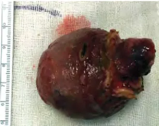
Laparoscopic wedge resection with a 1-cm rim of normal gastric tissue was performed for the complete removal of the polyp. At a macroscopic examination, it was found to be a 7.5×6.5×5.5 cm lesion (Figure 3). Its histopathology was consis-tent with the features of a gastrointestinal stromal tumor (GIST) with negative tumor margins. GISTs can originate from any-where along the gastrointestinal tract, but they are frequently located in the stomach (1). Gastric GISTs are generally inci-dentally found during upper gastrointestinal endoscopy (2). The vast majority of gastric GISTs are asymptomatic, unless the tumor is large enough to cause symptoms or has bleeding ul-cerations (2). Hyperplastic and pedunculated large gastric pol-yps located in the gastric antrum may cause gastric outlet syn-drome (3). Although GISTs are submucosal lesions, in our case, it mimicked a pedunculated polyp. This presentation is rare for
gastric GISTs. The endoscopic appearance of the lesion was not typical for a gastric GIST. The other interesting presentation of the tumor was its long pseudo-peduncle, which caused duo-denal obstruction.
Ethics Committee Approval: Ethics committee approval was received for this study from the ethics committee of Başkent University School of Medicine (Decision No: 162-4765).
Informed Consent: Written informed consent was obtained from the patient who participated in this study.
Peer-review: Externally peer-reviewed.
Author contributions: Concept - N.S., S.Ö., H.S.; Design - D.Ö.E., F.H., S.Ö.; Supervision - A.S.B., F.H., D.Ö.E.; Resource - N.S., S.Ö., F.H.; Materials - NS., D.Ö.E., S.Ö.; Data Collection and/or Processing - D.Ö.E., S.Ö., N.S.; Analysis and/or Interpretation - F.H., H.S., S.Ö.; Literature Search - N.S., D.ÖE., F.H.; Writing - N.S., F.H., H.S.; Critical Reviews - A.S.B., F.H., H.S. Conflict of Interest: No conflict of interest was declared by the au-thors.
Financial Disclosure: The authors declared that this study has re-ceived no financial support.
REFERENCES
1. Koo DH, Ryu MH, Kim KM, et al. Asian Consensus Guidelines for the Diagnosis and Management of Gastrointestinal Stromal
Tu-mor. Cancer Res Treat 2016; 48: 1155-66. [CrossRef ]
2. Quek R, George S. Gastrointestinal stromal tumor: a clinical
over-view. Hematol Oncol Clin North Am 2009; 23: 69-78. [CrossRef ]
3. Shaib YH, Rugge M, Graham DY, Genta RM. Management of gas-tric polyps: an endoscopy-based approach. Clin Gastroenterol
Hepatol 2013; 11: 1374-84.[CrossRef ]
420
Suna et al. Gastric outlet obstruction Turk J Gastroenterol 2017; 28: 419-20
Imag
e of the Is
sue
Figure 3. Laparoscopic wedge resection was performed for the complete removal of the giant polyp (7.5×6.5×5.5 cm at the macroscopic examination)