Comparison of intracameral dexamethasone and intracameral triamcinolone
acetonide injection at the end of phacoemulsifi cation surgery
Sirel Gür Güngör, Begüm Bulam, Ahmet Akman, Meriç Çolak
1Access this article online Website: www.ijo.in DOI: 10.4103/0301-4738.141045 PMID: *****
Quick Response Code:
Departments of Ophthalmology and 1Health Care Management,
Faculty of Medicine, Başkent University, Ankara, Turkey
C o r r e s p o n d e n c e t o : D r . S i r e l G ü r G ü n g ö r , Department of Ophthalmology, Faculty of Medicine, Baskent University, Fevzi Çakmak Caddesi, 06490, Bahçelievler, Ankara, Turkey. E-mail: [email protected]
Manuscript received: 11.09.13; Revision accepted: 17.06.14
Introduction
Although recent developments in cataract surgery have resulted in a decrease in the physical trauma related with the surgery, they have not eliminated the trauma- induced synthesis and release of inflammatory mediators.[1] Surgical
trauma elicits a cascade of ocular inflammatory reactions in eyes undergoing cataract surgery.[2] Uncontrolled
inflammation may cause complications such as cystoid macular edema, increased intraocular pressure (IOP), synechial formation, posterior capsule opacifi cation, and secondary glaucoma.[3]
There are several application/preferences about corticosteroid injections at the end of the phacoemulsifi cation surgery. Some surgeons apply such injections to suppress the infl ammation during the fi rst 24 hours, as well as, other surgeons apply nothing at all apart from topical steroids.[4,5]
Subconjunctival steroid injections are still one of the most prevalent methods to prevent postoperative inflammation, but it can be painful in cases with topical anesthesia and can
cause subconjunctival hemorrhage and chemosis. In our clinic, this method had been applied to suppress the infl ammation in the past. The triamcinolone acetonide (TA)-assisted anterior vitrectomy was described as an eff ective method to enable visualization and removal of the vitreous in complicated surgeries and in cases with vitreus loss.[6-8] Later, it was
observed that intracameral TA injection during/at the end of the surgery helped sustain a lesser degree of anterior chamber infl ammation and edema on the cornea on the postoperative fi rst day. Supporting articles[9,10] about the safety and effi ciency
of this method after cataract surgeries, encouraged us to change our routine to intracameral injection of 2 mg/0.05 ml TA as of today. However, the particulate structure of the TA and its tendency to increase the IOP in some patients, forced us to substitute it with intracameral 0.4 mg/0.1 ml dexamethasone in our clinic.
The aim of this clinical study was to compare the results of intracameral dexamethasone and intracameral TA in patients, who underwent uncomplicated phacoemulsifi cation surgery.
Materials and Methods
Sixty eyes of 60 patients underwent elective uncomplicated phacoemulsification and foldable intraocular lens implantations were enrolled in this prospective study. Institutional Review Board approval was obtained. All patients were informed about the design of the study and the procedure involved, and all gave writt en informed consent. A comprehensive questionnaire was completed, which included items on the patient’s age, medical and ocular
Purpose: To compare the results of intracameral dexamethasone and intracameral triamcinolone
acetonide injection in patients that underwent cataract surgery with phacoemulsification.
Materials and Methods: Sixty eyes of 60 patients that underwent cataract surgery with phacoemulsifi cation
were randomized into two groups. Preoperative visual acuity of all patients was 0.5 or lower and intraocular pressures were under 21mmHg. After surgery, eyes in group 1 (30 eyes) were injected with 0.4 mg/0.1 ml dexamethasone into the anterior chamber, and eyes in group 2 (30 eyes) were injected with 2 mg/0.05 ml triamcinolone acetonide into the anterior chamber. All eyes received standard postoperative prednisolone acetate and moxifloxacin eye drops. The biomicroscopic evaluation, visual acuity, and intraocular pressure measurements were done at baseline (preoperatively) and on postoperative days 1, 7 and 30.
Results: There were no statistically signifi cant diff erences in mean visual acuity, the amount of anterior
cells and fl are between the two groups (P ≥ 0.05). Mean intraocular pressure values at postoperative fi rst day were signifi cantly higher in group 2 than in group 1 (P = 0.009). The mean intraocular pressures on days 7 and 30 after surgery were not statistically diff erent between the two groups (P ≥ 0.05). Conclusions: Intracameral dexamethasone and intracameral triamcinolone acetonide were similarly effective in controlling postoperative infl ammation following phacoemulsifi cation. However, the intraocular pressures on postoperative fi rst day were higher in patients receiving intracameral triamcinolone acetonide. The highest intraocular pressure in triamcinolone acetonide group was 24 mmHg, and stabilized in a few days, therefore using triamcinolone acetonide may impose a minimal risk to patients. Nevertheless, intracameral dexamethasone seems to be a bett er alternative to apply at the end of surgery to suppress the infl ammation during the fi rst 24 hours.
Key words: Dexamethasone, phacoemulsifi cation, triamcinolone acetonide
Original Article
862 Indian Journal of Ophthalmology Vol. 62 No. 8
history. Inclusion criteria were the presence of a cataract that was suitable for phacoemulsifi cation, visual acuities 0.5 or lower and intraocular pressures of 21 mmHg or lower. Exclusion criteria were: Diabetes mellitus, current use of oral or topical anti-infl ammatory agents (steroidal or non-steroidal), history of steroid responsiveness, uveitis, glaucoma, pigment dispersion syndrome, pseudoexfoliation syndrome, age-related macular degeneration, corneal disease, and a history of cystoid macular edema. A detailed preoperative ophthalmic evaluation including slit-lamp examination, IOP measurement with Goldman applanation tonometry, central corneal thickness measurement with ultrasonic pachymetry and dilated fundus examination was performed.
All operations were performed by the same surgeon (AA) under topical anesthesia. Approximately 1-2 hours before surgery, phenylephrine 2.5% and tropicamide 1% eye drops were instilled. After topical anesthesia, a 2.8 mm clear corneal incision was made, after which sodium chondroitin sulphate 4%-sodium hyaluronate 2% (Viscoat, Alcon, Pharmaceuticals Ltd) was injected and 5.0 mm capsulorhexis was performed. The surgeon performed standard phacoemulsifi cation using the phaco-chop technique. The capsular bag was expanded with sodium hyaluronate 1% (Healon, Abbott Medical Optics), and a foldable intraocular lens was implanted in the capsular bag. The viscoelastic substance was removed vigorously from the bag, the capsular fornix, and the anterior chamber in a standard fashion using an irrigation/aspiration system.
At the end of the surgery, patients were randomly allocated to one of two groups. In group 1 (n = 30 eyes of 30 patients), dexamethasone (Dekort, Deva Holding Inc) 0.4 mg/0.1 ml was injected into the anterior chamber through a paracentesis using a 27-gauge cannula. In group 2 (n = 30 eyes of 30 patients), TA (Kenacort-A®; Bristol-Myers Squibb) 2 mg/0.05 ml was injected into the anterior chamber through a paracentesis using a 27-gauge cannula. In both groups, 0.1 ml moxifloxacin 0.5% was injected into the anterior chamber. After the postoperative examination (20-24 h later), moxifl oxacin 0.5% eye drop (Vigamox, Alcon, Pharmaceuticals Ltd) was prescribed five times a day for 1 week, and prednisolone acetate 1% eye drop (Pred Forte, Allergan, Pharmaceuticals Ltd) were prescribed fi ve times a day with a one drop/week taper over fi ve weeks.
Patients were examined on the postoperative days 1, 7 and 30. Postoperative evaluations included patient history regarding any ocular discomfort, Snellen visual acuity (VA), slit-lamp examination, IOP measurement and fundus examinations. Evaluation was based on effi cacy, safety and tolerance criteria. Subjective complaints were scored as a 0 for no complaints or 1 for symptoms of pain, blurry vision, redness, foreign body sensation, tearing or photophobia. The major effi cacy parameters assessed clinically on each visit were anterior chamber cells, anterior chamber fl are and conjunctival hyperemia. Anterior chamber cells were graded as: 0 = <5 cells; 1 = mild, 5-10 cells; 2 = moderate, 10-20 cells; 3 = marked, 21-50 cells; 4 = severe, >50 cells, and 5 = hypopyon. Aqueous fl are scale was scored as: 0 = none; 1 = mild (just detectable); 2 = moderate (iris details clear); 3 = marked (iris details hazy), and 4 = severe (heavy with fi brin deposits and clots). Anterior chamber cell and fl are scores were determined
using the narrowest slit beam (0.5 mm) at a height of 8 mm, with maximal luminance and magnifi cation of the slit-lamp.
Visual acuity in the study eye was measured using the Snellen VA chart and values were converted to logMAR for statistical analysis.
The preoperative IOP was measured using a Goldmann applanation tonometry, 1 day before surgery. The postoperative IOP was measured using the same Goldmann applanation tonometry 1 day, 7 day and 30 day after surgery.
All postoperative examinations were performed by the same surgeon (AA) in order to obtain consistent infl ammation grading scores, and all the scores were recorded for each visit and compared between the two treatment groups.
S t a t i s t i c a l a n a l y s i s wa s p e r f o r m e d u s i n g S P S S software (Statistical Package for the Social Sciences, version 9.0, SPSS Inc., Chicago, III, USA). Ordinal variables (anterior chamber cells and fl are) were evaluated by Mann–Whitney U-test. Group comparisons of the postoperative IOPs and VA were done using independent sample test. Mean IOP changes in each group from postoperative days 1, 7 and 30 were compared using paired t-tests. Age and sex were compared using the Chi-square test. A P > 0.05 was considered statistically signifi cant.
Results
Group 1 included 16 women and 14 men with an average age of 71 ± 9.4 years. Group 2 included 20 women and 10 men with an average age of 69.8 ± 10.5 years. The two groups were comparable with respect to age and sex. There were no signifi cant diff erences between the groups in age or sex (P > 0.05). There were no intraocular complications such as capsule rupture or zonular dialysis in any eye.
Preoperative mean VA values were similar in both groups (P > 0.05). There were no statistically significant differences in mean VA between the two groups at any postoperative visit (P > 0.05) [Table 1].
Subjective complaints of pain, blurry vision, redness, foreign body sensation, tearing and photophobia were in 5 patients in group 1 and in 6 patients in group 2 only on the postoperative fi rst day. There was no signifi cant diff erence in incidence of postoperative complaints in both groups (P = 0.56). There were no subjective complaints in both groups on postoperative days 7 and 30.
Injection of TA into the anterior chamber resulted in a ‘snow-globe effect’ of various densities at slit-lamp examination. Despite the suspension of TA crystals, it was easy to assess cell and fl are between crystals. The treatment modalities used in the two groups reduced anterior chamber cells and fl are equally and eff ectively, and no statistically signifi cant diff erences were observed at any postoperative visits (P > 0.05) [Table 2].
There was no signifi cant diff erence in corneal thickness between two groups at any postoperative visit (P > 0.05).
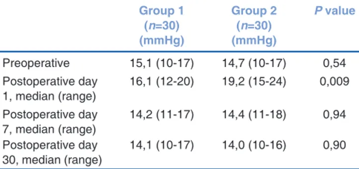
P r e o p e r a t i v e m e a n I O P v a l u e s w e r e s i m i l a r in both groups (P > 0.05). IOP values in group 1 were 16.1 mmHg (range: 12-20 mmHg) at postoperative fi rst day.
August 2014 Gungor, et al.: Comparison of intracameral dexamethasone and triamcinolone acetonide 863
IOP values in group 2 were 19.2 mmHg (range: 15-24 mmHg) at postoperative fi rst day. Mean IOP values on postoperative first day were significantly higher in group 2 than in group 1 (P = 0.009). There were no statistically signifi cant differences in IOP values between the two groups, on postoperative days 7 and 30 (P > 0.05) [Table 3].
Discussion
The intraocular injection of TA has been used for many years for the treatment of the posterior segment pathologies in which inflammation has a pivotal function. Oh et al.,[11] applied TA
intracamerally into rabbit eyes to investigate the eff ect of TA on the corneal endothelium and showed reduced microvilli, although no statistically signifi cant diff erences in endothelial counts and central corneal thickness were observed at 2 hours after the experimental procedure. Chang et al.,[12] showed
toxicity of TA on cultured endothelium in their experimental study. Despite the evidence of in vitro toxicity of intracameral TA on corneal endothelium, the use has been raised in practice to suppress postoperative infl ammation after cataract surgery. Gills and Gills[10] added TA to an anterior chamber
solution for controlling infl ammation after cataract surgery. As they did not fi nd the appropriate dose, they began the dosage conservatively, with 0.25 mg and gradually increased the doses to 3.0 mg and up to 4.0 mg in diabetes patients. The authors suggested that as the TA dose was gradually increased, the number of eyes requiring postoperative steroid treatment fell from 45% at the lowest dose to 2% at a dose of 1.8-2.1 mg.
The exact cleaning time of TA crystals from the eye is unknown. The TA crystals spread throughout the eye, the iris, the wound sites, the capsular bag, and into the vitreous. Much of the TA may progress through diff erent channels of access to the anterior chamber such as the trabecular meshwork and the iris itself.[10,13] Jonas[13] found detectable concentrations of TA
in aqueous humour samples obtained from eyes, which had undergone intravitreal 25 mg TA injection 6 months before sampling.
One of the potential side effects of corticosteroid administration by any route is the raised IOP. Intravitreal administration of TA for therapeutic indications is known to be associated with elevated IOP.[14,15] In the studies, in which
the clinical outcomes in patients who had TA assisted anterior vitrectomy after phacoemulsifi cation surgery complicated by posterior capsule rupture and vitreous loss was presented, clinically signifi cant IOP elevation occurred in a small number of patients.[6,16,17]
Karalezli et al.,[18] conducted a study to evaluate the eff ect
of 1mg intracameral TA on postoperative IOP after routine cataract surgery. The patients were randomized into two groups. Eyes in group 1 received an injection of 1 mg TA into the anterior chamber at the end of the surgery, but eyes in group 2 did not. The mean IOP values at postoperative 6 and 20-24 h were found slightly higher in group 1 than in group 2.
The high relative potency of dexamethasone may confer greater effi cacy than TA when given as a single injection. Rapid aqueous volume turnover and short half-life of intraocular dexamethasone, both in the order of several hours, would help minimize the risk of steroid-induced ocular hypertension.[19,20]
This study investigated whether dexamethasone injected intracamerally at the end of cataract surgery could safely and eff ectively reduce postoperative infl ammation compared to intracameral TA. The treatment modalities used in the two groups reduced anterior chamber cells and fl are equally and eff ectively, and no statistically signifi cant diff erences were observed at any postoperative visits (P > 0.05). There was no signifi cant diff erence in incidence of postoperative complaints in both groups (P = 0.56).
Chang et al.,[21] demonstrated that intracameral
dexamethasone can safely be given after surgery in eyes with diff erent types of glaucoma with minimal concern for postoperative IOP elevations. In our study, mean IOP values at postoperative fi rst day were signifi cantly higher in group 2 than in group 1 (P = 0.009). The highest IOP in the TA group was
Table 1: Visual acuity (logMAR) values for both treatment groups
Group 1 (n=30) Group 2 (n=30) P value
Preoperative, mean (range) 0,78 (0,40-1,00) 0,79 (0,40-1,00) 0,12 Postoperative day 1, mean (range) 0,16 (0,00-0,22) 0,18 (0,00-0,30) 0,60 Postoperative day 7, mean (range) 0,08 (0,00-0,22) 0,07 (0,00-0,18) 0,52 Postoperative day 30, mean (range) 0,07 (0,00-0,18) 0,07 (0,00-0,18) 0,54
logMAR = Logarithm of the minimum angle of resolution
Table 2: Comparison of infl ammation scores (anterior chamber cells, fl are) between the two groups
Group 1 (n=30) Group 2 (n=30) P value Cells
Postoperative day 1, median (range) 1,8 (0-2) 1,6 (0-2) 0,33 Postoperative day 7, median (range) 0,3 (0-1) 0,2 (0-1) 0,42 Postoperative day 30, median (range) 0 (0-0) 0 (0-0) 1,00 Flare
Postoperative day 1, median (range) 0,2 (0-1) 0,3 (0-1) 0,67 Postoperative day 7, median (range) 0 (0-0) 0 (0-0) 1,00 Postoperative day 30, median (range) 0 (0-0) 0 (0-0) 1,00
Table 3: Mean intraocular pressure values for both groups (in mmHg) Group 1 (n=30) (mmHg) Group 2 (n=30) (mmHg) P value Preoperative 15,1 (10-17) 14,7 (10-17) 0,54 Postoperative day 1, median (range) 16,1 (12-20) 19,2 (15-24) 0,009 Postoperative day 7, median (range) 14,2 (11-17) 14,4 (11-18) 0,94 Postoperative day 30, median (range) 14,1 (10-17) 14,0 (10-16) 0,90
864 Indian Journal of Ophthalmology Vol. 62 No. 8
24 mmHg, and stabilized in a few days. However, there were no statistically signifi cant diff erences in IOP values between the two groups at postoperative days 7 and 30 (P > 0.05). This might be because, we used very small amount of dexamethasone (0.4 mg/0.1 ml) and TA (2 mg/0.05 ml) intracamerally and carefully excluded patients with a known family history of glaucoma or any earlier ocular hypertensive response to systemic or topical corticosteroids from the study.
This is the fi rst study in the literature comparing the result of injection of intracameral dexamethasone and TA. Our study must be viewed in the light of some limitations. It was not a masked study: Surgery and observation were carried out by the same person and which otherwise might have aff ected the measured outcomes. We did not have an anterior chamber fl are cell meter, and so we used slit-lamp biomicroscopy to investigate the anterior chamber cells and flare. Treatment with corticosteroids may have raised the IOP in the patients, who had glaucoma or ocular hypertension. On the other hand, this study is a prospective, randomized, clinical trial, and the operations were performed by the same surgeon on the patients of similar age and further a sex-matched group strengthens the credibility of the fi ndings as well.
In conclusion, there are many surgeons to prefer perioperative corticosteroid injection. Intracameral corticosteroid usage may still be a bett er alternative, because of the adverse eff ects such as pain and hemorrhage due to subconjunctival steroid injections. This study demonstrates that intracameral dexamethasone and intracameral TA were similarly eff ective in controlling postoperative inflammation after uncomplicated cataract surgery with phacoemulsifi cation. However, the intraocular pressures on postoperative fi rst day were higher in patients receiving intracameral TA. This probably depends on the structure of particles of TA. Since the highest IOP in the TA group was 24 mmHg, and stabilized in a few days, in practical terms, using TA may impose a minimal risk to patients. This increase in IOP may be very important in a patient with glaucoma. Because of that intracameral dexamethasone may be a bett er alternative to apply at the end of surgery to suppress the infl ammation during the fi rst 24 hours.
References
1. Rowen S. Preoperative and postoperative medications used for cataract surgery. Curr Opin Ophthalmol 1999;10:29-35.
2. Dick HB, Schwenn O, Krummenauer F, Krist R, Pfeiffer N. Inflammation after sclerocorneal versus clear corneal tunnel phacoemulsifi cation. Ophthalmology 1999;107:241-7.
3. Yaylali V, Ozbay D, Tatlipinar S, Yildirim C, Ozden S. Efficacy and safety of rimexolone 1% versus prednisolone acetate 1% in the control of postoperative inflammation following phacoemulsification cataract surgery. Int Ophthalmol 2004;25:65-8. 4. Ahmed MS, Moly KN, Aziz MA. Use of povidone-iodine drop
instead of sub-conjunctival injection of dexamethasone and gentamicin combination at the end of phacoemulsifi cation cataract surgery. Mymensingh Med J 2010;19:232-5.
5. Dieleman M, Wubbels RJ, van Kooten-Noordzij M, de Waard PW. Single perioperative subconjunctival steroid depot versus postoperative steroid eyedrops to prevent intraocular infl ammation
and macular edema after cataract surgery. J Cataract Refract Surg 2011;37:1589-97.
6. Yamakiri K, Uchino E, Kimura K, Sakamoto T. Intracameral triamcinolone helps to visualize and remove the vitreous body in anterior chamber in cataract surgery. Am J Ophthalmol 2004;138:650-2. 7. Arbisser LB, Charles S, Howcroft M, Werner L. Management
of vitreous loss and dropped nucleus during cataract surgery. Ophthalmol Clin North Am 2006;19:495-506.
8. Gopal L, Bhende M, Sharma T. Vitrectomy for accidental intraocular steroid injection. Retina 1995;15:295-9.
9. Karalezli A, Borazan M, Akova YA. Intracameral triamcinolone acetonide to control postoperative inflammation following cataract surgery with phacoemulsification. Acta Ophthalmol 2008;86:183-7. 10. Gills JP, Gills P. Eff ect of intracameral triamcinolone to control
inflammation following cataract surgery. J Cataract Refract Surg 2005;31:1670-1.
11. Oh JY, Wee WR, Lee JH, Kim MK. Short-term eff ect of intracameral triamcinolone acetonide on corneal endothelium using the rabbit model. Eye (Lond) 2007;21:812-8.
12. Chang YS, Tseng SY, Teseng SH, Wu CL, Chen MF. Triamcinolone acetonide suspension toxicity to corneal endothelial cells. J Cataract Refract Surg 2006;32:1549-55.
13. Jonas JB. Concentration of intravitreally injected triamcinolone acetonide in aqueous humour. Br J Ophthalmol 2002;86:1066. 14. Jonas JB, Degenring RF, Kreissig I, Akkoyun I, Kamppeter BA.
Intraocular pressure elevation after intravitreal triamcinolone acetonide injection. Ophthalmology 2005;112:593-8.
15. Williams CP, Konstantopoulos A, Rowley SA, Luff AJ. Late intraocular pressure rise following intravitreal triamcinolone injection. Clin Exp Ophthalmol 2007;35:385-6.
16. Angunawela RI, Liyanage SE, Wong SC, Litt le BC. Intraocular pressure and visual outcomes following intracameral triamcinolone assisted anterior vitrectomy in complicated cataract surgery. Br J Ophthalmol 2009;93:1691-2.
17. Kasbekar S, Prasad S, Kumar BV. Clinical outcomes of triamcinolone-assisted anterior vitrectomy after phacoemulsifi cation complicated by posterior capsule rupture. J Cataract Refract Surg 2013;39:414-8.
18. Karalezli A, Borazan M, Kucukerdonmez C, Akman A, Akova YA. Effect of intracameral triamcinolone acetonide on postoperative intraocular pressure after cataract surgery. Eye (Lond) 2010;24:619-23.
19. Kwak HW, D’Amico DJ. Evaluation of the retinal toxicity and pharmacokinetics of dexamethasone after intravitreal injection. Arch Ophthalmol 1992;110:259-66.
20. Fraunfelder FT, Fraunfelder FW. Drug-induced Ocular Side Eff ects. 5th ed. Boston: Butt erworth-Heinemann; 2001.
21. Chang DT, Herceg MC, Bilonick RA, Camejo L, Schuman JS, Noecker RJ. Intracameral dexamethasone reduces infl ammation on the fi rst postoperative day after cataract surgery in eyes with and without glaucoma. Clin Ophthalmol 2009;3:345-55.
Cite this article as: Gungor SG, Bulam B, Akman A, Colak M. Comparison of intracameral dexamethasone and intracameral triamcinolone acetonide injection at the end of phacoemulsifi cation surgery. Indian J Ophthalmol 2014;62:861-4.
Source of Support: Nil. Confl ict of Interest: None declared.