Yazışma Adresi/Address for Correspondence: Dr. Nurhilal Buyukkurt, Baskent University School of Medicine, Department of Hematology, Adana, Turkey E-mail: [email protected]
Geliş tarihi/Received: 02.01.2017 Kabul tarihi/Accepted: 23.02.2017
ARAŞTIRMA / RESEARCH
Effects of bone marrow fibrosis and angiogenetic structure on
autologous hematopoietic stem cell engraftment
Kemik iliği fibrozis ve anjiogenezisinin hematopoetik kök hücre engraftmanı üzerine
etkisi
Nurhilal Büyükkurt
1, Güner Hayri Özsan
2, Sermin Özkal
3, Gülşah Seydaoğlu
4, İnci Alacacıoğlu
2,
Mehmet Ali Özcan
2, Fatih Demirkan
2, Özden Pişkin
2, Bülent Ündar
21Baskent University School of Medicine, Department of Hematology, Ankara, Turkey
2Dokuz Eylül University, School of Medicine, Department of Hematology, 3Department of Pathology, Izmir, Turkey 4Cukurova University School of Medicine, Department of Biostatistics, Adana, Turkey
Cukurova Medical Journal 2017;42(3):499-506
Abstract Öz
Purpose: Hematopoietic stem cell (HSC) engraftment is influenced by many factors. We investigated the effects of bone marrow fibrosis and angiogenetic structure on engraftment in patients with hematological malignancies. Materials and Methods: Data were collected from 34 patients (20 males and 14 females) who underwent autologous HSC transplantation. Bone marrow myelofibrosis was graded from 0 to 3, angiogenesis was quantified using a stereological method in the most recent bone marrow biopsy before the transplantation. Patients were categorized into two groups according to intensity of angiogenesis parameters.
Results: Half of the patients had fibrosis and majority had multiple myeloma (73.5%). Eleven patients had grade 1, six had grade 2 myelofibrosis. The engraftment day (ED) for platelets and erythrocytes was significantly different between the grade 2 fibrosis and non-fibrosis groups. VSD and NVES levels were significantly higher in the grades 1 and 2 fibrosis groups than the no fibrosis group. While the overall survival time was shorter in the grade 2 fibrosis group than the others, the difference was not statistically significant.
Conclusion: Bone marrow fibrosis was found to be independent risk factor. It may have a negative effect on platelet and erythrocyte engraftment time of autologous transplantation process but this effect does not influence survival.
Amaç: Birçok faktör hematopoetik kök hücre (HKH) engraftmanını etkiler. Bu çalışmada hematolojik kanseri olan hastalarda kemik iliğindeki myelofibrozis ve anjiogenezisin engraftman üzerine etkisi araştırılmıştır. Gereç ve Yöntem: Otolog kök hücre nakli yapılan 34 hasta (20 erkek, 14 kadın) verileri değerlendirildi. Nakilden önceki son kemik iliğindeki fibrozis 0-3 arasında derecelendirildi, anjiogenezis stereolojik metod ile ölçüldü. Hastalar anjiogenezis parametrelerinin yoğunluğuna göre de iki gruba ayrıldı.
Bulgular: Hastaların çoğunluğu (%73.5) multipl myelom idi ve yarısında fibrozis saptandı. On bir hasta derece 1, 6 hasta derece 2 fibrozise sahipti. Trombosit ve eritrosit engraftman günleri açısından derece 2 fibrozisi olan grupla fibrozis saptanmayan grup arasında istatistiksel anlamlı farklılık vardı. Derece 1 ve 2 fibrozis gruplarında fibrozisi olmayanlara göre VSD ve NVES düzeyleri anlamlı olarak yüksekti. Toplam yaşam derece 2 fibrozisi olan grupta daha düşük olmakla birlikte istatistiksel anlamlılık yoktu. Sonuç: Kemik iliği myelofibrozisinin bağımsız bir risk faktörü olduğu saptanmıştır. Bunun toplam yaşamı etkilemeden otolog nakil sürecinde trombosit ve eritrosit engraftmanı üzerine negatif etkisi olabilir. Fibrozisin derecesi ile doğru orantılı olarak artmış anjiogenezis arasındaki ilişkiyi açıklamak için ileri çalışmalar gereklidir. Key words: Bone marrow fibrosis, angiogenesis,
INTRODUCTION
One of the most important criteria for successful autologous stem cell transplantation is overcoming aplasia of bone marrow that occurs due to the conditioning regimen. Recovery of bone marrow and reconstitution of hematopoiesis with re-infused autologous stem cells is called engraftment.
The period of engraftment is expected to be 7–21 days after transplantation. If the duration is > 21 days, it indicates an engraftment delay. Engraftment failure is defined as > 42 days1-3. Bone marrow
consists of hematopoietic and stromal
compartments composed of reticular cells, adipocytes, and osteogenic bone cells near the surfaces of vascular endothelial cells, vascular
endothelial smooth muscle cells, and
macrophages4,5. The “hematopoietic stem cell
niche” was first defined by Schofield in 1978. Stem cells are located in this micro-anatomic region of bone marrow where they are blocked from differentiating or self-renewing. The stem cells are supported by adjacent stromal cells.
Hematopoietic stem cells (HSCs) and hematopoietic progenitor cells are distributed randomly in the bone marrow but are rather close to the bone endosteum6-8. Recent studies have demonstrated
that they accumulate around blood vessels9,10. The
differentiation and maturation processes of bone marrow HSCs were described by Shacney in 197511.
The area where HSCs are located is rich in vascularization and closer to the centre of bone marrow. Shacney also noted that undifferentiated cells are found throughout the endosteum.
The endosteal bone marrow sinusoidal network zone defines a different anatomical and functional structure and was adapted to the “vascular niche” concept. Ultrastructural studies have shown that differentiated HSCs are located closer to bone marrow microvascular structures compared to immature HSCs12,13. HSC engraftment is a complex
process that entails the collection of circulating bone marrow HSCs throughout the microvascular structure and subsequent trans-endothelial migration to the bone marrow hematopoietic cords. These events are referred to as HSC homing. Then, stem cells settle to a specific region known as the niche, which defines lodgment14.
These findings indicate that the interaction between progenitor cells and stromal cells is a critical
determinant of the course of HSC maturation. Many factors that affect engraftment kinetics have been examined in patients undergoing autologous peripheral blood stem cell transplantation; however, the debate over engraftment process continues. In the present study, we investigated the effects of bone marrow fibrosis and angiogenesis on engraftment.
MATERIAL AND METHODS
Patients
This study was planned as a single-center, cross-sectional and retrospective study. Patients who underwent autologous peripheral stem cell transplantation (PSCT) over 6 years were enrolled in this retrospective study. Study material was collected from the records starting on January 2003 and ending at December 2008. Pre-transplant bone marrow biopsies obtained from 34 consecutive patients (20 males and 14 females) were examined. Sections from paraffin blocks were placed on poly L-lysine slides, preserved in the Pathology Department archive, and were evaluated by two
hematopathologists. Marrow structure was
examined fibrotic and angiogenetic structure. Patients were grouped according to the degree of fibrosis.
Diagnosis, disease stage, treatment, response, total number of CD34+ peripheral stem cells (PSCs)
collected, mobilization regimen, pre-transplant disease status, and the last date of contact or date of death were recorded from the patient files. Neutrophil, platelet and erythrocyte engraftment day were noted.
Primary endpoint was defined as neutrophil, platelet and erythrocyte engraftment time associated with bone marrow structure. Secondary endpoint was the effect of marrow structure on survival.
Ethics committee approval was obtained from the Institutional Ethics Committee prior to the study. Informed consent was obtained from all individual participants included in this study.
Definitions
Neutrophil engraftment day (ED) was defined as the first day that the neutrophil count remained ≥ 500 × 106/L. Platelet engraftment had occurred
when the platelet count was > 20 × 109/L for 7 500
days without a transfusion. Erythrocyte engraftment required an interval of 3 weeks without a blood transfusion1.
Angiogenesis was evaluated by using the CD34 (NeoMarkers, Freemont, CA, USA) antigen streptavidin-avidin-immunoperoxidase method. Stereological measurements were performed to determine the vascular density visible with CD34. Number of vessels per unit stroma (NVES) and vascular surface density (VSD) per tumor tissue volume were calculated according to the following formulae15.
𝑉𝑉𝑉𝑉𝑉𝑉 =𝛴𝛴𝛴𝛴𝛴𝛴.2.121𝐼𝐼𝐼𝐼𝑡𝑡𝑡𝑡.𝐿𝐿𝑡𝑡 𝑁𝑁𝑉𝑉𝑁𝑁𝑉𝑉 =𝑁𝑁.121𝐼𝐼𝐼𝐼𝑡𝑡𝑡𝑡
The sections were fixed at 100× magnification for light microscopy and reflected on a camera (CCD; Sony, Tokyo, Japan) and monitor (Sony Trinitron). A 525× magnified image was also obtained. The image was placed on a transparent monitor composed of 121 points formed by 11 horizontal and 11 vertical test lines. The tumor tissue was contained within the borders of the transparent area, and the number of junction points where the vessels crossed test lines (In) were noted as the total vessel count (N) whether they crossed or not. Vv (str) indicates the stromal volume ratio above the test line and was determined by the point-counting method. Vv (str) is the point count per stroma
divided by 121. The same process was repeated in 10 randomly selected fields. Lr (constant number), 10.34; Istr, number of crossed horizontal and vertical lines in the specified stromal field.
Myelofibrosis was graded based on the European Consensus Report of Bone Marrow Fibrosis grading of 0–3 in sections treated with reticulin silver and Masson’s trichrome stains16. All patients received
2.4 g/m2 cyclophosphamide and a granulocyte
colony stimulating factor protocol to mobilize the stem cells. The conditioning regimen was melphalan for multiple myeloma and BEAM (carmustine, etoposide, ARA-C, and melphalan) for lymphoma. The patients were classified into three groups according to CD 34 level, fibrosis based on literature. Furthermore, patients were classified into two groups with regard to intensity of angiogenetic parameters.
Statistical analysis
Continuous variables between the groups were analysed using the Mann–Whitney U or Kruskal– Wallis tests. Correlations between continuous variables were detected using Spearman’s correlation test. Linear regression models were performed to determine the independent risk factors related with neutrophil, platelet, and erythrocyte engraftment days.
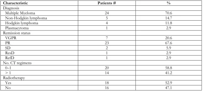
Table 1. Pre-transplantation characteristics of the patients
Characteristic Patients # % Diagnosis Multiple Myeloma 24 70.6 Non-Hodgkin lymphoma 5 14.7 Hodgkin lymphoma 4 11.8 Plasmacytoma 1 2.9 Remission status VGPR 7 20.6 PR 23 67.6 SD 2 5.9 ResD 1 2.9 RefD 1 2.9 No. CT regimens 0–1 20 58.8 > 1 14 41.2 Radiotherapy Yes 18 52.9 No 16 47.1
No. CT: number of prior chemotherapy regimens, VGPR: Very good partial remission, PR: partial remission, SD: stabile disease, ResD: residual disease, RefD: refractory disease
The predictors of time to transplantation and overall survival rate were analysed using the Kaplan–Meier method and compared to the Mantel log-rank test. Results are reported as median (range), number (n), and percent (%). A p-value < 0.05 was considered significant. Statistical analyses were performed using the SPSS v 20.0 software package (SPSS Inc., Chicago, IL, USA).
RESULTS
The median age of the patients was 50 years (range, 17–70 years). Most patients were diagnosed with multiple myeloma (73.5%), and disease status was partial remission in 67.6%. Of all patients, 55% had received only one line of chemotherapy (CT) before transplantation and 45% had received two or more lines of CT. The pre-transplantation characteristics of the patients are shown in Table 1. The patients were divided into three groups based on CD34+ cell
level. The CD34+ cell levels were < 5 × 10⁶/kg, 5–
9.9 × 10⁶/kg, and ≥ 10 × 106/kg in groups 1, 2, and
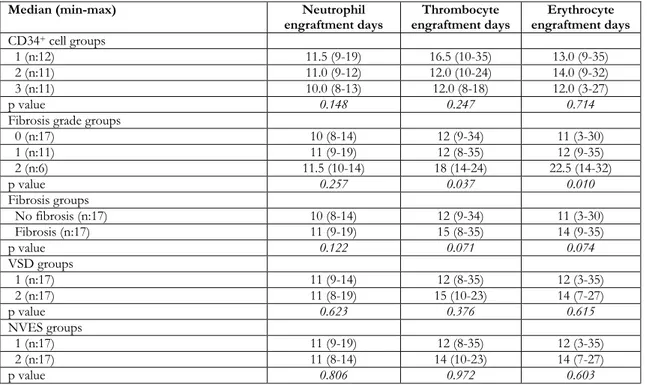
3 respectively. No differences were found in terms of the numbers of neutrophil, platelet, or erythrocyte EDs among these three groups (Table 2). There were no statistically significant differences among CD 34+groups with regard to age, disease
status, receiving radiotherapy, fibrosis and angiogenetic parameters (p>0,05 for all)
A correlation analysis was performed between for the numbers of neutrophil, platelet, and erythrocyte EDs in each CD34+ groups. As a result, no
significant correlation was detected between CD34+
cell level and the numbers of neutrophil or erythrocyte EDs. A trend to negative correlation was observed between CD34+ cell level and the
number of platelet EDs (r=−0.32, p = 0.016). The numbers of patients in fibrosis groups 0, 1 and 2 were 17 (50%), 11 (32.4%), and 6 (17.6%), respectively. No patient had grade 3 fibrosis. A significant difference was detected in the number of platelet and erythrocyte EDs between the groups (p=0.037 and p=0.010, respectively). The median numbers of platelet and erythrocyte EDs were similar in patients with grades 0 and 1, but these durations were significantly longer in the grade 2 fibrosis group than in the other groups. However, the number of neutrophil EDs was not different (p=0.257) among the three fibrosis groups. The median levels of the angiogenesis parameters (VSD and NVES) increased as the degree of fibrosis
increased. Significant differences were detected in the VSD and NVES among the fibrosis groups (p=0.034 and p=0.028, respectively). The median VSD and NVES levels were similar in patients with grades 1 and 2 fibrosis, but the median levels were significantly lower in grade 0. A post-hoc test was not applied between the fibrosis subgroups due to insufficient sample size.
No correlations were found between VSD, NVES, and the numbers of neutrophil, platelet, and erythrocyte EDs (r<0.20 for all). Angiogenesis was evaluated as number of micro vessel count per field (NVES) and vascular surface density (VSD) per tumor tissue volume. Median was detected as 35,4 (min:4,02 max:112,3) mm-2 for VSD and 92,8
(min:10,3- max:325,7) mm-1 for NVES. Patients
were categorized into two groups according to median levels of VSD and NVES. VSD levels of ≤35,4 mm-2 and >35,5 mm-2were classified as
Group 1 and Group 2, respectively. Similarly, NVES values of ≤92,9 mm2and ≥93 mm-2were classified
as Group 1 and Group 2, respectively. A statistically significant difference was not detected neither among VSD groups nor NVES groups in terms of neutrophil, platelet and erythrocyte engraftment days (Table 2).
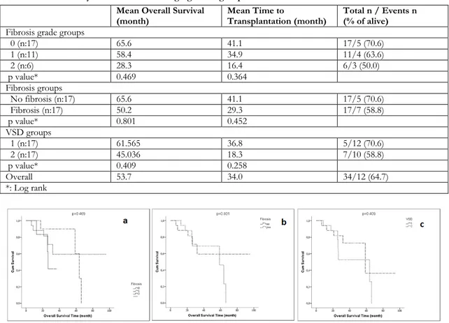
Mean overall survival was 53.7 months (95% confidence interval [CI], 40.5–66.9 months) and mean time to transplantation was 34.0 months (95% CI, 24.7–43.3 months) (Table 3). Overall survival was lower in grade 2 compared to grade 1 patients (28.3 vs. 58.4 months). Similar results were observed among patients with fibrosis grades 1 and 2 for time to transplantation (34 vs. 16 months, respectively) (Table 3, Figure 1). Cox regression models were not performed to determine the independent risk factors related with neutrophil, thrombocyte and erythrocyte engraftment days due to insufficient sample size.
Three linear regression models were performed: neutrophil , platelet, and erythrocyte engraftment days were used as dependent variable; VSD, CD 34+
cell amount measured as continuous variables; fibrosis and disease status at pre-transplantation were used as independent variables in the separately models. The results of linear regression models showed that fibrosis was found to be significantly independent factor related with platelet and erythrocyte engraftment days (p=0.037 and p <0.001 respectively).
Table 2. The neutrophil, thrombocyte and erythrocyte engraftment days in severalgroups
Median (min-max) Neutrophil
engraftment days engraftment days Thrombocyte engraftment days Erythrocyte CD34+ cell groups
1 (n:12) 11.5 (9-19) 16.5 (10-35) 13.0 (9-35)
2 (n:11) 11.0 (9-12) 12.0 (10-24) 14.0 (9-32)
3 (n:11) 10.0 (8-13) 12.0 (8-18) 12.0 (3-27)
p value 0.148 0.247 0.714
Fibrosis grade groups
0 (n:17) 10 (8-14) 12 (9-34) 11 (3-30) 1 (n:11) 11 (9-19) 12 (8-35) 12 (9-35) 2 (n:6) 11.5 (10-14) 18 (14-24) 22.5 (14-32) p value 0.257 0.037 0.010 Fibrosis groups No fibrosis (n:17) 10 (8-14) 12 (9-34) 11 (3-30) Fibrosis (n:17) 11 (9-19) 15 (8-35) 14 (9-35) p value 0.122 0.071 0.074 VSD groups 1 (n:17) 11 (9-14) 12 (8-35) 12 (3-35) 2 (n:17) 11 (8-19) 15 (10-23) 14 (7-27) p value 0.623 0.376 0.615 NVES groups 1 (n:17) 11 (9-19) 12 (8-35) 12 (3-35) 2 (n:17) 11 (8-14) 14 (10-23) 14 (7-27) p value 0.806 0.972 0.603
NVES: Micro vessel count per field, VSD: vascular surface density per tumor tissue volume.
While vascular surface density per tumor unit was found to be significantly independent factor for erythrocyte engraftment day (p=0.025), CD 34+ cell
amount was found to be significantly independent factor for platelet and neutrophil engraftment day (p=0.004 and p=0.020 respectively).
DISCUSSION
It is quite important to determine the hematopoietic engraftment rate and long-term continuity of engraftment following peripheral stem cell transplantation. Mortality and morbidity risks, transfusion and antibiotic needs, hospital stay duration, and economic costs of engraftment make this condition critical.
The number of CD34+ cells in a graft is one of the
factors affecting its kinetics. The required number of HSCs for autologous transplantation is 2.5–5×10⁶
CD34+/kg according to some authors17-20.
However, Maunier et al.21 found that the optimal
level was a minimum of 5×10⁶ CD34+/kg in a study
that investigated the influence of CD34+ cell
number (before cryopreservation and when melted)
on long-term hematologic reconstruction following autologous stem cell transplantation in patients with lymphoma. Furthermore, Hohaus et al.22 showed
that platelet engraftment is faster in patients with lymphoma, if the number of infused CD34+ cells is
> 5×10⁶/kg. Our results show a similar number of
EDs for neutrophils, platelets and erythrocytes regardless of the CD34+ cell count.
There are some evidences that marrow fibrosis can delay in stem cell engraftment time in patients with acute and chronic leukaemia. Soll et al.23
investigated the role of myelofibrosis in 203 patients with myelofibrosis and 203 controls. No difference in the number of neutrophil EDs was reported, but platelet engraftment was delayed 3 days in the myelofibrosis group. This result was a 7-day interval in the severe myelofibrosis group (grades 3–4) for platelet engraftment. Those authors also found a delay of 2 days for erythrocyte engraftment; however, the transfusion requirement was not different. In contrast, Scott et al.24 investigated
patients with acute myeloid leukaemia and advanced myelodysplastic syndrome with multi-lineage dysplasia (471 patients: 113 with myelofibrosis and
358 without fibrosis), and detected severe fibrosis in 28% of the patients. Neutrophil engraftment was observed in 98 of 113 patients, and the median ED
was day 28 (range, 10–80 days) in these patients and day 17 (range, 10–33 days) in the cases without fibrosis.
Table 3. Survival analyses in fibrosis and angiogenesis groups Mean Overall Survival
(month) Mean Time to Transplantation (month) Total n / Events n (% of alive) Fibrosis grade groups
0 (n:17) 65.6 41.1 17/5 (70.6) 1 (n:11) 58.4 34.9 11/4 (63.6) 2 (n:6) 28.3 16.4 6/3 (50.0) p value* 0.469 0.364 Fibrosis groups No fibrosis (n:17) 65.6 41.1 17/5 (70.6) Fibrosis (n:17) 50.2 29.3 17/7 (58.8) p value* 0.801 0.452 VSD groups 1 (n:17) 61.565 36.8 5/12 (70.6) 2 (n:17) 45.036 18.3 7/10 (58.8) p value* 0.409 0.258 Overall 53.7 34.0 34/12 (64.7) *: Log rank
These results indicate a significant delay in the fibrosis group (p≤0.0001). We found a delay in platelet and erythrocyte engraftment time in patients with fibrosis. Our results reveal that the engraftment delay becomes more prominent as fibrosis grade increases. Although angiogenesis processes of many diseases have been investigated, particularly those for solid tumours, their influence on transplantation kinetics have not been sufficiently evaluated. Some evidence of vascular support for the malignant process can be seen in the bone marrow of patients with hematologic cancers, as in other solid organ tumours 25,26. Angiogenesis was evaluated in the
bone marrow of patients with polycythemia vera, chronic myeloid leukaemia and myelofibrosis, and the results showed that neovascularisation increased in the patients with myeloproliferative diseases. The vascular density of bone marrow was statistically
significant in patients with myelofibrosis (5.9±2.1 and 14.4±5.5, respectively, p<0.001).
According to a novel pathogenic hypothesis for
myelofibrosis with myeloid metaplasia,
megakaryocytes and/or monocytes clonally increase due to abnormal levels of cytokines released from impaired bone marrow stroma 27 leading to collagen
fibrosis and new bone formation. This impaired stromal microenvironment also serves as a source of angiogenetic cytokines, such as increased expression of vascular growth factor by megakaryocytes28.Our
results show that only erythrocyte engraftment is influenced by the VSD. This result is consistent with the hypothesis mentioned above. We suggest that angiogenesis may help alleviate the negative effects of fibrosis, although no direct effect on engraftment was shown.
Figure 1: Kaplan Meier curves according to fibrosis grade groups (a), fibrosis groups (b) and VSD groups (c).
Regarding survival, one study investigated the influence of fibrosis on ED and overall survival in 19 patients with fibrosis who underwent autologous transplantation. There was no difference in overall survival between the groups with and without fibrosis and the results according to fibrosis grade were not evaluated in that study29. We showed no
statistically significant difference in survival by grade of fibrosis.
In conclusion, bone marrow fibrosis accompanying multiple myeloma and lymphoma was detected to be an independent risk factor which negatively affects platelet and erythrocyte engraftment time, but this effect does not influence overall survival. Preliminary findings in this study also revealed angiogenetic changes in the bone marrow occurred dependent from intensity of marrow fibrosis. The observation that VSD (part of angiogenetic structure) is an independent risk factor for erythrocyte engraftment may be an important preliminary data for the studies that would be conducted with patients group with larger sample size. For explaining the exact mechanism, future studies are needed.
Acknowledgements
This study was funded by Izmir Hematologic Diseases and Cancer Research and Fraternal Association.
REFERENCES
1. Ippoliti C, Przepiorka D, Giralt S, Andersson BS, Wallerstein RO, Gutterman J et al. Low-dose non-glycosylated rhGM-CSF is effective for the treatment of delayed hematopoietic recovery after autologous marrow or peripheral blood stem cell transplantation. Bone Marrow Transplant. 1993;11:55–9.
2. Crump M, Couture F, Kovacs M, Saragosa R, McCrae J, Brandwein J et al. Interleukin-3 followed by GM-CSF for delayed engraftment after autologous bone marrow transplantation. Exp Hematol. 1993;21:405–10.
3. Khwaja A, Goldstone AH, Linch DC. Delayed neutrophil recovery after BEAM chemotherapy and autologous bone marrow transplantation for lymphoma is not associated with increased mortality from infection. Bone Marrow Transplant. 1995;15:313–5.
4. Lichtman MA. The ultrastructure of the hemopoietic environment of the marrow: a review. Exp Hematol. 1981;9:391-410.
5. Weiss L. The hematopoietic microenvironment of
the bone marrow: an ultrastructural study of the stroma in rats. Anat Rec. 1976;186:161-84.
6. Fliedner TM, Graessle D, Paulsen C, Reimers K. Structure and function of bone marrow hemopoiesis: mechanisms of response to ionizing radiation exposure. Cancer Biother Radiopharm. 2002;17:405-26.
7. Calvi LM, Adams GB, Weibrecht KW, Weber JM, Olson DP, Knight MC et al. Osteoblastic cells regulate the haematopoietic stem cell niche. Nature. 2003;425:841-6.
8. Lord BI, Testa NG, Hendry JH. The relative spatial distributions of CFUs and CFUc in the normal mouse femur. Blood. 1975;46:65-72.
9. Kiel MJ,Yilmaz OH, Iwashita T, Yilmaz O, Terhorst C, Morrison SJ. SLAM family receptors distinguish hematopoietic stem and progenitor cells and reveal endothelial niches for stem cells. Cell. 2005;121:1109-21.
10. Shen Q, Goderie SK, Jin L, Karanth N, Sun Y, Abramova N et al. Endothelial cells stimulate self-renewal and expand neurogenesis of neural stem cells. Science. 2004;304:1338-40.
11. Shackney SE, Ford SS, and Wittig AB. Kinetic-microarchitectural correlations in the bone marrow of the mouse. Cell Tissue Kinet. 1975;8:505-16. 12. Shirota T and Tavassoli M,
Cyclophosphamide-induced alterations of bone marrow endothelium: implications in homing of marrow cells after transplantation. Exp Hematol. 1991;19:369-73. 13. Tavassoli M. Hemopoietic endothelium, incognito.
Exp Hematol. 1992;20:386-87.
14. Nilsson SK, Simmons PJ, and Bertoncello I. Hemopoietic stem cell engraftment. Exp Hematol. 2006;34:123-9.
15. Barth PJ, Weingartner K, Köhler HH, Bittinger A. Assesment of vascularization in prostatic carcinoma: a morphometric investigation. Hum. Pathol. 1996;27:1306-10.
16. Thiele J, Kvasnicka HM, Facchetti F, Franco V, van der Walt J, Orazi A. European consensus on grading bone marrow fibrosis and assessment of cellularity. Haematologica. 2005;90:1128-32.
17. Haas R, Witt B, Möhle R, Goldschmidt H, Hohaus S, Fruehauf S et al. Sustained long-term hematopoiesis after myeloablative therapy with peripheral blood progenitor cell support. Blood. 1995;85:3754-61.
18. Bensinger WI, Longin K, Appelbaum F, Rowley S, Weaver C, Lilleby K et al. Peripheral blood stem cells (PBSCs) collected after recombinant granulocyte colony stimulating factor (rhG-CSF): an analysis of factors correlating with the tempo of engraftment after transplantation. Br J Haematol. 1994;87:825-31. 19. van der Wall E, Richel DJ, Holtkamp MJ, , Slaper-Cortenbach IC, van der Schoot CE, Dalesio O et al. Bone marrow reconstitution after high-dose chemotherapy and autologous peripheral blood 505
progenitor cell transplantation: effect of graft size. Ann Oncol. 1994;5:795-802.
20. Weaver CH, Hazelton B, Birch R, Palmer P, Allen C, Schwartzberg L et al. An analysis of engraftment kinetics as a function of the CD34 content of peripheral blood progenitor cell collections in 692 patients after the administration of myeloablative chemotherapy. Blood. 1995;86:3961-9.
21. Mounier N, Larghero J, Manson J, Brice P, Madelaine-Chambrin I, Brière J et al. Long term hematologic recovery after autologous stem cell transplantation in lymphoma patients: impact of the number of prefreeze and post-thaw CD34+ cells. Bull Cancer. 2005;92:E31-8.
22. Hohaus S, Goldschimidt H, Ehrhardt R, Haas R. Successful autografting following myeloablative conditioning therapy with blood stem cells mobilized by chemotherapy plus rhG-CSF. Exp Hematol. 1993;21:508-14.
23. Soll E, Massumoto C, Clift RA, Buckner CD, Appelbaum FR, Storb R et al. Relevance of marrow fibrosis in bone marrow transplantation: a retrospective analysis of engraftment. Blood. 1995;86:4667-73.
24. Scott BL, Storer BE, Greene JE, Hackman RC, Appelbaum FR, Deeg HJ. Marrow fibrosis as a risk
factor for posttransplantation outcome in patients with advanced myelodysplastic syndrome or acute myeloid leukemia with multilineage dysplasia. Biol Blood Marrow Transplant. 2007;13:345-54.
25. Vacca A, Ribatti D, Presta M, Minischetti M, Iurlaro M, Ria R et al. Bone marrow neovascularization, plasma cell angiogenic potential, and matrix metalloproteinase-2 secretion parallel progression of human multiple myeloma. Blood. 1999;93:3064-73. 26. Perez-Atayde AR, Sallan SE, Tedrow U, Connors S,
Allred E, Folkman J. Spectrum of tumor angiogenesis in the bone marrow of children with acute lymphoblastic leukemia. Am J Pathol. 1997;150:815-21.
27. Reilly J.T Idiopathic myelofibrosis: pathogenesis, natural history and management. Blood Rev. 1997;11:233-42.
28. Pruneri G, Bertolini F, Soligo D, Carboni N, Cortelezzi A, Ferrucci PF et al. Angiogenesis in myelodysplastic syndromes. Br J Cancer. 1999;81:1398-401.
29. Suyanı E, Akı SZ, Yegin ZA, Ozkurt ZN, Altındal S, Akyürek N et al. The impact of bone marrow fibrosis on the outcome of hematopoietic stem cell transplantation. Transplant Proc. 2010;42:2713-9.