Abstract
Peste des petits ruminants (PPR) is an economically important contagious disease of small ruminants. PCR-based techniques have been successfully used for rapid diagnosis of PPR. The method used for isolation of RNA from tissue samples is an important concern when using reverse transcription-PCR (RT-PCR) methods for the detection of PPR virus (PPRV). In this study, a commercial kit for manual preparation and an automated processing technique for RNA extraction were compared in terms of performance. Thirty-two small ruminants, each from different flocks, with PPR suspect submitted to laboratory were chosen to compare manual and automated extraction methods for the detection of PPRV. Vero cells were used for PPRV isolation. One-step RT-PCR was used for the detection of PPRV RNA. From the 32 submitted samples, CPE was observed in 11 samples. PPRV nucleic acid was detected in 11 of 32 samples that were manually extracted, while viral RNA was detected in 9 of 32 extracts prepared by the robot. Two samples that were negative with automated extraction were weakly positive in manual extraction. RNA quality and quantity were assessed using a spectrophotometer. According to the results, difference in quantity among two methods was statistically significant (P<0.0001, two-tailed paired t-test), and manual extraction method is suitable for detection of low amounts of PPRV RNA in clinical samples.
Keywords: RNA purification, Manual, Automated, Quality, Quantity, RT-PCR
Peste Des Petits Ruminants Virus RNA’sının Tespitinde Manuel ve
Otomatik Nükleik Asit Ekstraksiyon Yöntemlerinin
Karşılaştırılması
Özet
Koyun ve keçi vebası (PPR), küçük ruminantların ekonomik açıdan önemli, bulaşıcı bir hastalığıdır. Günümüzde PCR tabanlı teknikler, PPR’ın hızlı tanısı için başarıyla kullanılmaktadır. PPR virusunun (PPRV) tespitinde reverse transkripsiyon PCR (RT-PCR) metotları kullanılır iken, doku örneklerinden RNA izolasyonu için kullanılan yöntem önem arz etmektedir. Bu çalışmada, RNA ekstraksiyonu için ticari manuel bir kit ve otomatik bir işleme tekniği performans açısından karşılaştırılmıştır. Laboratuvara PPR şüphesi ile gönderilen herbiri farklı sürüden otuz iki küçükbaş hayvan, PPRV tespitinde manuel ve otomatik ekstraksiyon yöntemlerinin karşılaştırılması için seçilmiştir. Vero hücreleri PPRV izolasyonu için kullanılmıştır. PPRV RNA’sının tespiti için one step RT-PCR metodu kullanılmıştır. Otuz iki örnekten, 11 âdetinde sitopatojenik efekt (CPE) gözlenmiştir. Manuel ekstraksiyon metodu ile 32 örneğin 11’inde PPRV nükleik asidi tespit edilirken, robot kullanılarak yapılan otomatik ekstraksiyon metodu ile 9 örnekte viral RNA tespit edilmiştir. Otomatik ekstraksiyon metodu ile negatif tespit edilen 2 örnek, manuel ekstraksiyon metodu sonucu zayıf pozitif olarak tespit edilmiştir. Manuel ve otomatik ekstraksiyon sonucu elde edilen RNA miktarı ve kalitesi spektrofotometre cihazı kullanılarak karşılaştırılmıştır. Elde edilen sonuçlara göre, iki metot arasında elde edilen RNA miktar farkı istatiksel olarak önemli bulunmuş (P<0.0001, iki - kuyruklu t - testi) olup, klinik örneklerdeki düşük miktardaki PPRV RNA’sını tespit etmek için manuel ekstraksiyon metodu daha uygundur.
Anahtar sözcükler: RNA purifikasyonu, Manuel, Otomatik, Kalite, Miktar, RT-PCR
Comparison of Manual and Automated Nucleic Acid Extraction
Methods for Detection of Peste Des Petits Ruminants Virus RNA
[1]Murat ŞEVİK
1
Oğuzhan AVCI
2Ömer Barış İNCE
3 [1]1 2 3
This study has been presented at 5 th European Congress of Virology, 11-14 September 2013, Lyon, France
Molecular Microbiology, Veterinary Control Institute, TR-42080 Meram, Konya - TURKEY
Department of Virology, Faculty of Veterinary Medicine, Selçuk University, TR-42000 Konya - TURKEY Agriculture and Rural Development Support Institution, TR-03000 Afyonkarahisar - TURKEY
İletişim (Correspondence)
+90 332 3224741
[email protected]INTRODUCTION
Peste des petits ruminants (PPR) is an acute and highly contagious viral disease of domestic and wild small ruminants that is characterized by fever, purulent ocular and nasal discharge, diarrhoea and enteritis [1]. Cattle,
buffaloes and camels can become infected, although they are not susceptible to clinical disease [2].
The causative agent, peste des petits ruminants virus (PPRV), belongs to the genus Morbillivirus within family
Paramyxoviridae along with rinderpest virus (RPV),
measles virus (MeV), canine distemper virus (CDV), and morbilliviruses of marine mammals [3,4]. Based on the
basis of partial sequence analysis of the fusion protein (F) and nucleoprotein (N) genes, PPRV can be grouped into four lineages [5-7]. PPR viruses belonging lineages I and II
have been found exclusively in west and central Africa [7,8].
Lineage III has been isolated from eastern Africa and Arabian Peninsula whereas lineage IV has been isolated in Asia, Middle East and northern Africa [6,9-13].
Serological assays can be used to detect the presence or absences of antiviral antibodies [14]. Neutralization and
isolation of virus in cell culture are time-consuming, and require special laboratory requirements, so they aren’t suitable for routine diagnosis [6]. Recent advances in
molecular biology have led to the development of reliable and faster diagnostic tests for diagnosis of PPR. Reverse transcription-PCR (RT-PCR) provides rapid, sensitive and reliable diagnosis of the disease [15,16]. Samples of nasal and
ocular discharge and anticoagulant-treated blood from live animals or lymph nodes, especially the mesenteric and bronchial nodes, lungs, spleen and intestinal mucosa from necropsied animals are used for diagnosis PPR [14].
The method used for isolation of RNA from tissue samples is an important concern when using reverse trans-cription-PCR (RT-PCR) methods for the detection of PPRV. The aim of this study was to compare the performance of a manual extraction method, QIAamp (Qiagen), and an automated extraction instrument, MagNA Pure LC (Roche Applied Sciences), with each other for the detection of low amounts of PPRV RNA in clinical samples. The time, effort, and reagent costs for both methods were analysed. Furthermore, various routine RT-PCR methods were tested and compared using RNA extracted with both methods.
MATERIAL and METHODS
Samples and Positive Control
During January-December 2012, 32 animals (20 sheep and 12 goats), each from different flocks with no vaccination history, suspected to have PPR were submitted to the Veterinary Control Institute, Konya, Turkey. Tissue samples
(lung, liver, spleen and mesenterial lymph node) were collected from 32 animals, aged between 1 and 24 months, were tested for PPR virus by RT-PCR. All tissue samples were kept at -85°C prior to sample preparation and the RT-PCR assays. Lyophilized freeze-dried live PPR vaccine (Nigeria75/1 vaccine strain) obtained from the Division of Virology, Etlik Central Veterinary Control and Research Institute, Ankara, Turkey, was used as the positive control.
Virus Isolation
Tissue samples of each animal were combined and homogenised in PBS containing antibiotics using tissue rupture (Qiagen, Valencia, CA) to give a 10% suspension. The suspensions were then centrifuged at 3.000 g for 15 min at 4°C. Supernatants were filtered (0.2-μm pore size) and then inoculated on to Vero cells, and maintained in Dulbecco’s minimum essential medium supplemented with 5% foetal bovine serum. The cultures were incubated at 37°C in 5% CO2 atmosphere and daily examined for
appearance of cytopathic effect (CPE). All materials were passaged in Vero cell cultures for three times. If CPE was not observed even after 3 blind passages, the sample was considered negative. Supernatants of CPE-positive cultures were examined for nucleic acid of PPRV using RT-PCR.
Analytical Sensitivity and Specificity Experiments Supernatants of CPE-positive cultures were collected and virus titres (PFU/ml) were determined on Vero cells in a standard plaque assay. Panels of PPRV were created by serial dilution (100 to 10-4 PFU/ml) in nuclease-free water
(Qiagen, Valencia, CA). Virus dilutions were extracted by each method and PPRV RNA was detected by RT-PCR. Nuclease-free water (negative control) was used per extraction method.
RNA Extraction Methods
Viral RNA was extracted from supernatants of CPE-positive cultures using two different methods. Manual RNA extraction of the supernatants was performed with the RNeasy Mini kit (Qiagen, Hilden, Germany) according to the manufacturer’s instructions (140 µl sample input, 50 µl output). The MagNA Pure LC 2.0 system (Roche Applied Science, Indianapolis, IN, USA) was applied for RNA extraction from supernatants using with the Magna Pure LC total nucleic acid isolation kit (Roche Applied Science, Indianapolis, IN, USA) following the manufacturer’s instructions (200 µl sample input, 100 µl sample output).
RNA Quantity and Purity
A spectrophotometer (NanoDrop ND-1000, Thermo Fisher Scientific, Wilmington, U.S.A.) was used for the RNA concentration of each sample after RNA extraction. The ratio of the absorbance at 260nm and 280 nm was used to assess purity of RNA. A ratio of ~ 2.0 was used as a standard for pure RNA. Additionally, the ratio of the absorbance at
260 nm and 230 nm was calculated to assess purity of RNA, and considered to represent pure RNA within the range of about 1.8-2.0 [17].
RT-PCR Methods
The quality of manually and robotically extracted RNA was tested in two different RT-PCR methods. One-step RT-PCR was performed with primers (PPRVF1b/PPRVF2d) which amplify a 448 bp fragment on fusion (F) protein gene sequence [5], and primers (N1/N2) which amplify a 463
bp fragment on nucleocapsid (N) protein gene sequence [6]
using One-step RT-PCR kit (Qiagen, Hilden, Germany). The assay was carried out in a 20 μl reaction mixture containing, 20 pmol of each primer, 4 μl of the 5Xone step RT-PCR buffer (Qiagen, Germany), 10 mmol dNTPs, 0.8 μl of One Step RT-PCR enzyme mix (Qiagen, Germany) and 3 microliters of the extracted RNA using a PTC 100 Thermal cycler (MJ Research Inc., USA). The amplification conditions used were reverse transcription step of 30 min at 50ºC and 15 min at 95ºC, followed by 40 cycles at 94ºC for 1 min, 50ºC for 1 min and 72ºC for 2 minutes and final extension step in 72ºC for 10 min. The PCR products were analysed on 1.5% agarose gel after electrophoresis at 90 V for 45 min.
Time and Cost Analysis
The cost analysis per sample for both manual and automated techniques was performed described by elsewhere [17]. The cost analysis per sample were included
the commercial kit, reagents, consumables, and the total time requirements for processing.
Statistical Analysis
Pairwise comparison of manual and automated techniques was performed by using two-tailed paired t-test. P˂0.05 was considered to be statistically significant. All statistical analysis was performed with GraphPad InStat version 3.10 (GraphPad Software, San Diego, CA, USA).
RESULTS
Virus Isolation
The PPRV isolates were successfully isolated in Vero cells at passage level one after 3-4 days of infection. From the 32 submitted animals, CPE was observed in tissue samples of 11 animals.
RNA Quantity and Purity
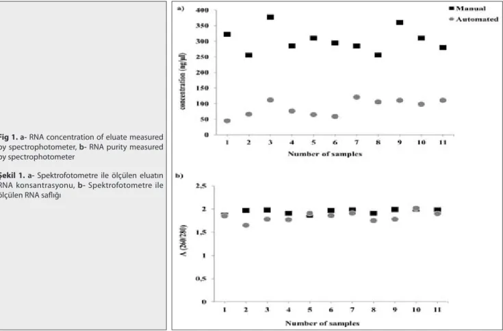
Supernatants of CPE-positive cultures (n=11) were used for the comparative analysis of RNA extraction and purification of the RNeasy Mini kit (Qiagen, Hilden, Germany) and the MagNA Pure LC 2.0 system (Roche Applied Science, Indianapolis, IN, USA). The quantity of RNA obtained with manual extraction was significantly (P<0.0001) higher than automated extraction. Samples that were manually extracted displayed a mean concentration of 303.3±38.7
Fig 1. a- RNA concentration of eluate measured
by spectrophotometer, b- RNA purity measured by spectrophotometer
Şekil 1. a- Spektrofotometre ile ölçülen eluatın
RNA konsantrasyonu, b- Spektrofotometre ile ölçülen RNA saflığı
ng/µl (range = 250-377.6 ng/µl) whereas extracts prepared by the automated extraction showed a mean concentration of 88±26.1 ng/µl (range 45-120.3 ng/µl; Fig. 1a).
Furthermore, the quality of extracts was assessed by comparing the 260/280 ratios of RNAs. The mean value of the manual extracts was 1.94±0.04 (range=1.87-2.0), and the automated extracts displayed a mean value of 1.78±0.23 (range=1.12–2.02; Fig. 1b).
The ratio of the absorbance at 260 and 280 nm (A260/280) was significantly different between manual and automated techniques (P=0.0327). Next, the ratio of the absorbance at 260 and 230 nm (260/230), used as a secondary measure of nucleic acid purity, ratio was determined. RNA extracted with both methods showed ratios below the optimum (manual: 1.16±0.09; automated: 1.7±0.4; P=0.0003).
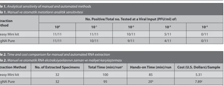
Analytical Sensitivity of PPRV RT-PCR after
Extraction of RNA by Manual and Automated Methods The sensitivity of PPRV detection by RT-PCR after extraction by manual and automated methods was compared (Table 1).
All CPE-positive samples (n=11) at 100 PFU/ml were
detected by RT-PCR after manual and automated extraction, but no replicates of PPRV at 10-4 PFU/ml were detected by
RT-PCR after extraction by all two methods. None of the negative controls was positive by RT-PCR after manual and automated extraction.
PPRV Detection in Clinical Specimens after Extraction by Manual and Automated Methods In order to compare the efficacies of PPRV RNA extraction from different clinical specimens, tissue samples (including lung, liver, spleen and mesenterial lymph node) were extracted by each method and PPRV RT-PCR was performed with using F and N gene specific primers. The
results of RT-PCR (based upon the PPRV F and N genes) were concordant in 30 of 32 samples that were extracted by all two methods (9 of 32 positive for PPRV RNA; 21 of 32 PPRV RNA were not detected). PPRV RNA was weakly detected in the two samples that were extracted manually whereas not detected by automated method.
Time and Cost Analysis
The amount of time required for sample processing and costs were compared for both manual and automated methods (Table 2).
The times required for extraction of a comparable number of samples by the manual RNeasy and MagNA Pure methods were equivalent. However, the actual hands-on time was less for the automated compared to the manual extraction method. MagNA Pure reagents for RNA extraction were more expensive than RNeasy Mini kit.
DISCUSSION
Peste des petits ruminants is one of the important viral disease of small ruminants, and has been detected in all regions of Turkey since it was first officially reported in 1999 [18,19]. Different types of tests are available for
diagnosis of PPR such as virus isolation, ELISA and RT-PCR. Virus isolation is not routinely available in diagnostic laboratories because of time consuming. RT-PCR methods are recommended by OIE for confirmation of clinical cases. Therefore, RT-PCR methods have been commonly used to diagnose PPR in many diagnostic laboratories in Turkey.
Extraction is the first step in RT-PCR methods. Extracted nucleic acid concentration and purity are important to obtain reliable results. In recent years, different manual and automated extraction methods are used for diagnosis of diseases. In this study, we assessed RNA extracts generated Table 1. Analytical sensitivity of manual and automated methods
Tablo 1. Manuel ve otomatik metotların analitik sensitivitesi
Extraction Method
No. Positive/Total no. Tested at a Viral Input (PFU/ml) of:
100 10-1 10-2 10-3 10-4
RNeasy Mini kit 11/11 11/11 10/11 5/11 0/11 MagNA Pure 11/11 10/11 9/11 4/11 0/11
Table 2. Time and cost comparison for manual and automated RNA extraction Tablo 2. Manuel ve otomatik RNA ekstraksiyonlarının zaman ve maliyet karşılaştırması
Extraction Method No. of Extracted Specimens Total Time (min)/runa Hands-on Time (min)/run Cost (U.S. Dollars)/Sample
RNeasy Mini kit 32 100 85 5.31
MagNA Pure 32 95 20b 7.89c
a Total time includes hands-on time, b Hands-on time for the MagNA Pure does not include time necessary to homogenize stored tissue samples, c List price for
by the manual (Qiagen, Hilden, Germany) and automated extraction (Roche Applied Science, Indianapolis, IN, USA) methods.
Our data demonstrate that manual and automated methods specificities were identical at 100%, and manual extraction’s analytical sensitivity of PPRV detection by RT-PCR was better than the automated extraction (Table
1). These results are consistent with those of Riemann
et al.[17] who reported that quantity and quality of the
generated DNAs were slightly higher using the manual extraction method. However, Knepp et al.[20] reported that
analytical sensitivity of enterovirus detection by RT-PCR is similar after RNA extraction by manual and automated methods. A possible explanation for this result may be the differences between kits which were used for manual extraction. In this study, we used RNeasy Mini kit (Qiagen, Hilden, Germany), but they used QIAamp Viral RNA kit (Qiagen, Hilden, Germany) for manual extraction. For both extraction methods no false positive results were obtained when negative controls were tested. These findings in agreement with previous reports [20-22], but seems contrary
to previous study that reported false positive results were obtained by using MagNA Pure system [23]. Possible
explanation for this result may be contamination happened during the pipetting steps of the extraction protocol.
Furthermore, in the present study the yield of manually prepared RNAs was 344% higher than the yield of automated extracts when the procedures were performed according to the supplier’s manual. Similarly, the purity of manually extracted RNAs was closer to the optimum value compared to RNAs produced by the MagNA Pure. However, the 260/230 ratios determined for both methods are far below the optimum. Remaining salts in the eluate usually account for these low values. Ionic strength is known to influence the absorbance of nucleic acids, especially the absorbance at 260 nm [24]. It may be explain why low
260/230 ratios obtained by MagNA Pure. Since Magna Pure LC total nucleic acid isolation kit contains high ionic strength buffers.
Discordant results of PPRV RT-PCR were observed from two samples that were previously demonstrated to contain PPRV by virus isolation. Two samples extracted with the MagNA Pure failed to generate fragments. The lack of concordance appears to correlate with the effectiveness of the extraction technique (as defined by sensitivity studies above). Studies with serially diluted PPRV demonstrated a trend of higher sensitivity after extraction by manual method.
In this study, PPRV RNA was detected by F and N gene based RT-PCR methods. It has been reported that N gene based primers are more sensitive than F gene based primers [6]. However, we obtained consentient results
between F and N gene based RT-PCR methods. All CPE-positive samples were found CPE-positive with both methods.
Also, no false positive results were obtained when negative controls were tested.
In our experience, when we used manual and automated methods for direct extraction from tissue samples, we obtained much and pure nucleic acid (especially RNA) concentrations by manual method than automated method. We suggest that magnetic beads in automated methods influence the concentration of nucleic acids. In automated methods, magnetic beads coated with nucleic acids (DNA or RNA), and these stuck nucleic acids can’t fully separated from magnetic beads. Therefore, obtained concentrations of nucleic acids are low.
A major concern in the implementation of manual methods to extract nucleic acid for use in amplification assays is the potential for contamination. We did not observe any contamination after extraction by RNeasy Mini kit. Likewise, MagNA Pure reduce the chances of contamination of samples. MagNA Pure provides an integrated tip guard to prevent dripping by the tips and UV sterilization between runs.
From a cost perspective, the MagNA Pure and RNeasy Mini kit extraction differed minimally at approximately $2.5/sample each. Less hands-on time and the fact that approximately two to three times the number of specimens can be processed at once make the MagNA Pure a real alternative for larger sample preparations, even though the cost per sample is higher than that for RNeasy extractions.
Nowadays, manual and automated extraction methods can be used for detection of PPRV RNA from field samples. Automated extraction methods minimize the potential sample contamination compared to the manual methods. Also, they demanded much less hands-on time than the manual methods. The first and most important step in molecular diagnosis of PPRV infection is the nucleic acid isolation. From our data, it was concluded that both RNA extraction methods (manual; 11/32, automated; 9/32) demonstrated similar performance, with no significant difference (P=0.7879). However, manual extraction performed slightly better analytical sensitivity, by 5%, than the automated extraction (Table 1). Accordingly, our results suggest that manual extraction is suitable for RNA extraction when small numbers of tissue samples needed to be examined.
REFERENCES
1. Lefevre PC, Diallo A: Peste des petits ruminants virus. Rev Sci Tech Off
Int Epiz, 9, 951-965, 1990.
2. Anderson J, McKay JA: The detection of antibodies against peste
des petits ruminants virus in cattle, sheep and goats and the possible implication to rinderpest control programme. Epidemiol Infect, 112, 225-231, 1994.
3. Gibbs EPJ, Taylor WP, Lawman MPJ, Bryant J: Classification of peste
des petits ruminants virus as a fourth member of the genus Morbillivirus.
4. Barrett T, Visser IKG, Mamaev L, Goatley L, Bressem MF, Van Osterhaus ADM: Dolphin and porpoise morbilliviruses are genetically
distinct from phocine distemper virus. Virology, 193, 1010-1012, 1993.
5. Ozkul A, Akca Y, Alkan F, Barrett T, Karaoglu T, Dagalp SB, Anderson J, Yesilbag C, Cokcaliskan C, Gencay A, Burgu I: Prevalence, distribution,
and host range of peste des petits ruminants virus, Turkey. Emerg Infect
Dis, 8, 708-712, 2002.
6. Kerur N, Jhala MK, Joshi CG: Genetic characterization of Indian peste
des petits ruminants virus (PPRV) by sequencing and phylogenetic analysis of fusion protein and nucleoprotein gene segments. Res Vet Sci, 85, 176-183, 2008. DOI: 10.1016/j.rvsc.2007.07.007
7. Munir M, Zohari S, Saeed A, Khan QM, Abubakar M, LeBlanc N, Kanu S, Sankoh FA, Berg M, Barrie ML, Ståhl K: Genetic characterization
of peste des petits ruminants virus, Sierra Leone. Emerg Infect Dis, 18, 193-195, 2012. DOI: 10.3201/eid1801.111304
8. Luka PD, Erume J, Mwiine FN, Ayebazibwe C, Shamaki D: Molecular
characterization and phylogenetic study of peste des petits ruminants viruses from North central states of Nigeria. BMC Vet Res, 7, 32, 2011. DOI: 10.1186/1746-6148-7-32
9. Shaila MS, Shamaki D, Forsyth MA, Diallo A, Goatley L, Kitching RP, Barrett T: Geographic distribution and epidemiology of peste des petits
ruminants viruses. Virus Res, 43, 149-153, 1996. DOI: 10.1016/0168-1702(96)01312-3
10. Wang Z, Bao J, Wu X, Liu Y, Li L, Liu C, Suo L, Xie Z, Zhao W, Zhang W, Yang N, Li J, Wang S, Wang J: Peste des petits ruminants virus in Tibet,
China. Emerg Infect Dis, 15, 299-301, 2009. DOI: 10.3201/eid1502.080817
11. Kwiatek O, Ali YH, Saeed IK, Khalafalla AI, Mohamed OI, Obeida AA, Abdelrahman MB, Osman HM, Taha KM, Abbas Z, Harrak M, Lhor Y, Diallo A, Lancelot R, Albina E, Libeau G: Asian Lineage of Peste des
Petits Ruminants Virus, Africa. Emerg Infect Dis, 17, 1223-1231, 2011. DOI: 10.3201/eid1707.101216
12. Munir M, Zohari S, Saeed A, Khan QM, Abubakar M, LeBlanc N, Berg M: Detection and phylogenetic analysis of peste des petits
ruminants virus isolated from outbreaks in Punjab, Pakistan. Transbound
Emerg Dis, 59, 85-93, 2012. DOI: 10.1111/j.1865-1682.2011.01245.x
13. De Nardi M, Lamin Saleh SM, Batten C, Oura C, Di Nardo A, Rossi D: First evidence of peste des petits ruminants (PPR) virus circulation
in Algeria (Sahrawi Territories): Outbreak investigation and virus lineage identification. Transbound Emerg Dis, 59, 214-222, 2012. DOI:
10.1111/j.1865-1682.2011.01260.x
14. OIE: Peste des petits ruminants. In, OIE (Ed): Manual of Diagnostic
Tests and Vaccines for Terrestrial Animals. Chapter 2.7.11. http://www. oie.int/fileadmin/Home/eng/Health_standards/tahm/2.07.11_PPR.pdf, Accessed: 06/09/2014.
15. Forsyth MA, Barrett T: Evaluation of polymerase chain reaction for
the detection and characterisation of rinderpest and peste des petits ruminants viruses for epidemiological studies. Virus Res, 39, 151-163, 1995. DOI: 10.1016/0168-1702(95)00076-3
16. Couacy-Hymann E, Roger F, Hurard C, Guillou JP, Libeau G, Diallo A: Rapid and sensitive detection of peste des petits ruminants virus by
a polymerase chain reaction assay. J Virol Methods, 100, 17-25, 2002. DOI: 10.1016/S0166-0934(01)00386-X
17. Riemann K, Adamzik M, Frauenrath S, Egensperger R, Schmid KW, Brockmeyer NH, Siffert W: Comparison of manual and automated
nucleic acid extraction from whole-blood samples. J Clin Lab Anal, 21, 244-248, 2007. DOI: 10.1002/jcla.20174
18. OIE: OIE Disease Information. 12, 137, 1999.
19. Sevik M: Molecular Detection of Peste des petits ruminants virus
from different organs/tissues of naturally infected animals. Kafkas Univ
Vet Fak Derg, 20, 165-168, 2014. DOI: 10.9775/kvfd.2013.9706
20. Knepp JH, Geahr MA, Forman MS, Valsamakis A: Comparison of
automated and manual nucleic acid extraction methods for detection of enterovirus RNA. J Clin Microbiol, 41, 3532-3536, 2003. DOI: 10.1128/ JCM.41.8.3532-3536.2003
21. Marshall JA, Bruggink LD: Laboratory diagnosis of norovirus. Clin
Lab, 52, 571-581, 2006.
22. Kurar E, Atlı MO, Guzeloglu A, Ozsensoy Y, Semacan A: Comparison
of five different RNA isolation methods from equine endometrium for gene transcription analysis. Kafkas Univ Vet Fak Derg, 16, 851-855, 2010.
23. Chranioti A, Aga E, Margari N, Kottaridi C, Pappas A, Panayiotides I, Karakitsos P: Performance evaluation of manual and automated
(MagNA pure) nucleic acid isolation in HPV detection and genotyping using Roche Linear Array HPV Test. Infect Dis Obstet Gynecol, 2011: 931281, 2011. DOI: 10.1155/2011/931281
24. Wilfinger WW, Mackey K: Chomczynski P: Effect of pH and ionic
strength on the spectrophotometric assessment of nucleic acid purity.