Olgu Sunumu
A Rare Cause Of Intrathorasic Mass In An
Asymptomatic Child
ASEMPTOMATİK BİR ÇOCUKTAKİ NADİR BİR İNTRATORASİK KİTLE NEDENİ
Duygu ÖLMEZ
1, Arzu BABAYİĞİT
1, Nevin UZUNER
1, Özlem TÜFEKÇİ
2, Handan ÇAKMAKÇI
3,
Dilek TEZCAN
1, Özkan KARAMAN
11 Department of Pediatrics Allergy, Dokuz Eylül University, Medical School 2 Department of Pediatrics, Dokuz Eylül University, Medical School 3 Department of Radiology, Dokuz Eylül University, Medical School
Duygu ÖLMEZ
Dokuz Eylül Üniversitesi Tıp Fakültesi
Çocuk Sağlığı ve Hastalıkları AD Allerji Bilim Dalı
35340 İnciraltı, İZMİR Tel: 0-232-4123660/ 3661 Fax: +90 232 2599723
e-mail: [email protected]
SUMMARY
Pulmonary sequestration is an uncommon usually cystic mass of nonfunctioning primitive tissue that does not communicate with the tracheobronchial tree that receives it’s blood supply from an anomalous systemic artery rather than the pulmonary arteries. Intralobar pulmonary sequestration is usually diagnosed later in childhood or adulthood when the patient presents with pulmonary infection. Many patients with extralobar pulmonary sequestration present during infancy with pulmonary respiratory distress and chronic cough. Here, we reported a case of extralobar pulmonary sequestration found coincidentally on chest radiograph in a nine year old girl during routine preoperative investigations for insertion of tympanostomy tube.
Key words: Pulmonary sequestration, extralobar, children ÖZET
Pulmoner sekestrasyon trakeobronşiyal ağaçla ilişkisi olmayan ve kan akımını pulmoner arterler yerine anormal bir sistemik arterden sağlayan, fonksiyon göstermeyen primitif dokudan oluşmuş, nadir görülen, genellikle kistik bir kitledir. İntralober pulmoner sekestrasyon genellikle geç çocukluk çağı veya erişkin çağda hasta enfeksiyon ile başvurduğunda tanınır. Ekstralober pulmoner sekestrasyonlu hastaların çoğu infant döneminde solunum sıkıntısı ve kronik öksürük ile başvurur. Burada, timpanostomi tüpü yerleştirilmek üzere rutin preoperatif tetkikler yapılırken akciğer filminde tesadüfen ekstralober pulmoner sekestrasyon saptanan dokuz yaşındaki bir kız olguyu sunduk. Anahtar sözcükler: Pulmoner sekestrasyon, ekstralober, çocuklar
Pulmonary sequestration is an uncommon usually cystic mass of nonfunctioning primitive tissue that does not communicate with the tracheobronchial tree and that receives its blood supply from an anomalous systemic artery (1).
Pulmonary sequestrations are divided into two types on the basis of their pleural coverings. Extralobar pulmonary sequestrations (ELS) are masses of lung parenchyma that have a distinct pleural covering
maintaining complete anatomical separation of the mass from adjacent normal lung tissue. In contrast, intralobar sequestrations (ILS) are masses of lung parenchyma that are contiguous with the adjacent normal lung (2). ILS accounts for 75% of sequestrations and ELS accounts for 25% (3). Incidence is equal in males and females in intralobar type, but ELS is found more commonly in males (80% of cases) (4). We report a rare case of ELS found accidentally on chest roentgenogram in a nine year old
© 2006
DEÜ
TIP FAKÜLTESİ DERGİSİ CİLT 20, SAYI 2, (MAYIS) 2006, S: 109 - 112109
A cause of intrathoracic mass in a child
girl during routine preoperative investigations for insertion of tympanostomy tube.
CASE REPORT
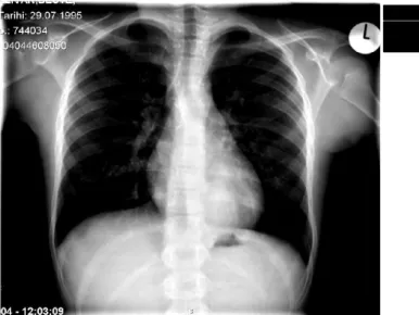
A nine year old girl was referred to our hospital with a complaint of hearing loss in the left ear. She had recurrent otitis media and tympanocentesis was performed to the left ear a year before. She had no complaints regarding lower respiratory system. On admission, physical examination findings were normal except the left serous otitis media that is unresponsive to medical treatment. Therefore, tympanostomy tube insertion was planned. During preoperative investigations, chest roentgenogram showed a dense opacity in the posterior basal segment of the left lower lobe (Fig 1). Complete blood count and biochemical analysis were normal. Contrast enhanced computerized tomography (CT) imaging of the lung revealed a mass of 3x4x4 cm consistent with ELS that is paravertebrally located in the left lower lobe. Arterial supply was directly from aorta (Fig 2). Surgical intervention was planned but consent could not be obtained from parents. The patient is stil on follow up without any symptoms for a year.
DISCUSSION
Pulmonary sequestration can be described as a segment of the lung that has no identifiable communication with the normal bronchial tree (2). It’s incidence is 0.5 - 6.4% (4). Many theories about the embriyology of pulmonary sequestration have been proposed. The theory most widely accepted suggests that ELS results from the formation of an accessory lung bud caudal to the normal lung buds (5,6).
ILS is usually diagnosed later in childhood or adulthood when the patient presents with pulmonary infection (4). A communication with other bronchi or lung parenchyma may exist allowing for pulmonary infection to occur. More than half the cases of ELS are diagnosed during infancy. Pulmonary infection is less common in
ELS and the child may be asymptomatic except for an intrathoracic mass as accurred in our case (7). ELS may also be diagnosed in the first six months of life with respiratory distress and feeding difficulties (3,8).
ILS are not associated with other cardio-pulmonary anomalies, but ELS may be found in association with cardiac, or more frequently, diaphragmatic anomalies in 50% of cases (9,10). ILS demonstrates no preferences for either lung whilst ELS is found in the left lung in 80% of cases (4). In our patient, lesion was detected in the left lower posterior lobe but there was no co-existing congenital anomaly.
In ELS, the systemic arterial supply is typically via the thoracic aorta or the abdominal aorta (>80%); however, the arterial supply can be via pulmonary (5%) and another systemic artery (15%). Venous drainage is usually via the azygos or hemiazygous veins or the inferior vena cava (>80%), although the subclavian and portal veins are rarer options. In approximately 25% of patients, venous drainage is at least partially via the pulmonary veins (2).
Figure 1. Chest radiograph showing dense opacity in posterior basal segment of the left lower lobe
Olgu Sunumu
Figure 2. Computerized tomography showing left paravertebral mass (ELS)When correlated with clinical presentation, chest radiographs can provide a reasonable diagnostic clue to pulmonary sequestration but most are too small to be detected with chest radiography (11). Contrast enchanced thorax CT is usually sufficient for the diagnosis of pulmonary sequestration but may not accurately identify the aberrant arterial supply and venous drainage (12,13). The multi-planar nature of magnetic resonance imaging demonstrates parenchymal lung changes as well as anatomic localisation of the systemic arterial supply and venous drainage of a pulmonary sequestration (14).When non-invasive imaging fails, conventional angiography still has a place in the evaluation of pulmonary sequestration (3,11). Diagnosis was established with contrast enhanced
thorax CT, there fore no further evaluation was necessary.
Some authors advocate that extirpation is the only reasonable approach for pulmonary sequestrations even in asymptomatic cases because of the risk of pulmonary infection, hemorrhage and malignant transformation (4,15). Management of an asymptomatic ELS is controversial. Resection of all ILS and subdiaphragmatic ELS have been recommended (16). Some auther advocate that an asymptomatic non-cystic ELS within the thorax can be simply observed (17). Anatomical resections like lobectomy, segmentectomy and in the case of ELS, sequestrectomy is possible. The crucial part of the surgical procedure is identification and control of the aberrant vascular supply (2).
© 2006
DEÜ
TIP FAKÜLTESİ DERGİSİ CİLT 20, SAYI 2, (MAYIS) 2006, S: 109 - 112111
A cause of intrathoracic mass in a child
Surgical intervention was planned for our patient because of the afore mentioned risks, but consent could not be obtained from parents.
REFERENCES
1.
Kravitz RM. Congenital malformations of the lung. Pedi-atr Clin North Am 1994; 41: 453-472.2.
Corbett HJ, Humphrey GM. Pulmonary sequestration. Paediatr Respir Rev 2004; 5: 59-68.3.
Savic B, Birtel FJ, Tholen W, Funke HD, Knoche R. Lung sequestration: report of seven cases and review of 540 published cases. Thorax 1979; 34: 96-101.4.
Halkic N, Cuenoud PF, Corthesy ME, et al. Pulmonary sequestration: a review of 26 cases. Eur J Cardiothorac Surg. 1998; 14: 127-133.5.
Clements BS, Warner JO. Pulmonary sequestrations and related bronchopulmonary-vascular malformations: no-menclature and classification bases on anatomical and embryological considerations. Thorax 1987;42: 401–408.6.
Langston C. New concepts in the pathology of congenital lung malformations. Semin Paediatr Surg 2003;12:17–37.7.
Krummel TM. Congenital malformations of the lowerrespiratory tract. In: Chernick V, Boat TF, Kendig EL, editors. Kendig’s Disorders of the Respiratory Tract In Children. 6th ed. Philadelphia: WB Saunders Company; 1998; 287-328.
8.
Stocker JT. Sequestrations of the lung. Semin Diagn Pathol 1986; 3: 106-121.9.
Avishai V, Dolev E, Weissberg D, Zajdel L, Priel IE. Extralobar sequestration presenting a massive hemotho-rax. Chest 1996; 109: 843-845.10.
Cukier A, Kavakama J, Teixeira LR, Terra-Filho M, Vargas FS. Scimitar sign with normal pulmonary venous drainage and systemic arterial supply. Chest 1994; 105: 294-295.11.
John PR, Beasley SW, Mayne V. Pulmonary sequestra-tion and related congenital disorders. A clinico-radio-logical review of 41 cases. Pediatr Radiol 1989; 20: 4-9.12.
Ko SF, Ng SH, Lee TY, et al. Noninvasive imaging ofbronchopulmonary sequestration. AJR Am J Roentgenol 2000; 17: 1005-1012.