Effect of cytokine genes in the pathogenesis and on the clinical parameters for the treatment of multiple myeloma
Tam metin
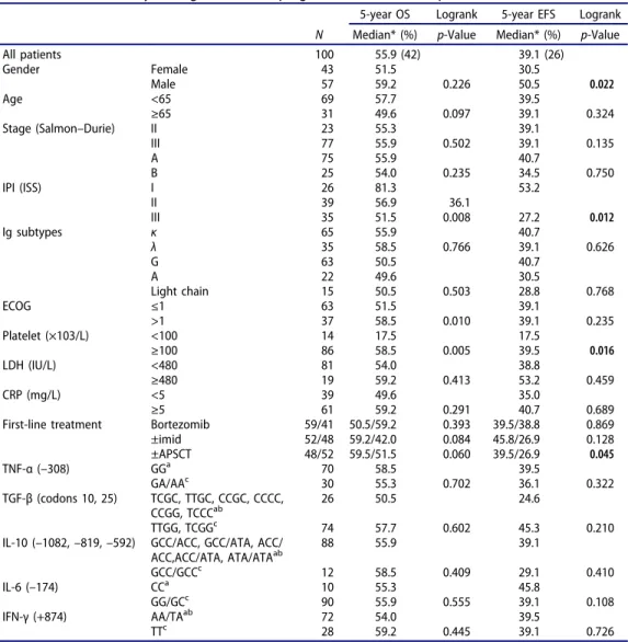
Şekil
Benzer Belgeler
In this study, there were no significant differences between various clinical states of HBV infection (asymptomatic car- rier or chronic infection) and anti-HBs positive
Bu çalışma kapsamında meslek yüksekokulu öğrencilerinin giysilerin yıkama ve bakım etiketleri ile ilgili görüşleri incelenmiş ve konu ile ilgili
Uygulama ile be- lirlediğiniz senaryolara göre dünya üzerinde hangi yere, hangi güçte bir bomba atıldığında ne kadar insanın ölece- ği, hangi bölgelerin
In this prospective study which was conducted to evaluate the effects of etanercept, a TNF-α inhibitor, on endothelial functions in patients with active RA, it was found
Background: This study aims to evaluate the effects of transforming growth factor-b3 and neutralizing antibody of transforming growth factor-b1 containing polymeric
This study aimed to assess inflammatory marker tumor necrosis factor-alpha (TNF-α), adiponectin (a modulator of anti-inflam- mation), and potential microvascular markers for
The Sequencing of the Insulin-like Growth Factor 1 and Fibulin 5 Gene Variants in the Pre and Post-menopausal Women with Stress Urinary Incontinence.. It can cause depression and
assessed the efficacy of biolo- gical agents in nail psoriasis in a prospective study allocated TNF inhibitor treatment for psoriasis or psoriatic arthritis with severe nail
