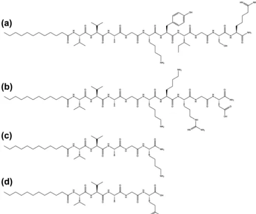
Bioactive self-assembled peptide nanofibers for corneal stroma regeneration
Tam metin
Şekil


Benzer Belgeler
*Students will express what they completed during their CAS activities to find links between the CORE subjects and their DP subjects *Understanding how to make inks to CAS
The next chapter presents a detailed review of literature on pragmatic competence in foreign language education, instruction of speech acts in EFL classes, limitations of
Corrosion behaviour of a new inhibitor (FT2000) is investigated in saline solution on a low carbon steel in the neutral aqueous media at 608C.. Effect of sulphate ion is
Su et al., A facile and sensitive fluorescent sensor using electrospun nanofibrous film for nitroaromatic explosive detection. Lin et al., Electrospun nanofibers of
Israel’s occupation of Palestinian and other Arab territories; the ‘unresolved’ status of Jerusalem; the ‘perceived’ acquiescence of Western governments in the Israeli
Modeling of Wire Electro Discharge Grinding (WEDG) Process ... General Effects of WEDG Process Parameters .... Spindle Speed ... Feed Length and Feed Rate ... Process
(3) As far as the biosensors in this work is considered, using dielectric layer walls for immobilization does not change the volume displaced even if the undercut is
