Case
repor
t
Congenital Hypoplasia of the Posterior Arch of the
Atlas: Case Report and Extensive Review of the
Literature
Atlasın Arka Yayının Doğumsal Gelişim Bozukluğu: Olgu Sunumu ve
Literatürün Geniş Gözden Geçirimi
Hakan SABUncUoGlU1, Selcuk oZDoGAn1, Demet kArADAG2, Erdener TImUrkAynAk2 1Ufuk University, School of Medicine, Department of Neurosurgery, Ankara, Turkey
2Ufuk University, School of Medicine, Department of Radiology, Ankara, Turkey
Correspondence address: Hakan SABuNCuOGLu / E-mail: [email protected]
ABSTRACT
Congenital hypoplasia of the posterior arch of the atlas (C1), a developmental failure of chondrogenesis, is a rare anomaly and may range from partial clefts to total agenesis of the posterior arch. Ossification of the posterior arch usually occurs between the 3rd and 5th years of life. The incidence of posterior arch anomalies of the atlas is between 0.69% and 2.95%. For the evaluation of the patient, cervical lateral plain radiography, 2D or 3D reconstructed CT and MRI are very useful and important tools in initial diagnosis. Surgery is the treatment of choice in symptomatic compression. Excision of the posterior arch is performed. during surgery. After the surgery, patients may be followed up for instability and treated as necessary. A patient, admitted to the emergency department with head and neck trauma after a traffic accident is presented in this article. C1 hypoplasia was determined after detailed imagining studies and the radiology department consulted. When upper cervical anomalies are found in a young patient, the patient should be evaluated in detail with advanced radiological studies to avoid misinterpretation as fractures, luxation, osteolysis or instability. Consulting a radiologist could help making an accurate diagnosis and deciding on current therapeutic interventions.
KeywOrds: Atlas, Hypoplasia, Posterior, Arch, Congenital, Anomaly
ÖZ
Kondrogenezdeki doğumsal bozukluk sonucu oluşan atlasın arka yayının gelişimsel eksikliği nadir bir anomalidir ve kısmi yarık oluşumundan atlasın arka yayının tam yokluğuna kadar gidebilir. Arka yayın kemikleşmesi 3-5 yaşlar arasında olur. Atlasın arka yayı anomalilerinin insidansı %0.69-2.95 arasındadır. Hastayı değerlendirmek için servikal lateral direkt grafi, 2 ve 3 boyutlu rekonstrüksiyonlu bilgisayarlı tomografi ve magnetik rezonans inceleme çok yararlı ve önemli tanı araçlarıdır. Semptomatik bası varlığında cerrahi bir tedavi seçeneğidir. Cerrahide arka yayın eksizyonu uygulanmaktadır. Cerrahi sonrasında hastalar instabilite için takip edilmeli ve gerektiğinde tedavi edilmelidir. Bu makalede, trafik kazası sonrası acil servise baş ve boyun travması ile gelen bir hasta sunulmaktadır. Detaylı radyolojik çalışmalar sonrası, atlas hipoplazisi saptanmış ve sonrasında hasta radyoloji bölümüne konsülte edilmiştir. Genç hastada üst servikal anomaliler saptandığında; kırık, luksasyon, osteoliz ve instabilite hasta ayrıntılı yanlış yorumlardan kaçınmak için hasta radyolojik incelemelerle ayrıntılı değerlendirilmelidir. Radyoloji uzmanına yapılan konsultasyon kesin tanıya ve doğru tedavi edici girişimlerin yapılması için karar verilmesine yardım olmaktadır.
AnAhtAr sÖZCÜKler: Atlas, Hipoplazi, Arka, Yay, Konjenital, Anomali
InTRoduCTIon
Congenital hypoplasia of the posterior arch of the atlas (C1), a developmental failure of chondrogenesis, is a rare anomaly and may range from partial clefts to total agenesis of the posterior arch (12,21). These abnormalities were considered by some to be a “benign variation” and indeed, almost all of them are discovered incidentally (3,4,11,13). In general, such abnormalities are asymptomatic but neurological symptoms may occur after minor cervical trauma in patients with isolated partial agenesis of the posterior arch of the C1 (2,12,13,28). Clinical features vary from mild neck pain to neurological
deficits occurring after minor cervical spine or head injury (29,31).
We report a patient with hypoplasia of the posterior arch of the C1 who was admitted to our emergency department for head trauma without any neurological deficit.
CASE REPoRT
A previously healthy, right handed, 26-year-old male patient was admitted to our emergency department with head trauma after traffic accident. The patient suffered from headache and neck pain. The general and cranial neurological examination
Sabuncuoglu H, et al: Posterior Arch of the Atlas
Figure 4: Three-dimensional reconstructed CT image of the C1 right posterolateral view. Note the hypoplasia of C1 (black arrow).
was unremarkable. There were no motor or sensory deficits. All reflexes were normal and plantar reflexes were downgoing. No clinical deterioration was found with neck flexion and extension. All the laboratory tests were normal. Lateral cervical spine radiograph revealed two hemiarches of C1 with a hypoplasia in the midline (Figure 1). Cervical 2D and 3D reconstructed computed tomography (CT) studies showed hypoplasia of the posterior arch of the C1 (Figure 2, 3A-B, 4). The patient was hospitalized for a night. A magnetic resonance
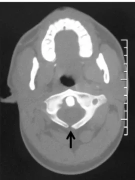
Figure 2: Axial CT image reveals hypoplasia of C1 (arrow).
Figure 3: Three-dimensional reconstructed CT image of C1: A) Upper B) Posterior view shows hypoplasia of C1. White arrow in Fig 3A and yellow arrow in Fig 3B.
A B
Figure 1: Lateral cervical spine radiograph shows two posterior hemiarch of C1 (arrows).
imaging (MRI) study was performed the following day and it also revealed the partial absence of the posterior arch of the C1 (Figure 5). MRI study did not show any neural alteration or other associated abnormalities, such as protrusion of disc material, compression of spinal cord, basilar invagination, Chiari malformation, a syringomyelic cavity or narrowing of the sagittal diameter at the level of C1(Figure 6). The patient was managed conservatively with medical therapy and a cervical collar and his neck pain subsided after a week. He
remained asymptomatic when he was last examined a year later.
dISCuSSIon
Understanding the embryology of the first and second vertebrae is the key to understanding developmental anomalies of the upper spine. The C1 derives from the primitive fourth occipital and first cervical sclerotomes. It has three primary ossification centers in the embryological period: an anterior center for formation of the anterior tubercle and two lateral centers from which the lateral masses and posterior arch form (23). In 7th week of gestation, the lateral centers extend dorsally to form the posterior arch (Figure 7). An additional center may be present posteriorly in the midline, forming the posterior tubercle of the C1 in second year of life in about 2% of the population (3,6,7,15,17,27,29). During maturation, the anterior arch of the C1 derives from usually one midline, but occasionally two or more ossification centers are seen radiologically in the first year of life. Ossification of the posterior arch usually proceeds perichondrally from two centers in the lateral masses, towards the midline ,and fusion occurs between the 3rd and 5th years of life; incomplete fusion of the two hemiarches may be normal in the first 5-10 years (9). However, incomplete fusion may persist in 3 to 5% of patients. The anterior center usually fuses with the two lateral centers between 5 and 9 years age (23). In rare cases, a separate ossification center forms in the midline and fuses secondarily with the lateral masses forming the posterior arch; a posterior midline ossified tubercle develops if this center fails to fuse with the hemiarches (9). At least two different anomalies can develop during the ossification: 1) median clefts of the posterior arch of C1, and 2) varying degrees of posterior arch dysplasia (6,17,29).
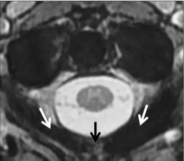
Figure 5: Axial T2-weighted MR image. The hypoplasia (black arrow) and two posterior hemiarch (white arrows) of C1 is hardly seen on the MR image. The spinal canal and cord appears normal.
Figure 6: Sagittal T2-weighted MR image. There is no spinal cord compression.
Figure 7: The lateral ossification centers of atlas vertebrae (arrows) extend dorsally to form the posterior arch in an 8-week-old human embryo. These centers fuse on the midline posteriorly to form the posterior arch of atlas (with the permission of M. Hill, Ph.D.: http://embryology.med.unsw.edu.au/wwwhuman/ lowpower/HumC/C3L.htm)
Sabuncuoglu H, et al: Posterior Arch of the Atlas
Different theories have been proposed to explain congenital defects of the C1 posterior arch (2,11,18,22,23,24,31). However, the exact mechanism remains obscure. Congenital absence or hypoplasia of the posterior arch of the C1 may be associated with several disorders, such as the Arnold Chiari malformation, gonadal dysgenesis and the Klippel-Feil, Down and Turner syndromes respectively (3,15,16,29). Martich et al. found hypoplasia of the posterior arch in 26% of 38 children with Down syndrome. If hypoplasia of the C1 is found in a young patient, the authors recommend ruling out Down syndrome, gonadal dysgenesis, achondroplasia or Turner syndrome (4,10,14,29,30). It is commonly accepted that defects of the posterior arch of the C1 are due to a lack of chondrification rather than a primary disturbance of ossification (2,9). The reported incidence in a large study of 1613 autopsies with regard to presence of congenital aplasia is 4% for the posterior arch and 0.1% for the anterior arch (16). It has been reported that hypoplasia of the posterior arch of C1 may increase the risk of atlantoaxial subluxation in about 26% of children aged 2-3 years (14,16). Patient presented in the literature are predominantly children or women in the second or third decade of life (1,16,24). In Martich’s study, clefts of the posterior arch were found in 45 of 1613 cadaveric dissections, 97% of which were median clefts (14). Şenoğlu et al. reported their findings which are obtained from 1104 patients by cervical spine CT scans: 37 patients had posterior arch defects, and of these, 29 (2.6%) had median clefts. Therefore, type A defects represented 78.4% of all posterior arch defects. Furthermore, they determined such defects in 2 (1.2%) of 166 dried C1 specimens and 1 (1.19%) of 84 fresh human cervical segments (23). The exact incidence of posterior arch anomalies of C1 is unknown but according to Currarino’s study, the incidence is 0.69% (2). In Şenoglu’s study, the overall incidence (CT scan + dried specimens + fresh specimens) of the posterior atlantal arch in 1354 evaluated cases is 2.95% (40 cases). On the other hand, the anterior arch defects are even less common than posterior and the rate is reported as 0.1% in several studies (7,10,14,23,29).
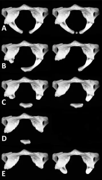
A complete classification of congenital anomalies of the posterior arch of the C1 is proposed by Currarino et al (2). Plaut and Lawrence & Anderson first published an article about a developmental abnormality of C1 in 1937. A total of 41 articles have been published about posterior defects of C1 in the literature so far (Table I). In 1975, Richardson et al. were the first to propose a mechanism by which neurological deficits occur. They theorized that the isolated posterior fragment moved anteriorly and traumatized the dorsal spinal cord when, with extension, the distance between the occiput and the spinous process of the axis caused inward buckling of the ligaments (11,20). According to the classification of Currarino et al. there are five types of anomalies (Figure 8) (Table II): Type A defects are defined as failure of the posterior midline with a small gap remaining (that is, failure of the two lateral ossification centers to unite posteriorly in the midline); type B, unilateral clefts, ranging from a small defect to a complete absence of one hemiarch; type C, bilateral defects with preservation
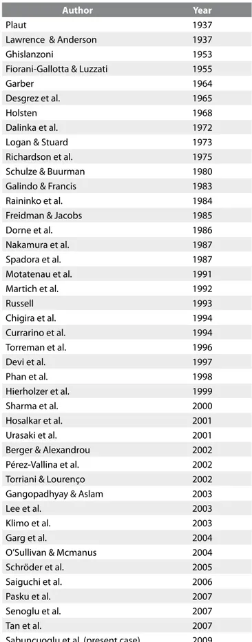
Table I: Whole Literature Analysis of C1 Hypoplasia Between Years 1937 and 2009
Author Year
Plaut 1937
Lawrence & Anderson 1937
Ghislanzoni 1953
Fiorani-Gallotta & Luzzati 1955
Garber 1964
Desgrez et al. 1965
Holsten 1968
Dalinka et al. 1972
Logan & Stuard 1973
Richardson et al. 1975
Schulze & Buurman 1980
Galindo & Francis 1983
Raininko et al. 1984
Freidman & Jacobs 1985
Dorne et al. 1986 Nakamura et al. 1987 Spadora et al. 1987 Motatenau et al. 1991 Martich et al. 1992 Russell 1993 Chigira et al. 1994 Currarino et al. 1994 Torreman et al. 1996 Devi et al. 1997 Phan et al. 1998 Hierholzer et al. 1999 Sharma et al. 2000 Hosalkar et al. 2001 Urasaki et al. 2001
Berger & Alexandrou 2002
Pérez-Vallina et al. 2002
Torriani & Lourenço 2002
Gangopadhyay & Aslam 2003
Lee et al. 2003
Klimo et al. 2003
Garg et al. 2004
O’Sullivan & Mcmanus 2004
Schröder et al. 2005
Saiguchi et al. 2006
Pasku et al. 2007
Senoglu et al. 2007
Tan et al. 2007
of the most dorsal part of the arch; type D , absence of the posterior arch with persistent posterior tubercle and type E, the complete absence of the posterior arch and posterior tubercle (2). The frequency of only type A is nearly 3-4% of individuals and these make up more than 90% of all posterior arch defects of C1 (2). This type is difficult to diagnose from lateral films of the cervical spine, and its diagnosis cannot be made with certainly in the first 5 to 10 years of life when two hemiarches of C1 may still be unfused normally (2). Patients with types C and D, have a free-floating posterior tubercle at the apex of the arch. It is hypothesized that the patient has both error of chondrification as well as the rare fourth ossification center to form this anomaly (11). A free-floating posterior tubercle is a problem in patients with C1 hypoplasia. This defect seems to suggest that the presence of an isolated posterior bony fragment somehow predisposes the patients to neurological morbidity. Richardson et al. tried to explain the symptoms of intermittent quadriplegia with compression of the cord by the inward mobility of the isolated posterior bony fragment during extension of the cervical spine (Figure 9) (20,24). Syrinx formation can be seen in these patients. This hypothesis supports the notion that the cord impingement occurred during extension, when the cord is expected to move superiorly in relation to the posterior arch. On the basis of the literature, the presence of an posterior isolated bony fragment is a potential risk factor for neurological morbidity rather than as an inconsequential normal variant (24). Curarino et al, have also subdivided the patients into five clinical groups: 1) asymptomatic incidental findings, 2) neck pain or stiffness after trauma to the head or neck, 3) chronic symptoms referable to the neck, 4) various chronic neurological problems, and 5) acute neurological symptoms after minor cervical trauma (2,16).
Figure 8: The figure shows the types of Curarino’s classification in hypoplasia of C1.
Figure 9: Spinal cord compression by the inward mobility of the isolated posterior bony fragment (arrows) can lead to some neurological symptoms in the patient. The presence of an posterior isolated bony fragment is a potential risk factor for neurological morbidity.
Table II: Currarino’s Morphologic Classification for C1 Hypoplasia
Type description
A Failure of posterior midline fusion of the two hemiarches
B Unilateral defect
C Bilateral defects
D Absence of posterior arch, with persistent posterior tubercle
Sabuncuoglu H, et al: Posterior Arch of the Atlas
Cervical lateral plain radiography, 2D or 3D reconstructed CT and MRI are very useful and important tools for evaluation of the patient. Congenital anomalies of the craniovertebral junction are common incidental findings on cervical plain radiography; however, some may cause confusion when routine radiographs show apparently abnormal findings. These anomalies should to be kept in mind to avoid misinterpretation as fractures, luxation or osteolysis (2,5,8,9,19). Cervical lateral plain graphy with flexion and extension views may help to detect cases with mechanical instability where there is increased risk of spinal cord injury and determine the mobility of the isolated tubercle (21,24,26). CT also provides excellent contrast between the nonossified and ossified portions of the arch of C1, which can enable precise determination of the extent of the abnormality (29). Additionally CT scans can be particularly useful for smaller defects and 2D or even 3D reconstruction might display better the topography of the upper cervical spine, especially to clinicians (9). MRI study has advantages in evaluating the spinal cord, adjacent neural structures and paraspinal soft tissue (29). Although MRI has not been performed in most of the case reports due to absence of neurological symptoms, an MRI study is needed in some symptomatic cases to determine the secondary changes within the spinal cord (2,16,29). These changes possibly represent focal myelomalacia, cord edema or a presyrinx state (12). It is possible that cord compression could have been directly visualized had the MRI study been performed with the neck extended (24).
Our patient may be classified as type A, clinical subgroup 1, according to Currarino’s morphologic and clinical classification (2). Absence of ossification of the posterior arch of the C1 is asymptomatic and is generally detected as an incidental finding like our patient, although it may cause neck pain in some patients (29). Autopsies and surgical explorations have shown that dense fibrous bands bridge the bony gaps and account for good stability of the upper cervical spine. In the latter, hypoplasia of the posterior arch of C1, which may increase the risk of atlantoaxial subluxation, was seen in 26% of 38 children aged 2-3 years (16,29). Compensatory hypertrophy of the anterior arch of C1 and of the spinous process of C2 is usually found (15,29). Other coexisting abnormalities, including clefts of the anterior arch, atlantoaxial rotator subluxation, anterior atlantoaxial subluxation, downward projection of the posterior border of the foramen magnum and cervical myelopathy are reported in the literature (29). This anomaly can also stimulate basilar invagination, Jefferson’s fracture and occipitalization of the C1 (15,25,29).
Congenital hypoplasia of the C1 with posterior tubercle can be managed conservatively or by surgical resection. Patients with this anomaly should be advised to avoid contact sports. Currarino et al., describing a variety of neurological problems, observed that is often not clear whether the symptoms are in fact attributable to C1 defect, but recommended that certain sports should be avoided if such defects are shown (2,9). Due to previous case reports and review of the literature,
type C and D should undergo surgery at an early stage to prevent cumulative damage to the cord (24). Surgery is the treatment of choice in symptomatic compression. Excision of the posterior arch is curative. Patients may be followed up for instability and treated as necessary (4). Posterior fusion is a common surgical procedure in cases of atlantoaxial instability, provided the posterior elements of both vertebrae are intact. Posterior fusion involves the occiput and lower spinal segments, limiting the mobility of the upper cervical spine, if the posterior arch of C1 is abnormal (29).
In conclusion, most congenital anomalies of upper spine are determined incidentally in asymptomatic patients. Once it has been found in young patient, the patient should be evaluated in detail with advanced radiological studies to avoid misinterpretation as fractures, luxation, osteolysis or instability. Consulting a radiologist could help making an accurate diagnosis and deciding on current therapeutic interventions.
REFEREnCES
1. Atasoy C, Fitoz S, Karan B, Erden I, Akyar S: A rare cause of cervical spinal stenosis: Posterior arch hypoplasia in a bipartite atlas. Neuroradiol 44:253-255, 2002
2. Currarino G, Rollins N, Diehl JT: Congenital defects of the posterior arch of the atlas: A report of seven cases including an affected mother and son. AJNR Am Neuroradiol 15:249-254, 1994 3. Dalinka MK, Rosenbaum AE, Van Houten F: Congenital absence
of posterior arch of atlas. Radiology 103:581-583, 1972
4. Devi BI, Shenoy SN, Panigrahi MK, Chandramouli BA, Das BS, Jayakumar PN: Anomaly of arch of atlas – a rare cause of symptomatic canal stenosis in children. Pediatr Neurosurg 26:214-218, 1997
5. Dorne HL, Just N, Lander PH: CT recognition of anomalies of the posterior arch of the atlas vertebrae: Differentiation from fracture. AJNR 7:176-177, 1986
6. Duong DH, Chadduck WM: Reconstruction of the hypoplastic posterior arch of the atlas with calvarial bone grafts for posterior atlantoaxial fusion: Technical report. Neurosurgery 35(6): 1168-1170, 1994
7. Garg A, Gaikwad SB, Gupta V, Mishra NK, Kale SS, Singh J: Bipartite atlas with os odontoideum. Spine 29:35-38, 2004
8. Gehweiler JA, Daffner RH, Roberts L: Malformations of the atlas vertebra simulating the Jefferson fracture. AJR 140:1083-1086, 1983
9. Hierholzer H, Isalberti M, Hosten N, Stroszczynski C, Gandini G, Felix R: A rare, complex developmental anomaly of the atlas: embryological and radiological considerations. Neuroradiology 41:901-903, 1999
10. Hodak JA, Mamourian A, Dean BL: Radiologic evaluation of the craniovertebral junction, in Dickman CA, Spetzler RF, Sonntag VKH, eds. Surgery of the Craniocervical Junction. New York: Thieme,1998:81-102
11. Klimo P Jr, Blumenthal DT, Couldwell WT: Congenital partial aplasia of the posterior arch of the atlas causing myelopathy: Case report and review of the literature. Spine 28:224-228, 2003 12. Lee HC, Hsieh PC, Cho DY: Atlas hypoplasia combined with
atlantoaxial subluxation is a cause of non-traumatic cervical myelopathy. Mid Taiwan J Med 8:99-104, 2003
13. Logan WW, Stuard ID: Absent posterior arch of the atlas. Am J Roentgenol Radium Ther Nucl Med 118:431-434, 1973
14. Martich V, Ben-Ami T, Yousefzadeh DK, Roizen NJ: Hypoplastic posterior arch of C-1 in children with Down syndrome: A double jeopardy. Radiology 183:125-128, 1992
15. Menezes AH: Hypoplasia of C-1 in children with down syndrome. J Neurosurg (6 Suppl Pediatrics) 107:455-456, 2007
16. Pasku D, Katonis P, Karantanas A, Hadjipavlou A: Congenital posterior atlas defect associated with anterior rachischisis and early cervical degenerative disc disease: A case study and review of the literature. Acta Orthop Belg 73(2):282-285, 2007
17. Phan N, Marras C, Midha R, Rowed D: Cervical myelopathy caused by hypoplasia of the atlas: Two case reports and review of the literature. Neurosurgery 43(3):629-633, 1998
18. Piper JG, Traynelis VC: Congenital malformations of the craniovertebral junction, in Dickman CA, Spetzler RF, Sonntag VKH, eds. Surgery of the Craniocervical Junction. New York: Thieme,1998:123-149
19. Polga JP, Cramer CG: Cleft of anterior arch of atlas simulating odontoid fracture. Radiology 113:341-342, 1974
20. Richardson EG, Boone SC, Reid RL: Intermittent quadriparesis associated with a congenital anomaly of the posterior arch of the atlas. J Bone Joint Surg Am 57:853-854, 1975
21. Sagiuchi T, Tachibana S, Sato K, Shimizu S, Kobayashi I, Oka H, Fuji K, Kan S: Lhermitte sign during yawning associated with congenital partial aplasia of the posterior arch of the atlas. AJNR 27:258-260, 2006
22. Schulze PJ, Buurman R: Absence of the posterior arch of the atlas. AJR Am J Radiol 134:178-180, 1980
23. Senoglu M, Abbasi SS, Theodore N, Bambakidis NC, Crawford NR, Sonntag VKH: The frequency and clinical significance of congenital defects of the posterior and anterior arch of the atlas. J Neurosurg Spine 7:399-402, 2007
24. Sharma A, Gaikwad SB, Deol PS, Mishra NK, Kale SS: Partial aplasia of the posterior arch of the atlas with an isolated posterior arch remnant: finding in three cases. AJNR Am J Neuroradiol 21: 1167-1171, 2000
25. Smoker WR: Craniovertebral junction: Normal anatomy, craniometry and congenital anomalies. Radiographics 14(2): 255-277, 1994
26. Tan EC, Soon HC, Kevin M, Se To BC: Congenital absence of the posterior arch of the atlas – A strange but benign anomaly. Malaysian Orthopaedic Journal 1(2):30-32, 2007
27. Thompson GH, Likavec MJ, Archibald I, Rush T: Atlantoaxial rotatory subluxatiaon, congenital absence of the posterior arch of the atlas, and cerebral palsy: An unusual triad. J Pediatr Orthop 5(2):232-235, 1985
28. Torreman M, Verhagen IT, Sluzewski M, Kok AJ, Van Rooij WJ: Recurrent transient quadriparesis after minor cervical trauma associated with bilateral partial agenesis of the posterior arch of the atlas. J Neurosurg 84:663-665, 1996
29. Torriani M, Lourenco JL: Agenesis of the posterior arch of the atlas. Rev Hosp Clin Fac Med Sao Paulo 57:73-76, 2002
30. Urasaki E, Yasukouchi H, Yokota A: Atlas hypoplasia manifesting as myelopathy in a child . Neurol Med Chir 41:160-162, 2001 31. Vallina JRP, Galan IR, Ruisanchez AC, Arango GOR, Muniz CL,
Martinez JMF: Congenital anomaly of craniovertebral junction: Atlas-Dens fusion with C1 anterior cleft. Journal of Spinal Disorders and Techniques 15(1):84-87, 2002