Reprod Dom Anim. 2020;55:421–428. wileyonlinelibrary.com/journal/rda © 2020 Blackwell Verlag GmbH
|
4211 | INTRODUCTION
Embryo transfer is widely used to improve reproductive perfor-mance, to increase the number of animals with high genetic charac-teristics and to form high-yielding herds (Bó et al., 2012; Hasler, 2010, 2014). In large-scale bovine embryo transfer programmes, many fac-tors have been identified affecting the success and widespread use
of this application (Ferraz et al., 2016; Hasler, McCauley, Lathrop, & Foote, 1987; Vieira et al., 2014). In particular, the differences in superovulation response (Bó & Mapletoft, 2014) and the variation in pregnancy rates (Ferraz et al., 2016) adversely affect success of the embryo transfer programmes.
Transfer location in uterine horn, cervix transfer score (time for cervix passing or embryo transfer), developmental stage and quality Received: 19 November 2019
|
Accepted: 7 January 2020DOI: 10.1111/rda.13623
O R I G I N A L A R T I C L E
Evaluation of the factors that affect the pregnancy rates during
embryo transfer in beef heifers
Hasan Alkan
1| Tahir Karaşahin
2| Şükrü Dursun
3| Fatma Satılmış
1|
Hüseyin Erdem
1| Mehmet Güler
11Department of Obstetrics and
Gynaecology, Faculty of Veterinary Medicine, Selcuk University, Konya, Turkey
2Department of Physiology, Faculty of
Veterinary Medicine, Aksaray University, Aksaray, Turkey
3Department of Obstetrics and
Gynaecology, Faculty of Veterinary Medicine, Aksaray University, Aksaray, Turkey
Correspondence
Hasan Alkan, Department of Obstetrics and Gynaecology, Faculty of Veterinary Medicine, Selcuk University, Konya, Turkey. Email: [email protected]
Abstract
The aim of this study was to evaluate the effects of the transfer side, transfer loca-tion, cervix transfer score, type and diameter of corpus luteum (CL) during embryo transfer on pregnancy rates in beef heifers. Progesterone-based synchronization and superovulation protocol were applied to Simmental cows used as donors (n = 168). Uterine flushings were performed on day 7 following artificial insemination. Obtained Code I (excellent or good) and II (fair) quality embryos were transferred to recipient beef heifers (n = 561). During embryo transfer, side of transfer (right or left), transfer location (the cranial or middle third of uterine horn), cervix transfer score (easy, mod-erate or difficult) and type (CLa, CLb and CLc) and diameter of CL were determined. Pregnancy rates following the transfer of Code I and II embryos were 44.66% and 33.07%, respectively (p < .05). The rates of pregnancy after transfers to the right and left uterine horn were 37% and 42.2%, respectively (p > .05). The pregnancy rates were 41.2%, 34.9% and 30.3% for cervix transfer scores as easy, moderate and dif-ficult, respectively (p > .05). Pregnancy rates after transfer to the cranial third and middle third were 41.06% and 29.67%, respectively (p < .05). According to types of CL, pregnancy rates were 31.7%, 40.4% and 45.3% for CLa, CLb and CLc, respec-tively (p < .05). Moreover, it was found that as the CL diameter increased, the preg-nancy rates increased. As a result, it was concluded that there was no effect of side of transfer and cervix transfer score, but embryo quality, transfer location, type and diameter of CL had significant effects on the pregnancy rate during embryo transfer in beef heifers.
K E Y W O R D S
of embryos, season, parity of recipient (heifer or cow), fresh or frozen transfer, and nutritional status are among the factors affecting preg-nancy rates in previously reported embryo transfer studies ( Bényei et al., 2006; Ferraz et al., 2016; Leroy et al., 2005; Roper et al., 2018). Many factors that affect the pregnancy rates after embryo transfer cause an increase in the application costs and lead to discussions about the success of the programme. If higher pregnancy rates are achieved by recipients, more efficient use of embryos would occur and financial savings could be realized through reductions in costs associated with the transfer of embryos and maintenance of non-pregnant recipients (Looney, Nelson, Schneider, & Forrest, 2006). The aim of this study was to determine the effects of transfer location and side, cervix transfer score, type and diameter of corpus luteum (CL) during embryo transfer on pregnancy rates in beef heifers.
2 | MATERIALS AND METHODS
The study was conducted with the approval and permission of the Ethics Committee of Selcuk University Faculty of Veterinary Medicine, Experimental Animals Production and Research Center (Approval Number: 2018/99). This study was conducted between the months of September and December.
2.1 | Donors and recipients
In this study, 2.5- to 4-year-old, body condition score of 2.5–3.5, and at postpartum 60–150 days, Simmental breed cows (n = 168) were used as donors. Beef heifers, 16–20 months of age, were used as re-cipients (n = 561). Genital organs of donor cows and recipient heifers were examined rectally and ultrasonographically, and cows/heifers with corpus luteum on ovaries and without any problems in uterus and cervix were selected as donors and recipients.
2.2 | Production of embryos
The progesterone-based protocol was applied to the donors for the synchronization of oestrus. The progesterone source (1.38 g, Eazi-Breed CIDR, Zoetis) was placed intravaginally, and a GnRH analogue (10 μg, Buserelin Acetate, Receptal, MSD) was injected intramuscularly (day 0). The donors were treated with 500 μg FSH (Stimufol, Reprobiol) intramuscularly as 8 decreasing doses starting from day 7 to 10. On
day 9, PGF2α (25 mg, Dinoprost Tromethamin, Dinolytic, Zoetis) was
injected intramuscularly in the morning and progesterone source was removed in the evening. Cows were then artificially inseminated at 36 and 48 hr after the progesterone source was removed. On day 7 after artificial insemination, non-surgical uterine flushing procedure was per-formed during upper epidural anaesthesia. The flushed uterine mate-rial was taken onto petri dishes and scanned under a stereomicroscope (Leica S8 APO). The assessment of embryo quality was made according to morphological integrity. Code I (excellent or good) corresponded to
very low levels of irregularity between the cells, a ratio of >85% viable embryonic cells and a round and unfolded zona pellucida. Code II (fair) is characterized by a medium level of irregularity between the cells and a viable cell ratio of 50%. Code III (poor) is characterized by irregularities in the form of the embryo and a viable cell ratio of 25%. Code IV (dead or degenerated) is embryos with oocytes or dead cells with uncom-pleted division. According to these criteria, Code I and Code II embryos were considered to be of transferrable quality (Bó & Mapletoft, 2013).
2.3 | Synchronization of recipients
The recipient heifers were synchronized with Ovsynch protocol.
Firstly, Kamar heatmount detector (Kamar®, INC., Steamboat Springs,
CO) was attached to the rump region on all recipients. GnRH (day 0),
PGF2α (day 7) and second GnRH (day 9) were administered
intramus-cularly. After the second injection of GnRH, heifers were monitored for
1–3 days and heifers with colour change in Kamar® recorded as
stand-ing oestrus. The heifers were examined again rectally and
ultrasono-graphically on the day when the colour change is detected in Kamar®.
Heifers with oestrous findings (cervical mucus, uterine tone and graff follicle) were identified as possible recipient candidates.
2.4 | Embryo transfer to the recipients
On the day of embryo transfer, candidate heifers with previously detected oestrous findings were re-examined by ultrasonography (6.0 MHz linear probe, Falcovet, Pie Medical, Netherlands). During this examination, heifers with showed oestrus previously and had a CL larger than 15 mm in ovary were used as recipients (n = 561). Transfer of embryos was performed during upper epidural anaesthesia (4 ml of 2% lidocaine solution). A total of 561 embryos classified as Code I (excellent or good) and II (fair) were transferred by an experienced veterinarian. Transfers were made in the ipsilateral horn with the ovary that had a CL. The side of transfer (right or left) was recorded. Diameter of CL and type of CL (Purcell, Beal, & Gray, 2005; Scenna et al., 2007; Spell, Beal, Corah, & Lamb, 2001) were defined (Corpus luteum type: CLa = asym-metric, non-embedded in ovary and soft texture, CLb = non-embedded in ovarium, classic mushroom-like and with cavity and CLc: prominent papillae, embedded in the ovary and compact texture). During the em-bryo transfer, three groups were formed according to cervix transfer score: easy (<60 s), moderate (60–80 s) and difficulty (>80 s). In addition, the location was recorded for each transfer (cranial third or middle third of the uterine horn ipsilateral to the ovary with a CL).
2.5 | Statistical analyses
For the analysis of the data, SPSS 25 (IBM Corp. Released 2017. IBM SPSS Statistics for Windows, Version 25.0, Armonk, NY: IBM Corp.) statistical package program was used. For discrete and continuous variables, descriptive statistics (mean, standard deviation, median,
minimum value, maximum value and percentile) were given. In addition, the homogeneity of the variances, which is one of the prerequisites of parametric tests, was checked through Levene's test. The assumption of normality was tested via the Shapiro–Wilk test. To compare the dif-ferences between three and more groups, one-way analysis of vari-ance was used when the parametric test prerequisites were fulfilled, and the Kruskal–Wallis test was used when such prerequisites were not fulfilled. The Bonferroni correction method, which is a multiple comparison test, was used to evaluate the significant results concern-ing three and more groups. Chi-square test was used for determinconcern-ing the relationships between two discrete variables. When the expected sources were less than 20%, values were determined through the Monte Carlo simulation method in order to include such sources in analysis. Contingency coefficient was used to determine the degree of relationship. The relationship between two continuous variables was assessed by the Pearson correlation coefficient, and by the Spearman correlation coefficient when the parametric test prerequisites were not met. In this study, binary logistic regression analysis was used to reveal the model of the relationship between independent variables and de-pendent variables. For the significance level of the tests, p < .05 and p < .01 were accepted.
3 | RESULTS
In this study, 298 and 263 Code I and II quality embryos were obtained, respectively. These embryos were freshly transferred to recipient heif-ers (n = 561). Pregnancy rates following the transfer of Code I and II quality embryos were 44.66% and 33.07%, respectively (p < .05).
Following the transfer of Code I embryos, the probability of pregnancy (odds ratio, OR) was found to be 1.39 (Table 1). There was no differ-ence in the pregnancy rate between the right and left uterine horn fol-lowing the transfer of 561 embryos (p > .05). However, the probability of pregnancy (OR, 1.27) was higher following the transfers to the left horn of the uterus (Table 1). The diameter and type of CL were found to have an effect on pregnancy rates (Table 1). There was a positive corre-lation between CL diameter and pregnancy rate (Table 2) and that the rate of pregnancy was higher in recipient heifers with larger diameter of CL (Table 3). The mean CL diameter was 2.11 ± 0.46 cm in pregnant heifers and 1.96 ± 0.34 cm in non-pregnant heifers (p < .05). The effect of the diameter of the corpus luteum on estimated probability of preg-nant (using data from pregnancy diagnosis on Day 30) in beef heifers was shown in Figure 1.
It was determined that heifers with CLc (OR: 1.67) and CLb (OR: 1.49) were more likely to become pregnant than those with CLa (Table 1; p < .05). When CL diameter and type were evalu-ated together, it was determined that CL diameter had no effect on pregnancy rate in heifers with CLc. In heifers with CLb type, the pregnancy rate increased as CL diameter increased. For CLa, the pregnancy rate increased when CL diameter was >2.50 cm (Table 4).
TA B L E 1 Pregnancy rates on the day 30 and odds ratio based on the transfer side, the cervix transfer score, the transfer location and type of CL
Not pregnant Pregnant Total
Pregnancy
rate (%) Odds ratio
95% confidence
interval p
Embryo quality
Code I (Excellent or good) 165 133 298 44.66 1.39 1.090–1.670 <.05
Code II (Fair) 176 87 263 33.07 Reference –
Transfer Side
Left 137 100 237 42.2 1.27 0.550–1.113 >.05
Right 204 120 324 37.0 Reference –
Cervix Transfer Score
Easy 254 178 432 41.2 1.49 0.438–1.355 >.05
Moderate 41 22 63 34.9 1.29 0.377–1.186
Difficulty 46 20 66 30.3 Reference
Transfer Location
Cranial third 277 193 470 41.06 1.37 0.473–1.266 <.05
Middle third 64 27 91 29.67 Reference –
Type of CL
CLa 129 60 189 31.7 Reference – <.05
CLb 102 69 171 40.4 1.43 0.923–2.220
CLc 110 91 201 45.3 1.67 1.100–2.548
TA B L E 2 Correlation between diameter of CL during embryo transfer and pregnancy rate on the day 30
Pregnancy rate
Diameter of CL (cm) 0.161*
It was determined that cervix transfer scores had no statistical ef-fect on pregnancy rates. However, it was found that the probability of pregnancy was higher when the cervix transfer score was easy (OR: 1.43) and moderate (OR: 1.29) compared to the difficult (Table 1).
The region of the uterine horn where the embryo was trans-ferred had an effect on the pregnancy rate (Table 1). Higher rate of pregnancy was detected when embryos were placed in cranial third (OR: 1.37) of the uterine horn (p < .05).
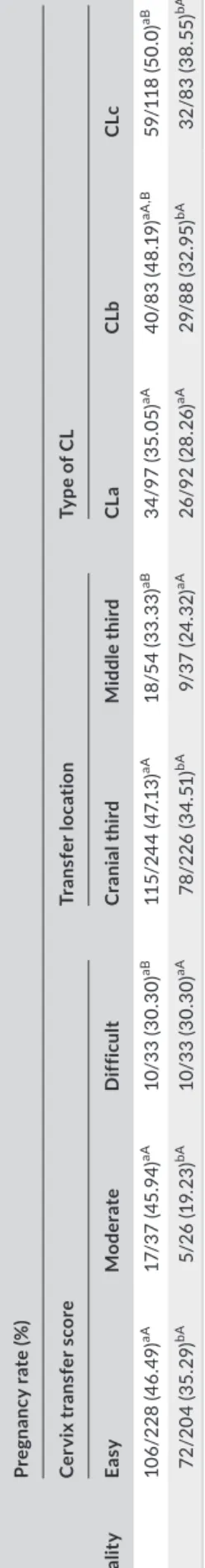
Pregnancy rates based on the relationship between embryo quality and cervix transfer score, transfer location and type of CL are given in Table 5. The rates of pregnancy were highest in recipients with cervix transfer score easy (46.49%) and moderate (45.94%) during the trans-fer of Code I quality embryos (p < .05). In addition, it was found that difficult passage of cervix during the transfer of Code I quality embryos decreased the pregnancy rates (p < .05). However, it was determined that the cervix transfer score had no effect on the pregnancy rate during the transfer of Code II quality embryos (p > .05). Moreover, the pregnancy rates following the transfer of Code I quality embryos to cranial third of uterine horn were found to be highest (47.13%, p < .05).
When pregnancy rates according to embryo quality and CL type were evaluated, the highest rates were obtained following the transfer of Code I quality embryos to heifers with CLb and CLc type corpus luteum (48.19% and 50.0%, respectively). Pregnancy rates according to the relationship between embryo quality and CL diam-eter are given in Table 6. It was ddiam-etermined that CL diamdiam-eter had no effect on pregnancy rates following the transfer of Code I quality embryos (p > .05). Following the transfer of Code II quality embryos,
pregnancy rates were higher in recipients with CL diameter larger than 2.5 cm compared to those with CL diameter less than 2.5 cm (p < .05). However, no statistically significant difference was found between the rates of pregnancy following transfer of Code I and II quality embryos to recipients with CL diameter larger than 2.5 cm (44.11% and 50.0%, respectively).
Pregnancy rates according to the interaction between cervix transfer score and transfer location are given in Table 7. When heifers with easy cervix transfer score were transferred to cranial third (43.50%) of the uterine horn, pregnancy rates were higher than those transferred to middle third (25.45%) (p < .05). In addi-tion, it was found that as the cervix transfer score became diffi-cult, the pregnancy rates decreased even if the embryo transfer location was cranial third (easy: 43.50%, moderate: 36.17% and difficult: 26.08%).
4 | DISCUSSION
The effects of certain factors on pregnancy rate following transfer of fresh in vivo produced bovine embryos were evaluated in the present study. To eliminate the variation due to animals, only beef heifers were used as recipients, each heifer was transferred only once, and exactly the same synchronization protocol was applied to all recipient heifers. In this study, it was determined that the side of transfer (right or left horn) and cervix transfer score (difficult, mod-erate or easy) had no statistically significant effect on the pregnancy Diameter of CL
(cm) Pregnant Not pregnant Total PR (%) p
1.5–2.00 58 115 173 33.52a <.01
2.00–2.50 134 194 328 40.85ab
>2.50 28 32 60 46.66b
a,b indicate differences in the columns, p < .05.
TA B L E 3 Pregnancy rates on the day 30 based on diameter of CL during embryo transfer
F I G U R E 1 Effect of diameter of the corpus luteum on estimated probability of conception (using data from pregnancy diagnosis on day 30) in beef heifers
T A B LE 4 Pr eg na nc y r at e o n t he d ay 3 0 b as ed o n d ia m et er a nd t yp e o f C L d ur in g e m br yo t ra ns fe r D ia m et er o f C L ( cm ) Ty pe o f C L CL a CL b CL c Pre gn an t N ot pr eg na nt To ta l PR ( % ) Pre gn an t N ot pr eg na nt To ta l PR ( % ) Pre gn an t N ot pr eg na nt To ta l PR ( % ) 1. 5–2 .0 0 18 42 60 30.0 aA 11 41 52 21 .1 5 aA 29 32 61 47. 64 aB 2. 00 –2. 50 34 76 11 0 30. 90 aA 50 56 10 6 47. 16 bB 50 62 11 2 44. 64 aB >2 ,5 0 8 11 19 42 .1 0 aA 8 5 13 61 .5 3 bA 12 16 28 42 .8 5 aA N ote : a-b: d iff er en ce s i n t he c ol um ns , A-B d iff er en ce s i n t he r ow s, p < .05 . T A B LE 5 Pr eg na nc y r at e o n t he d ay 3 0 b as ed o n r el at io ns hi p b et w ee n e m br yo q ua lit y a nd c er vi x t ra ns fe r s co re , t ra ns fe r l oc at io n a nd t yp e o f C L d ur in g e m br yo t ra ns fe r Emb ry o qu al it y Pr eg na nc y r at e ( % ) C er vi x t ra ns fe r s co re Tr an sf er loc at ion Ty pe o f C L Ea sy Mo der at e D iff icu lt C ra ni al thir d Mid dl e third CL a CL b CL c C od e I 10 6/ 22 8 ( 46 .4 9) aA 17/ 37 (4 5. 94 ) aA 10 /3 3 ( 30 .3 0) aB 11 5/ 24 4 ( 47. 13 ) aA 18 /5 4 ( 33 .3 3) aB 34 /97 (3 5. 05 ) aA 40 /8 3 ( 48 .1 9) aA ,B 59 /1 18 (50 .0) aB C od e I I 72 /2 04 ( 35 .2 9) bA 5/ 26 (1 9. 23 ) bA 10 /3 3 ( 30 .3 0) aA 78 /2 26 ( 34 .5 1) bA 9/ 37 (2 4. 32 ) aA 26 /9 2 ( 28 .26 ) aA 29 /8 8 ( 32 .9 5) bA 32 /8 3 ( 38 .5 5) bA N ote : a-b: d iff er en ce s i n t he c ol um ns , A-B d iff er en ce s i n t he r ow s, p < .05 .
rates following embryo transfer. However, the pregnancy rates (easy 41.2%, moderate 34.9% and difficult 30.3%) and the odds ratios (easy 1.49 and moderate 1.29, difficult reference) were higher in the recipients with easy access to the cervix during embryo transfer. In addition, difficult passage of cervix during transfer of Code I quality embryos decreased the rates of pregnancy (easy: 46.49%, moderate: 45.94%, difficult: 30.30%). Nevertheless, in heifers with an easy cer-vix transfer score, the highest pregnancy rates were found when the embryos were transferred to the cranial third of the uterine horn. Also, as cervix transfer score became easier, the rate of pregnancy increased in heifers with transfer location was cranial third. Aguiar, Araújo, Tirloni, and Martins (2013) and Lopes, Balbinot, Fonseca, Araújo, and Martins (2015) reported that the pregnancy rates in cows with difficult cervical passage (>80 s) during embryo transfer were lower compared to those with easy cervical passage (Aguiar et al., 2013; Lopes et al., 2015). In a previous study with Angus cross beef cows, it was reported that pregnancy rates were lower after embryo transfer when it was difficult to perform (transfer with maximum dif-ficulty, with extreme manipulation of the genital tract, with embryo deposited in the upper half of the uterine horn) (Kasimanickam et al., 2018). Roper et al. (2018) reported that longer duration of the transfer had a negative effect on pregnancy rate and that the preg-nancy rate decreased especially when the transfer took longer than 14 min. Excessive uterine manipulation during embryo transfer, dif-ficult passage of the cervix and prolonged application leads to the
release of chemical inflammatory mediators such as PGF2α. It was
reported that PGF2α may cause pregnancy losses by preventing the
survival and development of the embryo (Purcell et al., 2005; Scenna et al., 2005; Schrick, Inskeep, & Butcher, 1993).
In the present study, it was found that the quality of em-bryo during transfer had an effect on pregnancy rate (PR, Code I 44.66% and Code II 33.07%, p < .05). Only Code I and II quality embryos are considered as transferable (Bó & Mapletoft, 2013). Previous studies have reported that the quality of embryos af-fects pregnancy rates in embryo transfer programmes in cattle (Bényei et al., 2006; Hasler et al., 1987; Rodrigues et al., 2018). Ferraz et al. (2016) found that pregnancy rates following the transfer of Code I and II quality embryos were 42.2% and 32.8%, respectively. Bényei et al. (2006) and Rodrigues et al. (2018) re-ported that the rate of pregnancy following the transfer of Code I quality embryos was higher than Code II. As a result of this study, it was found that pregnancy rate decreases as embryo quality
decreases. The reason for this was thought to be the lower rate of viable cells with decreasing embryo quality. However, Code I embryos have a higher chance of survival than Code II quality embryos. Because Code I quality embryos are known to be more resistant to mortality. In addition, well-developed embryos pro-duce more interferon tau during the maternal recognition period of pregnancy and thus continue to develop by preventing PGF2a release (Carter et al., 2008; Mann, Fray, & Lamming, 2006; Mann & Lamming, 1999). Therefore, it is considered that the rates of pregnancy following the transfer of better-quality embryos was higher.
In this study, we determined that the region where the embryo was placed in the uterus (cranial or middle third of the uterine horn) had an effect on the pregnancy rates (cranial 41.06% and middle 29.67%, p < .05). In addition, when Code I quality embryos were transferred to cranial third of the uterine horn, the highest pregnancy rates were found. Previous studies reported that the embryo transfer to the cra-nial third of the embryo during the embryo transfer increased the preg-nancy rate (Beal, Hinshaw, & Whitman, 1998; Bó et al., 2012; Roper et al., 2018; Scenna et al., 2005). Normally the embryo reaches from the oviduct into the uterus approximately 6–8 days after fertilization (Bilodeau-Goeseels & Kastelic, 2003). Therefore, in uterine flushings on day 7, embryos are mostly collected from the cranial part of the uterine horn (Gordon, 2004; Niemann & Wrenzycki, 2012; Sponchiado et al., 2019). Therefore, it is recommended to transfer the embryos to the cranial part of the uterine horn. This practice is thought to increase pregnancy rates (Hasler, 2010; Siedel & Siedel, 1991).
According to the results of the present study, it was determined that diameter and type of CL had an effect on the pregnancy rates after embryo transfer. It was showed that the pregnancy rate in-creased as CL diameter inin-creased. The pregnancy rate was higher in the recipients with CLc type corpus luteum compared to recipients with other type corpus luteum. Furthermore, the highest pregnancy rates were found when Code I quality embryos were transferred to heifers with CLb and CLc type corpus luteum (CLb and CLc, 48.19% and 50.0%, respectively). Many studies have also reported that recip-ient cows with larger CL have higher pregnancy rates after embryo transfer (Ambrose et al., 1999; Baruselli et al., 2010; Vieira et al., 2014). Yoshida et al. (2012) reported that cows with a functional and larger CL after artificial insemination are more likely to become pregnant. However, there are also other reports that CL diameter does not have any effect on pregnancy rates (Bényei et al., 2006; Spell et al., 2001). TA B L E 6 Pregnancy rate (PR) on the day 30 based on diameter
of CL and embryo quality during embryo transfer Diameter of CL (cm) Embryo quality (PR, %) Code I Code II 1.5–2.00 32/88 (36.36)aA 26/85 (32.94)aA 2.00–2.50 86/176 (48.86)aA 48/152 (31.57)aB >2.50 15/34 (44.11)aA 13/26 (50.0)bA
Note: a-b: differences in the columns, A-B differences in the rows, p < .05.
TA B L E 7 Pregnancy rate (PR) on the day 30 based on cervix transfer score and transfer location during embryo transfer
Cervix transfer score
Transfer location (PR, %)
Cranial third Middle third
Easy 164/377 (43.50)aA 14/55 (25.45)aB
Moderate 17/47 (36.17)a,bA 5/16 (31.25)aA
Difficult 12/46 (26.08)bA 8/20 (40.0)aA
Previous studies have reported that larger CL structures maintain higher circulating progesterone concentrations in dioestrus (Ambrose et al., 1999; Baruselli et al., 2010). There is also a positive correlation between plasma progesterone concentration and the diameter of CL/ luteal tissue volume (Spell et al., 2001). Furthermore, Ambrose et al. (1999) defined a positive correlation between CL type and plasma progesterone concentrations. Although plasma progesterone concen-trations were not measured in this study, higher pregnancy rates in the recipient heifers with a more compact and larger CL may be due to higher circulating progesterone concentrations as stated above. Therefore, we concluded that embryo transfer after defining the CL diameter and type may provide advantages to further increase preg-nancy rates.
5 | CONCLUSION
The results of the present study showed that transfer side and cervix transfer score did not have a statistically significant effect on preg-nancy rates in beef heifers. However, embryo quality, the transfer location, diameter and type of CL were found to be effective on the pregnancy rates. Therefore, it can be concluded that the type and diameter of CL can be considered as an additional criterion for the selection of recipient animals.
ACKNOWLEDGEMENTS
The present study was supported by General Directorate of Agricultural Enterprises (TİGEM). The authors are thankful to the staff of Gozlu Agricultural Enterprises for assistance during the study.
CONFLIC T OF INTEREST
None of the authors have any conflict of interest to declare. AUTHOR CONTRIBUTIONS
H. Alkan and H. Erdem planned/designed the study and drafted the manuscript. H. Alkan, H. Erdem, T. Karasahin, S. Dursun, F. Satilmis and M. Guler took part in fieldwork. H. Alkan assisted the data analysis. All authors have read and approved the final version of the manuscript.
DATA AVAIL ABILIT Y
Data available on request from the authors. The data that support the findings of this study are available from the corresponding au-thor upon reasonable request.
ORCID
Hasan Alkan https://orcid.org/0000-0001-8332-5334
Tahir Karaşahin https://orcid.org/0000-0003-2358-0389
Şükrü Dursun https://orcid.org/0000-0002-2453-3464
Fatma Satılmış https://orcid.org/0000-0002-9877-8405
Hüseyin Erdem https://orcid.org/0000-0002-1416-5354
Mehmet Güler https://orcid.org/0000-0002-8040-9345
REFERENCES
Aguiar, T. S., Araújo, C. V., Tirloni, R. R., & Martins, L. R. (2013). Effect of meloxicam on pregnancy rate of recipient heifers following transfer of in vitro produced embryos. Reproduction in Domestic Animals, 48, 984–988. https ://doi.org/10.1111/rda.12197
Ambrose, J. D., Drost, M., Monson, R. L., Rutledge, J. J., Leibfried-Rutledge, M. L., Thatcher, M. J., … Thatcher, W. W. (1999). Efficacy of timed embryo transfer with fresh and frozen in vitro produced embryos to increase pregnancy rates in heat-stressed dairy cattle.
Journal of Dairy Science, 82, 2369–2376. https ://doi.org/10.3168/jds.
S0022-0302(99)75487-1
Baruselli, P. S., Ferreira, R. M., Filho, M. F. S., Nasser, L. F. T., Rodrigues, C. A., & Bó, G. A. (2010). Bovine embryo transfer recipient synchronisa-tion and management in tropical environments. Reproducsynchronisa-tion, Fertility
and Development, 22, 67–74. https ://doi.org/10.1071/RD09214
Beal, W. E., Hinshaw, R. H., & Whitman, S. S. (1998). Evaluating embryo freezing method and the site of embryo deposition on pregnancy rate in bovine embryo transfer. Theriogenelogy, 49(1), 241. https :// doi.org/10.1016/S0093-691X(98)90594-5
Bényei, B., Komlósi, I., Pécsi, A., Pollott, G., Marcos, C. H., de Oliveira Campos, A., & Lemes, M. P. (2006). The effect of internal and external factors on bovine embryo transfer results in a tropical environment.
Animal Reproduction Science, 93, 268–279. https ://doi.org/10.1016/j.
anire prosci.2005.07.012
Bilodeau-Goeseels, S., & Kastelic, J. P. (2003). Factors affecting em-bryo survival and strategies to reduce emem-bryonic mortality in cat-tle. Canadian Journal of Animal Science, 83, 659–671. https ://doi. org/10.4141/A03-029
Bó, G. A., & Mapletoft, R. J. (2013). Evaluation and classification of bo-vine embryos. Animal Reproduction, 10(3), 344–348. Retrieved from http://www.cbra.org.br/porta l/downl oads/publi cacoe s/anima lrepr oduct ion/issue s/downl oad/v10n3/ p344-348(AR628 ).pdf
Bó, G. A., & Mapletoft, R. J. (2014). Historical perspectives and recent re-search on superovulation in cattle. Theriogenology, 81, 38–48. https ://doi.org/10.1016/j.theri ogeno logy.2013.09.020
Bó, G. A., Peres, L. C., Mapletoft, R. J., Pincinato, D., Cutaia, L. E., & Baruselli, P. S. (2012). Treatments for the synchronisation of bovine recipients for fixed-time embryo transfer and improvement of preg-nancy rates. Reproduction, Fertility and Development, 24, 272. https :// doi.org/10.1071/RD11918
Carter, F., Forde, N., Duffy, P., Wade, M., Fair, T., Crowe, M. A., … Lonergan, P. (2008). Effect of increasing progesterone concentration from Day 3 of pregnancy on subsequent embryo survival and devel-opment in beef heifers. Reproduction, Fertility and Develdevel-opment, 20, 368–375. https ://doi.org/10.1071/rd07204
Ferraz, P. A., Burnley, C., Karanja, J., Viera-Neto, A., Santos, J. E. P., Chebel, R. C., & Galvão, K. N. (2016). Factors affecting the success of a large embryo transfer program in Holstein cattle in a commercial herd in the southeast region of the United States. Theriogenology, 86, 1834–1841. https ://doi.org/10.1016/j.theri ogeno logy.2016.05.032 Gordon, I. (2004). Reproductive technologies in farm animals (1st ed.).
Wallingford, UK: CABI Publishing. Retrieved from http://weekly. cnbne ws.com/news/artic le.html?no=124000
Hasler, J. F. (2010). Bovine embryo transfer: Are efficiencies improving? In Applied Reproductive Strategies Conference Proceedings.
Hasler, J. F. (2014). Forty years of embryo transfer in cattle: A review focusing on the journal Theriogenology, the growth of the industry in North America, and personal reminisces. Theriogenology, 81, 152– 169. https ://doi.org/10.1016/j.theri ogeno logy.2013.09.010 Hasler, J. F., McCauley, A. D., Lathrop, W. F., & Foote, R. H. (1987). Effect
of donor-embryo-recipient interactions on pregnancy rate in a large-scale bovine embryo transfer program. Theriogenology, 27, 139–168. https ://doi.org/10.1016/0093-691X(87)90075-6
Kasimanickam, R. K., Hall, J. B., Estill, C. T., Kastelic, J. P., Joseph, C. A., Abdel-Aziz, R. L., & Nak, D. (2018). Flunixin meglumine improves
pregnancy rate in embryo recipient beef cows with an excitable temperament. Theriogenology, 107, 70–77. https ://doi.org/10.1016/j. theri ogeno logy.2017.10.043
Leroy, J. L. M. R., Opsomer, G., De Vliegher, S., Vanholder, T., Goossens, L., Geldhof, A., … Van Soom, A. (2005). Comparison of embryo qual-ity in high-yielding dairy cows, in dairy heifers and in beef cows.
Theriogenology, 64, 2022–2036. https ://doi.org/10.1016/j.theri ogeno logy.2005.05.003
Looney, C. R., Nelson, J. S., Schneider, H. J., & Forrest, D. W. (2006). Improving fertility in beef cow recipients. Theriogenology, 65, 201– 209. https ://doi.org/10.1016/j.theri ogeno logy.2005.09.023 Lopes, L. M. J., Balbinot, M., Fonseca, B. A., de Araújo, C. V., & Martins, L.
R. (2015). Pregnancy rates and serum 13,14-dihydro-15-keto-PGF2α concentrations in recipient Nelore heifers treated with meloxicam after the transfer of invitro-produced embryos. Theriogenology, 84, 553–558. https ://doi.org/10.1016/j.theri ogeno logy.2015.04.010 Mann, G. E., Fray, M. D., & Lamming, G. E. (2006). Effects of time of
pro-gesterone supplementation on embryo development and interfer-on-τ production in the cow. The Veterinary Journal, 171, 500–503. https ://doi.org/10.1016/j.tvjl.2004.12.005
Mann, G. E., & Lamming, G. E. (1999). The influence of progesterone during early pregnancy in cattle. Reproduction in Domestic Animals,
34, 269–274. https ://doi.org/10.1111/j.1439-0531.1999.tb012 50.x
Niemann, H., & Wrenzycki, C. (2012). Animal Biotechnology 1, Reproductive
Biotechnologies.
Purcell, S. H., Beal, W. E., & Gray, K. R. (2005). Effect of a CIDR insert and flunixin meglumine, administered at the time of embryo trans-fer, on pregnancy rate and resynchronization of estrus in beef cattle.
Theriogenology, 64, 867–878. https ://doi.org/10.1016/j.theri ogeno
logy.2004.12.015
Rodrigues, M. C. C., Bonotto, A. L. M., Acosta, D. A. V., Boligon, A. A., Corrêa, M. N., & Brauner, C. C. (2018). Effect of oestrous synchrony between embryo donors and recipients, embryo quality and state on the pregnancy rate in beef cattle. Reproduction in Domestic Animals,
53(1), 152–156. https ://doi.org/10.1111/rda.13084
Roper, D. A., Schrick, F. N., Edwards, J. L., Hopkins, F. M., Prado, T. M., Wilkerson, J. B., … Smith, W. B. (2018). Factors in cattle af-fecting embryo transfer pregnancies in recipient animals. Animal
Reproduction Science, 199, 79–83. https ://doi.org/10.1016/j.anire
prosci.2018.11.001
Scenna, F. N., Hockett, M. E., Towns, T. M., Saxton, A. M., Rohrbach, N. R., Wehrman, M. E., & Schrick, F. N. (2005). Influence of a prosta-glandin synthesis inhibitor administered at embryo transfer on preg-nancy rates of recipient cows. Prostaglandins Other Lipid Mediators,
78, 38–45. https ://doi.org/10.1016/j.prost aglan dins.2005.02.003
Scenna, F. N., Munar, C. J., Mujica, I., Martin, E., Lafarga, P., Rajala-Schultz, P., & Schuenemann, G. M. (2007). Factors affecting pregnancy rate following timed embryo transfer program in cattle under field condi-tions. Reproduction, Fertility and Development, 21(1), 168–173. https ://doi.org/10.1071/RDv21 n1Ab145
Schrick, F. N., Inskeep, E. K., & Butcher, R. L. (1993). Pregnancy rates for embryos transferred from early postpartum beef cows into re-cipients with normal estrous cycles. Biology of Reproduction, 49, 617– 621. https ://doi.org/10.1095/biolr eprod 49.3.617
Siedel, G. E., & Siedel, S. M. (1991). Training manual for embryo transfer
in cattle (FAO Animal Production and Health). Retrieved from http://
www.fao.org/docre p/004/T0117 E/T0117 E00.htm
Spell, A. R., Beal, W. E., Corah, L. R., & Lamb, G. C. (2001). Evaluating recipient and embryo factors that affect pregnancy rates of em-bryo transfer in beef cattle. Theriogenology, 56, 287–297. https ://doi. org/10.1016/s0093-691x(01)00563-5
Sponchiado, M., Gonella-Diaza, A. M., Rocha, C. C., Turco, E. G. L., Pugliesi, G., Leroy, J. L. M. R., & Binelli, M. (2019). The pre-hatch-ing bovine embryo transforms the uterine luminal metabolite com-position in vivo. Scientific Reports, 9, 1–14. https ://doi.org/10.1038/ s41598-019-44590-9
Vieira, L. M., Rodrigues, C. A., Mendanha, M. F., Sá Filho, M. F., Sales, J. N. S., Souza, A. H., … Baruselli, P. S. (2014). Donor category and seasonal climate associated with embryo production and survival in multiple ovulation andembryo transfer programs in Holstein cattle.
Theriogenology, 82, 204–212. https ://doi.org/10.1016/j.theri ogeno
logy.2014.03.018
Yoshida, T., Seki, M., Watanabe, N., Furuta, H., Yoshimura, I., Osada, M., … Ushijima, H. (2012). Relation of reproductive perfor-mances and rectal palpation for luteum function of heifers 7 days
after estrus. Animal Science Journal, 83, 207–212. https ://doi.
org/10.1111/j.1740-0929.2011.00935.x
How to cite this article: Alkan H, Karaşahin T, Dursun Ş, Satılmış F, Erdem H, Güler M. Evaluation of the factors that affect the pregnancy rates during embryo transfer in beef
heifers. Reprod Dom Anim. 2020;55:421–428. https ://doi.