Turkish Propolis and Its Nano Form Can Ameliorate the Side Effects of Cisplatin, Which Is a Widely Used Drug in the Treatment of Cancer
Tam metin
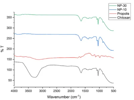
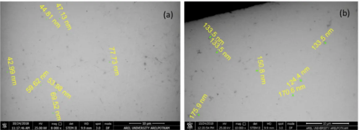
Şekil
Benzer Belgeler
In this study, demographic data were collected for each participant, including the Fitzpatrick skin type, the duration and severity of acne, the most recently prescribed treat-
Sevda Şener’in “Nâzım H ikm et’in Oyun Yazarlığı” adlı inceleme si geçtiğimiz günlerde okurla buluştu. Kitap, Nâzım’ın tiyatrosuna toplu bir bakışı
No significant difference was found between those living in the street and those living with their families in terms of the average number of offenses committed under and without
Orhan Karaveli, “Sakallı Celâl” (Pergamon Ya yını) adlı yapıtı neden, niçin, hangi amaçla yazdı, daha doğrusu iki yıl süren araştırmalarının sonu
In the present study, just as mentioned above, four patients in propofol group had deep hypotension and two of them; both was 80 years old, needed sedation termination and
The Mann-Whitney U test was used to investigate the relationship between valproate duration, dose, serum level and anemia, leukopenia, thrombocytopenia and macrocytosis,
He firmly believed t h a t unless European education is not attached with traditional education, the overall aims and objectives of education will be incomplete.. In Sir
Especially in male patients, I observed that when the treatment is given in summer, decreasing the drug dose to less than 0.5 mg/kg/day may increase patient tolerance..
