Effect of Chant Training on the Morphology of the Lateral
Thyrohyoid Ligament: A Biometric and Acoustic Assessment
*Meliha Berdan,†Emine Petekkaya, and*Ahmet Hilmi Y€ucel,*Adana, andyIstanbul, TurkeySummary: Objective. The purpose of this study was to determine the effects of chant training both on the morphologic structure of the lateral thyrohyoid ligament (LTL) and on the acoustic characteristics of the voice. Methods. Three groups of people participated in the study. Group I was new to chant training, group II had completed or was still continuing chant training, and group III, the control group, did not have any chant train-ing. For all participants, laryngeal cervical magnetic resonance imaging was performed to measure the right and the left LTLs. Additionally, vocal acoustic analyses were performed and compared with the anatomic morpho-metric measurements. Appropriate statistical assessments were performed to evaluate the measurements.
Results. The length of the LTL was greater in men, and thisfinding supports the gender-specific differences in laryn-geal structures. Anatomic differences between groups showed that 8 months of training was not sufficient to trigger morphologic changes. The left and right LTLs were asymmetric, but thisfinding was not statistically significant. Conclusions. Analyses revealed that vocal training causes morphologic changes in anatomic structures, which affect vocal quality.
Key Words: Right lateral thyrohyoid ligament−Left lateral thyrohyoid ligament−Metric analysis−Acoustic analysis−MRI.
INTRODUCTION
Voice plays a key role in everyday communication and social interactions. Singing is one of the most important dimensions of musical arts. Having just a“good voice” is not adequate for singing. Having both a healthy body and larynx is necessary for a high-quality voice. Many laryngeal structures change with voice training. Ligaments and membranes, which are the intrinsic elements of the larynx, contribute to laryngeal move-ments to produce voice. The thyrohyoid membrane, a wide fibroelastic lamina that resides between the thyroid cartilage and the hyoid bone, connects the larynx to the hyoid bone. This membrane allows the hyoid bone and the thyroid cartilage to move together during vocal production and swallowing. Singing changes the morphology of laryngeal ligaments and other anatomic laryngeal structures. The thyrohyoid mem-brane takes its banded shape by thickening at both sides of the larynx. This band reaches from the superior horns of the thy-roid cartilage to the greater horns of the hyoid bone. This por-tion is called the lateral thyrohyoid ligament (LTL).
The morphometric changes in the laryngeal anatomic struc-tures involved in speaking and singing are particularly impor-tant for the laryngologist to evaluate. The laryngologist will diagnose any phonatory diseases, determine the roles of these structures in phonation control, and plan surgery accord-ingly.1,2 Computerized tomography and magnetic resonance imaging (MRI) are widely used for the assessment of the larynx morphometry and in the follow-up of phonatory diseases.3
Metric analysis of these structures is preferred because stacking various morphologic forms, which are determined by morpho-metric measurements in imaging studies, over each other elimi-nates the need for a common reference plane. Metric analysis aims to define the anatomic structures quantitatively.4In the present study, we evaluated the metric analysis of the LTL using MRI and also obtained acoustic measures of the voice to determine the changes that voice training may have on laryn-geal anatomic structures.
METHODS
The present study included 16 men and 16 women between 17 and 35 years of age. Participants who previously took or were still taking vocal training at the time of the study were recruited from a group of opera and chant students. Those participants without voice training were students from the medical school. Group I was new to chant training, Group II had completed or was still continuing chant training, and Group III, the control group, did not have any chant training. Female and male distri-butions among the study groups were six of six in Group I,five offive in Group II, and five of five in the control group. The participants were matched according to gender, weight, and education level. The mean height of the women and men were 164.4 + 6.1 cm and 175.6 + 4.9 cm in the control group, 167.7 + 7.0 cm and 179.8 + 3.8 cm in Group I, and 164.8§ 3.9 cm and 178.2§ 5.1 cm in Group II, respectively. After the participants were informed about the study, laryngeal examinations were done in the Ear, Nose and Throat Department of the Faculty of Medicine of Cukurova University. Participants without any pathology regarding voice diseases were included in the study. One participant with a nodule on the left plica vocalis was referred for surgery and was excluded from the study.
Laryngeal cervical MRI examinations of 16 partici-pants were conducted at the Private Cukurova Guven Surgery Medical Center. The participants were instructed
Accepted for publication April 25, 2018.
From the*Department of Anatomy, Faculty of Medicine, Cukurova University, Adana, Turkey; and theyDepartment of Anatomy, Faculty of Medicine, Beykent University, Istanbul, Turkey.
Address correspondence and reprint requests to Emine Petekkaya, Department of Anatomy, The Faculty of Medicine, University of Beykent, Campus of Beylikd€uz€u, 34520, B€uy€uk¸cekmece, Istanbul, Turkey. E-mail:[email protected]
Journal of Voice, Vol. 33, No. 5, pp. 802.e17−802.e23 0892-1997
© 2018 The Voice Foundation. Published by Elsevier Inc. All rights reserved.
to take a normal breath during the MRI procedure. The study was approved by the Ethical Commission for Clini-cal Research in the MediClini-cal Faculty of Cukurova Univer-sity. The length of the LTL, which resides between the hyoid bone and the superior horn of the thyroid cartilage, was measured morphometrically between end points on the MRI by using Adobe Photoshop CS2 (Version 9.0) (Adobe, San Jose, CA) (Figures 1−6). Then, jitter, shim-mer, and fundamental frequencies of the voices of the par-ticipants were measured using Dr. Speech 6.2 ((Tiger DRS, Inc., Seattle, WA) voice analysis software at the Ear-Nose-Throat Department of Cukurova University Faculty of Medicine, Balcali Hospital. For the sample recordings, the participants produced an“a” vocalization for 5 seconds in a comfortable pitch. These procedures were performed in each group. The measurements were repeated after 8 months in each group.
Statistical analysis
Statistical comparisons between gender groups were performed with the Mann-Whitney U test, and comparisons between and within thefirst and the last measurements were performed with the Wilcoxon test by using the SPSS Statistical Software for Windows (Version 13.0) (IBM, Armonk, NY).
RESULTS
The mean ages of the women and the men were 20.4§ 1.1 years and 21.8§ 2.9 years in the control group, 18.7 § 1.8 years and 19.8§ 1 years in Group I, and 21.0 § 3.5 years and 30.0§ 6.9 years in Group II, respectively. Four men in the control group and three males and one female in Group II were smokers. As the number of the par-ticipants was low, smoking was not considered as an exclu-sion criterion (Table 1). Lengths of the LTLs were obtained from the cervical MRIs and measured biometrically on the first and the final images (Table 2). Results were compared within and between groups using the first and the last images (Tables 3−5). Both biometric measurements and acoustic vocal measurements between the initial and the final assessments within the control group did not reveal a significant difference. Because the participants in the control group had no voice training, we hypothesized that these similarities reflect the laryngeal anatomy when no vocal training has taken place.
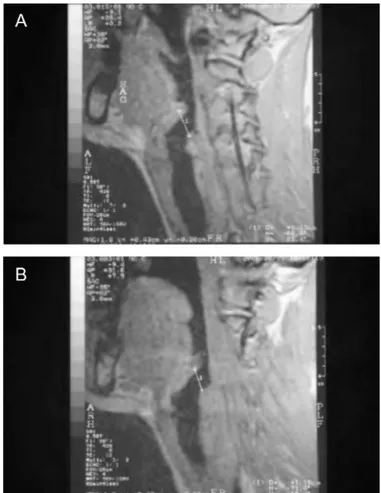
Comparisons of the results between the control group and Group I revealed a gender effect. The initial and the final measurements of the right LTL (P = 0.004) and the initial measurement of the left LTL were found to be shorter in the male participants in Group I than in the male participants in the control group (P< 0.05) (Table 3). Comparison of the FIGURE 1. MRI of a male participant in the control group shows
(A) the measurement of the right lateral thyrohyoid ligament and (B) the measurement of the left lateral thyrohyoid ligament.
FIGURE 2. MRI of a female participant in the control group shows (A) the measurement of the right lateral thyrohyoid ligament and (B) the measurement of the left lateral thyrohyoid ligament.
final measurement of the left LTL was not statistically sig-nificantly different.
The comparisons of female participants in the control group and in Group I revealed that initial andfinal jitter measure-ments were significantly lower in Group I than in the control group (P< 0.05). The within-group comparisons in these groups of female participants also showed that fundamental frequencies were significantly increased in the final measure-ments compared with the initial assessmeasure-ments (P = 0.028) (Tables 3−5). When it was considered that participants in Group I were at the beginning of their voice training, we con-cluded that this indicated the onset of changes in ligament size and vocal production. Nevertheless, our results also suggested that 8 months of vocal training was not sufficient for opera-chant training. The first measurement averages of the right LTL (mm) in the women in the control group and in Group I were statistically similar (P = 0.931). The comparisons between Groups I and II revealed that the lengths of the left LTL were significantly decreased in the final assessments of the female participants in Group II (P< 0.05) (Table 3). In addition, the female participants in Group II had significantly decreased fundamental frequencies in their final assessments. (P < 0.05) (Table 3). Because women in this group were taking voice training for a longer period, we hypothesize that the differen-ces were a result of the voice training. The comparisons
between the initial and thefinal assessments of the male partic-ipants in Group II revealed no statistically significant differ-ence (P> 0.05). Although men and women were taking the same training, we hypothesize that the lack of difference in the initial and the final measurements of the right and the left LTLs, and the acoustic parameters (including fundamental frequency, jitter, and shimmer) were due to the 60% smoking rate in the male participants in this group. Smoking negatively affects the voice training of these participants.
DISCUSSION
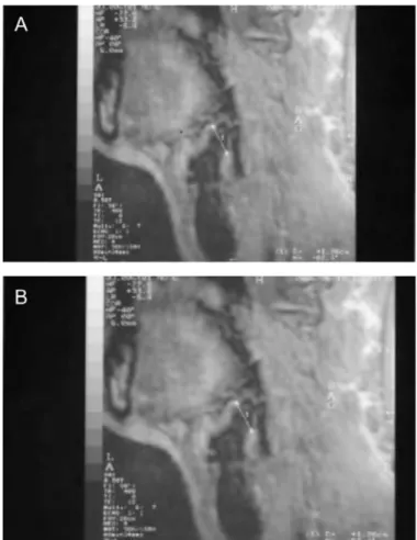
Laryngologists rely on accurate knowledge of the laryngeal anatomy for diagnoses and treatments of pathologies associ-ated with voice disorders.1It is particularly important for the laryngologist to be aware of and to take into account gender-specific differences and morphologic characteristics associated with anatomic changes in relation to voice training.5Unlike individuals who have not received vocal training, singers learn to coordinate many physiological processes by making physio-logical adaptations between the articulatory and laryngeal structures during chant training. Singers learn to stabilize vocal output by using their extrinsic laryngeal muscles. They use the intrinsic laryngeal muscles for adduction, abduction, and tension of the vocal chords.1 The larynx is in a lower FIGURE 3. MRI of a male participant in Group I shows (A) the
measurement of the right lateral thyrohyoid ligament and (B) the measurement of the left lateral thyrohyoid ligament.
FIGURE 4. MRI of a female participant in Group I shows (A) the measurement of the right lateral thyrohyoid ligament and (B) the measurement of the left lateral thyrohyoid ligament.
position during singing. Lowering the position of the larynx, especially for classical male singers, is considered a typical movement.6 Stepp et al7 reported that nonsingers tended to ascend their larynx, but singers tended to keep their larynx below the resting height.
Ligaments and cartilages are also modified as much as the muscles during vocal adjustments. Anatomic structures behave as a separate small anatomic complex distinct from the muscles.8 When the thyroid cartilage begins its descent, the thyrohyoid membrane descends to a new position as well. Because the thyroid cartilage moves together with the hyoid bone, it stretches the thyrohyoid membrane. Therefore, com-pliance-related changes in these ligaments are expected with regard to their usage.1Sonninen et al9evaluated the positional changes in the laryngeal structures according to the tone, reg-ister, and song mode and reported that the hyoid bone also adjusts to make vertical and sagittal directional changes when the pitch increases or decreases. According to Sonninen et al,9 the hyoid bone moves in a posterior-inferior direction by descending, especially during the production of certain fre-quencies. The hyoid-thyroid distance was reportedly decreased during high pitches in all singing modes. The head and neck also change their positions during vocal productions and so does the hyoid bone. The hyoid bone takes its position accord-ing to the movements of the mandible and the vocal functions of the larynx. Age and gender also contribute to the influence
of the hyoid bone and other laryngeal structures. Sprinzl et al1 reported that there are certain differences between the dimen-sions of the cartilages and ligaments of larynxes of men and women. Sprinzl et al reported the length of the LTL of men and women as 18.1§ 1.6 mm and 14.4 § 1.2 mm, respectively. Loth et al4reported the distances between the greater horn of the hyoid and the superior horn of the thyroid cartilage in 104 computed tomography scans of individuals between 19 and 62 years of age as 23.83 and 24.64 mm in women and 25.37 and 23.98 mm in men for the left and the right lateral thyro-hyoid distances, respectively. Loth et al4obtained these meas-urements between coordinates of the greater horn of the hyoid and the superior horn of the thyroid cartilage from computed tomography scans of adults using the Procrustes superimposi-tion and the principal component analysis methods. These subjects had no prior voice training. Findings in our study included initial andfinal measurements of 12.0 § 1.8 mm and 11.1§ 1.8 mm for the right LTL and 12.3 § 1.8 mm and 11.6§ 1.8 mm for the left LTL in women. These values were 22.1§ 2.0 mm and 20.2 § 4.0 mm for the right LTL and 21.9§ 2.6 mm and 21.2 § 4.1 mm for the left LTL in men.
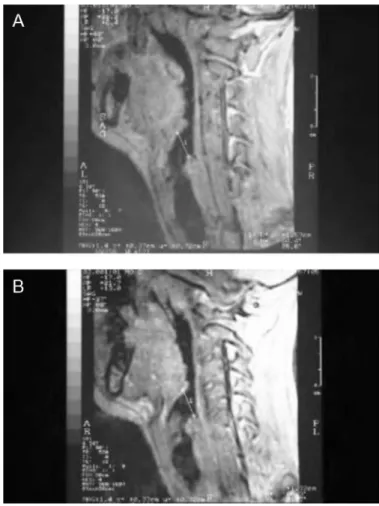
Similar to the morphometric measurements of Sprinzl et al,1 we found that the LTLs of the men were longer than those of the women. Loth et al4have reported the asymmetry in the FIGURE 5. MRI of a male participant in Group II shows (A) the
measurement of the right lateral thyrohyoid ligament and (B) the measurement of the left lateral thyrohyoid ligament.
FIGURE 6. MRI of a female participant in Group II shows (A) the measurement of the right lateral thyrohyoid ligament and (B) the measurement of the left lateral thyrohyoid ligament.
cartilage structures of the adult larynx and concluded that the left thyroid and cricoid lamina distances were higher. In our study, we have only observed an asymmetry in the left LTLs of the men in Group II and the women in Group I, but this finding was not statistically significant.
Hollien10suggested that excellent singing can be achieved by either talent or training. Sulter et al11reported that there were findings reflecting some native or acquired laryngeal functions in individuals who took training compared with individuals without any training. Besides being chosen due to talent, students in the conservatory advance their voice
quality by training. Mendes et al12followed up students tak-ing voice traintak-ing for four semesters and reported that the perturbation measurements, including jitter, shimmer, and harmonic-to-noise ratio, decreased with respect to increased duration of training. Lundy et al13 have stated that a decreased jitter, shimmer, and noise harmonic rates might be indicators of advanced voice quality in the chant. Our study revealed that chant training might have significant effects on voice quality. Our study revealedfindings suggest-ing that increased duration of chant trainsuggest-ing had significant effects on voice quality. Specifically, the effects of training TABLE 1.
Smoking Rates and Percentages in the Groups
Control Group Group I Group II
Variables Count % Count % Count %
Smoking None 6 60.0 12 100,0 6 60.0
Existing 4 40.0 0 0,0 4 40.0
TABLE 2.
Initial and Final Measurements of the Right and the Left Thyrohyoid Ligaments in the Study Groups
Control Group Group I Group II
Female Male Female Male Female Male
(n = 5) (n = 5) (n = 6) (n = 5) (n = 5) (n = 6)
Variables Mean§ SD Mean § SD Mean § SD Mean § SD Mean § SD Mean § SD
Right lateral thyrohyoid ligament (mm) Initial 12.2§ 1.8 21.6§ 2.3 12.0§ 0.6 12.2§ 1.8 21.6§ 2.3 12.0§ 0.6 Final 12.3§ 1.8 21.9§ 2.6 12.4§ 0.7 12.3§ 1.8 21.9§ 2.6 12.4§ 0.7 Left lateral thyrohyoid ligament (mm) Initial 11.4§ 1.8 20.4§ 3.8 13.7§ 2.9 11.4§ 1.8 20.4§ 3.8 13.7§ 2.9 Final 11.6§ 1.8 21.2§ 4.1 14.8§ 3.9 11.6§ 1.8 21.2§ 4.1 14.8§ 3.9
Abbreviation: SD, standard deviation.
TABLE 3.
Comparison of Right and Left Thyrohyoid Ligament Lengths and Acoustic Analysis Values in Independent Groups Control Group versus Group I Control Group versus Group II Group I versus Group II
Female Male Female Male Female Male
(n = 5) (n = 5) (n = 6) (n = 5) (n = 5) (n = 6)
Variables P Value P Value P Value P Value P Value P Value
Right lateral thyrohyoid ligament (mm) Initial 0.931 0.004* 0.222 0.548 0.126 0.537
Final 0.126 0.009* 0.310 0.421 0.052 0.329
Left lateral thyrohyoid ligament (mm) Initial 0.931 0.030* 0.222 0.421 0.126 0.662
Final 0.082 0.082 0.310 0.310 0,044* 0.662
Jitter (%) Initial 0.030* 0.537 0.008* 1.000 0.429 0.429
Final 0.017* 0.662 0.032* 1.000 0.429 0.931
Shimmer (%) Initial 0.082 0.792 0.016* 0.310 0.126 0.247
Final 0.126 0.537 0.032* 0.841 0.792 0.931
Fundamental frequency (Hz) Initial 1.000 0.931 0.690 0.548 0.931 0.429
Final 0.931 0.537 0.310 1.000 0,045* 0.792
* Mann-WhitneyU test P < 0.05.
on vocal acoustic parameters were evidenced by the decreases in the fundamental frequency of the women in Group II who took voice training. Nevertheless, the lack of such a difference in the men in the same group may be asso-ciated with factors such as cigarette smoking, which might result in perturbations of the voice. Mendes et al12suggested that 2 years of voice training might be insufficient for signifi-cant changes, and age factor might cause deviations in fun-damental frequencies. The wide age range of the men in Group II can be interpreted as a negative factor.
Despite the small sample size and the presence of smoking participants, the present study showed the significant effects of training on intrinsic laryngeal structures and voice qual-ity, which suggest the need for further studies on large sam-ples. Moreover, our recommendation for a similar research is that it is important to instruct individuals not to swallow during magnetic resonance scans, as swallowing does not allow positional changes in the hyoid.
CONCLUSIONS
Men and women have different laryngeal anatomic struc-tures. Aging and voice training result in morphologic changes in these structures. Anatomic differences between
groups showed that 8 months of training was not sufficient to trigger these morphologic changes. The left and the right LTLs were asymmetric, but thisfinding was not statistically significant. The length of the LTL was greater in men, and thisfinding supports the gender-specific differences in laryn-geal structures. Improved findings in the acoustical meas-ures in the group who took voice training suggest that chant training may be a means of improving voice quality.
Acknowledgment
We thank the Scientific Research Project (BAP) for supporting this study.
REFERENCES
1. Sprinzl GM, Eckel HE, Sittel C, et al. Morphometric measurements of the cartilaginous larynx: an anatomic correlate of laryngeal surgery. Head Neck. 1999;21:743–750.
2. Eckel HE, Sittel C, Zorowka P, et al. Dimensions of the laryngeal framework in adults. Surg Radiol Anat. 1994;16:31–36.
3. Jotz GP, Stefani MA, Pereira da Costa Filho O, et al. A morphometric study of the larynx. J Voice. 2014;28:668–672.
4. Loth A, Corny J, Santini L, et al. Analysis of hyoid−larynx complex using 3D geometric morphometrics. Dysphagia. 2015;30:357–364.
5. Howard DM. Acoustics of the trained versus untrained singing voice. Curr Opin Otolaryngol Head Neck Surg. 2009;17:155–159.
TABLE 4.
Initial and Final Measurement Averages of Acoustic Analysis Values According to Groups
Control Group Group I Group II
Female Male Female Male Female Male
(n = 5) (n = 5) (n = 6) (n = 5) (n = 5) (n = 6)
Variables Mean§ SD Mean§ SD Mean§ SD Mean§ SD Mean§ SD Mean§ SD
Jitter (%) Initial 0.24§ 0.03 0.22§ 0.10 0.16§ 0.05 0.19§ 0.09 0.13§ 0.03 0.23§ 0.08 Final 0.21§ 0.04 0.18§ 0.02 0.15§ 0.04 0.19§ 0.05 0.15§ 0.02 0.19§ 0.06 Shimmer (%) Initial 2.45§ 0.69 1.57§ 0.43 1.80§ 0.54 1.42§ 0.51 1.31§ 0.43 1.82§ 0.30 Final 1.80§ 0.39 1.30§ 0.50 1.37§ 0.67 1.54§ 0.38 1.07§ 0.42 1.72§ 1.01 Fundamental frequency (Hz) Initial 242.40§ 15.61 149.93 § 44.20 242.44 § 19.55 135.53 § 20.46 245.00 § 9.28 126.54§ 14.68 Final 262.20§ 35.42 133.01 § 23.37 268.40 § 14.19 124.57 § 16.26 243.36 § 21.23 130.27 § 17.37
Abbreviation: SD, standard deviation.
TABLE 5.
Comparison of Left and Right Thyrohyoid Ligament Lengths and Acoustic Analysis Values Within Gender Groups (Initial and Final)
Group I Group II Control Group
Female Male Female Male Female Male
(n = 5) (n = 5) (n = 6) (n = 5) (n = 5) (n = 6)
Variables P Value P Value P Value P Value P Value P Value
Right lateral thyrohyoid ligament (mm) Initial and final 0.180 0.317 1.000 0.593 0.180 0.317
Left lateral thyrohyoid ligament (mm) Initial and final 0.109 1.000 0.655 0.465 0.285 0.893
Jitter (%) Initial and final 0.588 0.752 0.041* 0.500 0.461 0.416
Shimmer (%) Initial and final 0.249 0.753 0.225 0.500 0.225 0.345
Fundamental frequency (Hz) Initial and final 0.028* 0.173 0.893 0.500 0.345 0.345
6. Mainka A, Poznyakovskiy A, Platzek I, et al. Lower vocal tract mor-phologic adjustments are relevant for voice timbre in singing. PLoS ONE. 2015;10:1–19.
7. Stepp CE, Heaton JT, Stadelman-Cohen TK, et al. Characteristics of phonatory function in singers and non-singers with vocal fold nodules. J Voice. 2011;25:714–724.
8. Bosma JF, Bartner H. Ligaments of the larynx and the adjacent phar-ynx and esophagus. Dysphagia. 1993;8:23–28.
9. Sonninen A, Hurme P, Laukkanen AM. The external frame function in the control of pitch, register, and singing mode: radiographic obser-vations of a female singer. J Voice. 1999;13:319–340.
10. Hollien H. That golden voice—talent or training? J Voice. 1993;3:195– 205.
11. Sulter AM, Schutte HK, Miller DG. Standardized laryngeal videostro-boscopic rating: differences between untrained and trained male and female subjects, and effects of varying sound intensity fundamental fre-quency, and Ag. J Voice. 1996;10:175–189.
12. Mendes AP, Brown WS, Rothman HB, et al. Effects of singing training on the speaking voice of voice majors. J Voice. 2004;18: 83–89.
13. Lundy DS, Roy S, Casiano RR, et al. Acoustic analysis of the singing and speaking voice in singing students. J Voice. 2000;14:490–493.