CLINICAL DENTISTRY AND RESEARCH 2018; 42(3): 120-126 Original Research Article
Correspondence Betül Aycan Alim, DDS
Department of Endodontics, Faculty of Dentistry, Bezmialem Vakıf University, Istanbul, Turkey
Phone: 0 507 2399507 Fax: 0 212 533 2326 E-mail: [email protected]
Betül Aycan Alim, DDS
Specialist Endodontist, Department of Endodontics, Faculty of Dentistry, Bezmialem Vakıf University, Istanbul, Turkey
Fatıma Betül Baştürk, DDS, PhD
Associate Professor, Department of Endodontics, Faculty of Dentistry, Marmara University, Istanbul, Turkey
Yıldız Garip Berker, DDS, PhD
Professor, Department of Endodontics, Faculty of Dentistry, Kent University, Istanbul, Turkey
Figen Eren Giray, DDS, PhD
Assistant Professor, Department of Pedodontics, Faculty of Dentistry, Marmara University, Istanbul, Turkey
EVALUATION OF DISTAL CANAL CONFIGURATIONS OF THE
MANDIBULAR FIRST MOLAR TEETH BY MICRO-COMPUTED
TOMOGRAPHY
ABSTRACT
Background and Aim: The aim of this study was to evaluate the morphological aspects of the root canal anatomy of the distal roots of mandibular first molars using micro computed tomography.
Materials and Methods: The distal roots of sixty mandibular first molar teeth were visualized two-dimensionally by micro computed tomography. The views of all roots at 2, 5, and 8 mm were measured in terms of major diameter (dmax), minor diameter (dmin) and total area of root canal (A) pixels by x400 magnification. The roundness of a discrete two-dimentionally object is defined as 4.A/(p.[dmax]²), where A is the area and dmax is the major diameter. The aspect ratio is a measure of shape, and it is achieved by dividing the value of the major diameter by the minor diameter.
Results: The roundness value at 2 mm were statistically significantly higher than the roundness value at 5 mm and 8 mm. The aspect ratio at 8 mm was found statistically significantly higher than other groups.
Conclusions: The increased rate of flattened canals was observed in a coronal direction from the apex. The distal root canals were more round at the apical third.
Keywords: Distal Canal Configuration, Mandibular Molar, Micro Computed Tomography
Submitted for Publication: 05.28.2018 Accepted for Publication : 11.10.2018
Sorumlu Yazar Betül Aycan Alim
Bezmialem Vakıf Üniversitesi, Diş Hekimliği Fakültesi, Endodonti Anabilim Dalı, Fatih, İstanbul, Türkiye. Telefon: 0 507 2399507 Faks: 0 212 533 2326 E-posta: [email protected]
Betül Aycan Alim
Dr., Bezmialem Vakıf Üniversitesi, Diş Hekimliği Fakültesi, Endodonti Anabilim Dalı, İstanbul, Türkiye
Fatıma Betül Baştürk
Doç.Dr., Marmara Üniversitesi, Diş Hekimliği Fakültesi, Endodonti Anabilim Dalı, İstanbul, Türkiye
Yıldız Garip Berker
Prof.Dr., Kent Üniversitesi, Diş Hekimliği Fakültesi, Endodonti Anabilim Dalı, İstanbul, Türkiye
Figen Eren Giray
Dr. Öğr.Üyesi, Marmara Üniversitesi, Diş Hekimliği Fakültesi, Pedodonti Anabilim Dalı, İstanbul, Türkiye
MANDIBULAR BİRİNCİ MOLAR DİŞLERİN DİSTAL KANAL
KONFİGURASYONLARININ MİKRO-BİLGİSAYARLI TOMOGRAFİ
KULLANILARAK DEĞERLENDİRİLMESİ
ÖZ
Amaç: Bu çalışmanın amacı, mikro bilgisayarlı tomografi kullanılarak mandibular birinci molar dişlerin kök kanal anatomisinin morfolojik yönlerini değerlendirmektir.
Gereç ve Yöntem: 60 adet mandibuler birinci molar dişin distal kökleri mikro-bilgisayarlı tomografi kullanılarak iki boyutlu olarak görüntülenmiştir. Tüm köklerin 2, 5 ve 8 mm’deki kesit görüntüleri üzerinde majör çap (dmax), minör çap (dmin) ve kök kanalının toplam alanı (A) x400 büyütme altında hesaplanmıştır. Elde edilen kök kanal görüntüsünün yuvarlaklık oranı, 4.A/(p.[dmax]²) formulü kullanılarak değerlendirilmiştir. En boy oranı ise, major çap değerinin minor çapa bölünmesiyle elde edilmiştir. Kök kanallarının yuvarlaklık ve en/boy oranlarının istatiksel analizi, tek yönlü ANOVA testi kullanılarak yapılmıştır (p<0.05).
Bulgular: Kök kanalının 2 mm’ deki yuvarlaklık değeri, 5 ve 8 mm mesafedeki yuvarlaklık değerinden istatistiksel olarak anlamlı derecede yüksek bulunmuştur. En boy oranı ise, 8 mm mesafeden alınan kesit görüntülerinde, diğer gruplara göre istatistiksel olarak anlamlı derecede yüksek bulunmuştur. Sonuçlar: Kök kanalı içinde apeksten koronal yöne doğru gittikçe yuvarlaklık değerinin azaldığı ve kanalın uzun-düz bir şekle dönüştüğü gözlenmiştir. Mandibular birinci molar dişlerin distal kök kanallarının apikal üçlüde daha yuvarlak olduğu belirlenmiştir.
Anahtar Kelimeler:Distal Kanal Konfigürasyonu, Mandibular Molar, Mikro Bilgisayarlı Tomografi Yayın Başvuru Tarihi : 28.05.2018
Yayına Kabul Tarihi : 10.11.2018
CLINICAL DENTISTRY AND RESEARCH INTRODUCTION
The success of endodontic treatment depends on removal of pulp tissue and necrotic material, biomechanical preparation, disinfection and obturation of the root canal.1-3 The rate of the root canal treatment failure was reported as 14% due to leakage, even with radiographically satisfactory fillings.4, 5 Thus, hermetic obturation of the root canal system is critical in endodontic treatment.6
The root canal morphology can be examined noninvasively and precisely with the availability of micro computed tomography (micro-CT). Internal and external anatomy could be demonstrated simultaneously or separately. Images could be assessed qualitatively and quantitatively.7-10 With voxel sizes as small as 19 μm, 3D images can be produced and analysed to determine the number and configuration of canals and foramina.11 Micro-CT system is also useful in quantitatively measuring the mineral concentration of bones and teeth with an accuracy of better than 1% and a resolution between 5 and 30 μm.10 The presence of accessory canals in micro-CT images of maxillary and mandibular roots showed a statistically significant correlation with the stereomicroscopic images, which was used as a gold standard.12
Generally, the morphology of the root canals varies greatly in shape and transversal cross-sections in different groups of teeth.13,14 The mandibular first molar exhibits a complex and distinct range of variations in the morphology of the root canal system.15-19 They usually have 2 roots, but, occasionally, they have 3, with 2 or 3 canals in their mesial roots and 1, 2, or 3 canals in their distal roots.16,17,20 When only one distal canal is present, it is usually oval-shaped buccolingually, and untreated surface areas can to be as much as 59%–79% when rotary instruments were used for the shaping procedure.21
The aim of this study was to evaluate the morphological aspects of the root canal anatomy of the distal roots of mandibular first molars using micro-CT.
MATERIALS AND METHODS
After ethics committee approval (Marmara University Institute of Health Sciences Ethics Committee, Protocol #22-2016), sixty extracted human mandibular first molars were selected. Adherent soft tissue was removed by an ultrasonic device. Teeth with resorption, cracks, fracture or restoration at the root surface were excluded. Teeth with mature apices and a root length more than 10 mm were
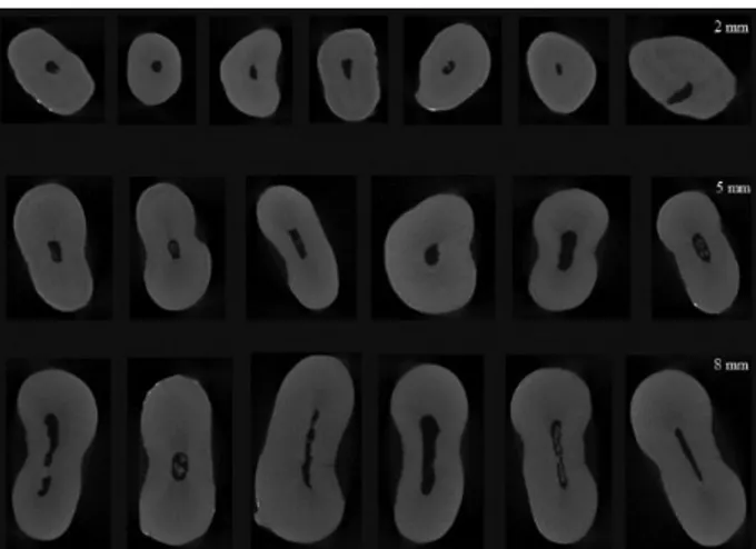
included. The selected teeth were then washed under tap water for 30 seconds and stored in 10% formalin solution. Apical (2 mm), middle (5 mm) and coronal (8 mm) sections of the distal root canals were scanned two-dimensionally using Micro-CT (Scanco Medical, μCT 50) at 70 kVp, 114 μA, FOV 20.5 mm, voxel size 20 μm, integration time 600 ms settings. X-rays were filtered with a 0.5 mm Al filter for changes in the sensitivity of the polychromatic radiation (Figure 1).
All images were transferred to Image J (Image J 1.42m, NIH, USA) program for sectional analysis. Area, roundness, major diameter, minor diameter, and aspect ratio of the root canals were calculated in pixels by x400 magnification. The roundness of a discrete 2D object is defined as 4.A/(p.[dmax]²), where A is the area and dmax is the major diameter. The value of roundness ranges from 0 to 1, with 1 expressing the perfect circle. The major diameter was defined as the distance between the 2 most distant pixels in that object. The minor diameter was defined as the longest chord through the object that can be drawn in the orthogonal direction to that of the major diameter.14,22,23 The aspect ratio is a measure of shape, and it is achieved by dividing the value of the major diameter by the minor diameter. An oval canal has an aspect ratio higher than 1 and lower than 2, a long oval canal higher than 2 but lower than 4, and a flattened canal higher than 4. Root canals with aspect ratios lower than 1 were classified as nonoval.24 For the statistical analysis, SPSS (Statistical Package for Social Sciences) for Windows 21.0 program was used. In the comparison of quantitative data, if there were more than two groups, One way ANOVA test was used to compare
Figure 1. The images of different root canals at 2 mm, 5 mm and 8 mm.
the normally distributed parameters. Tukey test was used to determine the group that caused the difference. The results were evaluated at a 95% confidence interval and a significance level of p <0.05.
RESULTS
The roundness value was calculated at 2, 5 and 8 mm distances, as 0.47, 0.36 and 0.29 respectively (Table 1). The roundness value at 2 mm was significantly higher than those at 5 mm (p=0,015) and 8 mm (p=0,000). There was no statistically significant difference between the roundness values at 5 mm and 8 mm (p=0,184).
The aspect ratio of root canals was calculated according to the dmax/dmin formula and was found statistically significant (p>0.05). The aspect ratio at 2 mm was significantly lower than the those at 5 mm (p=0,042) and 8 mm (p=0,000) (Table 2).
The mean value of aspect ratio at 2 mm was 2.83 and it was considered to be “long oval”. The mean value of aspect ratio at 5 mm distance was 4.28 and the root canals were defined “flat”. The mean value of aspect ratio at 8 mm distance was calculated as 6.24 and the root canals at this distance were expressed more “flat” than the root canals in the other sections (Figure 2).
We found that a single canal at all sections of the distal root was present in 73.33% of the mandibular first molars evaluated (n=44) (Figure 3).
DISCUSSION
The study of the dimensions of the internal anatomy of teeth was a challenge.25,26 Although conventional radiographs used during clinical procedures can show important details such as the number of root canals, the severity of the root canal curvature, or the presence of calcifications, periapical x-rays are two dimensional images and do not provide enough details of the internal anatomy. For this reason, anatomical studies are necessary to guide clinicians to achieve better cleaning of the root canals.19 Over the years, numerous root canal configurations have been identified resulting in the proposal of classifications and modifications. Although Vertucci classification is the one of most commonly used methods for the evaluation of root canal morphology,27 roundness and aspect ratio of the root canals were evaluated in this in vitro study using different formulas.
The mandibular first molar is recognized as exhibiting a complex and distinct range of variations in the morphology of the root canal system.15-19 When only 1 distal canal is present, it is usually oval-shaped buccolingually, and untreated surface areas can be as high as 59%–79% when rotary instruments are used for the shaping procedure.21 In this study, 73.33% of all the distal roots of mandibular first molars that we evaluated had a single canal at all sections (n=44). The studies which evaluated the morphology of the distal roots of mandibular first molars reported that a
Table 1. The value of roundness ranges at 2, 5 and 8 mm. (1: perfect circle) (Sd: standart deviation)
Roundness value Mean Sd p
2 mm 0,47 0,20
0,000*
5 mm 0,36 0,20
8 mm 0,29 0,21
*p<0,05
Table 2. The value of aspect ratio at 2, 5 and 8 mm. (oval canal: between 1 and 2, long oval: between 2 and 4, flattened canal: >4, non-oval canal: <1), (Sd: standart deviation)
Aspect ratio Mean Sd p
2 mm 2,83 1,79
0,000*
5 mm 4,28 2,70
8 mm 6,24 4,85
CLINICAL DENTISTRY AND RESEARCH
single canal was present in 66.62% of Korean population,28 49.8% of Turkish population,29 %70 of Belgian and Chilean population,30 65.9% of Chinese Subpopulation,31 61.3% of Iranian population.32 These results were similar with ours. The difference between the results was thought to be due to the fact that our study was only a cross-sectional analysis.
Filpo-Perez et al.33 evaluated the morphologic aspects of the distal roots of mandibular first molars using Micro-CT. They reported roundness values of 0.47 and 0.35 at 2 mm and 5 mm, respectively. This result coincides with our findings. Abou-Rass et al.34 used a term called “danger zone” to describe the thin inner walls of the root canals which are vulnerable to stripping by injudicious filing. The disparity between the mesial/distal and buccal/lingual dimensions of the canals in both roots shows that these canals are not round. This means that most instruments will inevitably widen the mesial/distal aspect of the canal (toward the “danger zone” in the mandibular first molar) more than the buccal lingual/aspect. If an endodontic file was introduced into a canal towards these dimensions, it could lead to a reduction in the thickness of the already-thin furcal aspect of the root while possibly leaving some of the buccal or lingual walls uninstrumented. For this reason, as reported
by previous authors,35-37 coronal enlargement may be needed to adequately clean and shape these dimensions; however, the thinness of dentin toward the furcation must be respected in order to avoid perforation in this area. CONCLUSIONS
The distal roots of mandibular first molars showed a high rate of single root canals. The increased rate of flattened canals was observed in a coronal direction from the apex. The distal root canals were more round at the apical third. ACKNOWLEDGES
The cost of this study was funded by Marmara University Scientific Research Project Commission (Grant number: SAG-C-DUP-131016-0450). All the imaging processes were carried out at the Application Center of Ege University Central Testing and Analysis Laboratory.
REFERENCES
1. Sundqvist G, Figdor D, Persson S, Sjogren U. Microbiologic analysis of teeth with failed endodontic treatment and the outcome of conservative re-treatment. Oral Surg Oral Med Oral Pathol Oral Radiol Endod 1998; 85: 86-93.
2. De Moor RJG, Martens LC. Apical microleakage after lateral condensation, hybrid gutta-percha condensation and Soft-Core obturation: an in vitro evaluation. Endod Dent Traumatol 1999; 15: 239-243.
3. Schafer E, Nelius B, Burklein S. A comparative evaluation of gutta-percha filled areas in curved root canals obturated with different techniques. Clin Oral Invest 2012; 16: 225–230.
4. Kersten HW, Wesselink PR, Thoden Van Velzen SK. The diagnostic reliability of the buccal radiograph after root canal filling. Int Endod J 1987; 20: 20-24.
5. Buckley M, Spangberg L. The prevalence and technical quality of endodontic treatment in an American subpopulation. Oral Surg Oral Med Oral Pathol 1995; 79: 92.
6. Dewsnup N, Pileggi R, Haddix J, Nair U, Walker C, Varella CH. Comparison of bacterial reduction in straight and curved canals using erbium, chromium: yttrium-scandium-gallium-garnet laser treatment versus a traditional irrigation technique with sodium hypochlorite. J Endod 2010; 36: 725-728.
7. Dowker SE, Davis GR, Elliott JC. X-ray micro- tomography: nondestructive three-dimensional imaging for in vitro endodontic studies. Oral Surg Oral Med Oral Pathol Oral Radiol Endod 1997; 83: 510–516.
Figure 2. The images of different sections of a distal root canal (2 mm: oval, 5 mm: long oval, 8 mm: flattened canal)
Figure 3. The images of different sections of a distal root canal (2 mm: two root canals, 5 mm: two root canals, 8 mm: single root canal)
8. Rhodes JS, Ford TR, Lynch JA, Liepins PJ, Curtis RV. Micro-computed tomography: a new tool for experimental endodontology. Int Endod J 1999; 32: 165–170.
9. Jung M, Lommel D, Klimek, J. The imaging of root canal obturation using micro-CT. Int Endod J 2005; 38: 617-26.
10. Swain MV, Xue J. State of the Art of Micro-CT Applications in Dental Research. Int J Oral Sci 2009; 1: 177-88.
11. Nielsen BR, Alyassin A, Peters DD, Carnes DL, Lancaster J. Microcomputed tomography: an advanced system for detailed endodontic research. J Endod 1995; 21: 561–568.
12. Acar B, Kamburoğlu K, Tatar İ, Arıkan V, Çelik HH, Yüksel S et al. Comparison of micro-computerized tomography and cone beam computerized tomography in the detection of accessory canals in primary molars. Imaging Sci Dent 2015; 45: 205-211.
13. Wu MK, R’Oris A, Barkis D, Wesselink PR. Prevalence and extent of long oval canals in the apical third. Oral Surg Oral Med Oral Pathol Oral Radiol Endod 2000; 89: 739–743.
14. Versiani MA, Cristescu RC, Saquy PC, Pecora JD, de Sousa-Neto MD. Enamel pearls in permanent dentition: case report and micro-CT evaluation. Dentomaxillofac Radiol 2013; 42: 20120332. 15. Vertucci FJ. Root canal anatomy of the human permanent teeth. Oral Surg Oral Med Oral Pathol 1984; 58: 589–599. 16. Gulabivala K, Aung TH, Alavi A, Ng YL. Root and canal morphology of Burmese mandibular molars. Int Endod J 2001; 34: 359–370.
17. Gulabivala K, Opasanon A, Ng YL, Alavi A. Root and canal morphology of Thai mandibular molars. Int Endod J 2002; 35: 56– 62.
18. Sert S, Bayirli GS. Evaluation of the root canal configurations of the mandibular and maxillary permanent teeth by gender in the Turkish population. J Endod 2004; 30: 391–398.
19. Villas-Boas MH, Bernardineli N, Cavenago BC, Marciano M, Del Carpio-Perochena A, de Moraes IG et al. Micro-computed tomography study of the internal anatomy of mesial root canals of mandibular molars. J Endod 2011; 37: 1682–1686.
20. Gu Y, Lu Q, Wang H, Ding Y, Wang P, Ni L. Root canal morphology of permanent three-rooted mandibular first molars-part I: pulp floor and root canal system. J Endod 2010; 36: 990–994.
21. Paque F, Peters OA. Micro-computed tomography evaluation of the preparation of long oval root canals in mandibular molars with the Self-Adjusting File. J Endod 2011; 37: 517–521.
22. Leoni GB, Versiani MA, Pecora JD, Dami~ao de Sousa-Neto M. Micro–computed tomographic analysis of the root canal morphology of mandibular incisors. J Endod 2014; 40: 710–716.
23. Ordinola-Zapata R, Bramante CM, Villas-Boas MH, Cavenago BC, Duarte MH, Versiani MA. Morphologic microcomputed tomography analysis of mandibular premolars with three root canals.J Endod 2013; 39: 1130-1135.
24. Jou YT, Karabucak B, Levin J, Liu D. Endodontic working width: current concepts and techniques. Dent Clin North Am 2004; 48: 323–335.
25. Kuttler Y. Microscopic investigation of root apexes. J Am Dent Assoc 1955; 50: 544–552.
26. Kerekes K, Tronstad L. Morphometric observations on the root canals of human molars. J Endod 1977; 3: 114–118.
27. Vertucci FJ. Root canal anatomy of the human permanent teeth. Oral Surg, Oral Med, Oral Pathol 1984; 58: 589-599. 28. Kim S, Kim BS, Woo J, Kim Y. Morphology of mandibular first molars analyzed by cone-beam computed tomography in a korean population: Variations in the number of roots and canals. J Endod 2013; 39: 1516–1521.
29. Nur BG, Ok E, Altunsoy M, Aglarci OS, Colak M, Gungor E. Evaluation of the root and canal morphology of mandibular permanent molars in a south-eastern Turkish population using cone-beam computed tomography. Eur J Dent 2014; 8: 154-159. 30. Torres A, Jacobs R, Lembrechts P, Brizuela C, Cabrera C, Concha G et al. Characterization of mandibular molar root and canal morphology using cone beam computed tomography and its variability in Belgian and Chilean population samples. Imaging Sci Dent 2015; 45: 95-101.
31. Zhang X, Xiong S, Ma Y, Han T, Chen X, Wan F et al. A Cone-Beam Computed Tomographic Study on Mandibular First Molars in a Chinese Subpopulation. PLoS ONE 2015; 10: e0134919. 32. Akhlaghi NM, Khalilak Z, Vatanpour M, Mohammadi S, Savadkouhi ST, Pirmoradi S et al. Root Canal Anatomy and Morphology of Mandibular First Molars in a Selected Iranian Population: An In Vitro Study. Iran Endod J 2016; 1: 87–91.
33. FilpoPerez C, Bramante CM, VillasBoas MH, Húngaro Duarte MA, Versiani MA, OrdinolaZapata R. Microcomputed tomographic analysis of the root canal morphology of the distal root of mandibular first molar. J Endod 2015; 41: 231236.
34. Abou-Rass M, Frank AL, Glick DH. The anticurvature filing method to prepare the curved root canal. J Am Dent Assoc 1980; 101: 792–794.
CLINICAL DENTISTRY AND RESEARCH
35. Berutti E, Fedon G. Thickness of cementum/dentin in mesial roots of mandibular first molars. J Endod 1992; 18: 545–548. 36. Lim SS, Stock CJR. The risk of perforation in the curved canal: anticurvature filing compared with the stepback technique. Int Endod J 1987; 20: 33–39.
37. Harris SP, Bowles WR, Fok A, McClanahan SB. An Anatomic Investigation of the Mandibular First Molar Using Micro–Computed Tomography. J Endod 2013; 39: 1374–1378.