https://doi.org/10.1007/s11240-018-01542-x ORIGINAL ARTICLE
The association of fraser photinia and its beneficial bacterium (PGB_
invit) provided in vitro storage without subculture
Irmak Şah1 · Hülya Akdemir1 · Ergun Kaya2 · Özlem Akkaya1 · Yelda Özden Çiftçi1
Received: 2 October 2018 / Accepted: 20 December 2018 / Published online: 2 February 2019 © Springer Nature B.V. 2019
Abstract
Endophytes play crucial roles due to their beneficial influence on plant development, growth, fitness, and diversification. Due to these important capabilities, they have received attention from the scientific community and many papers have been pub-lished recently about their beneficial role in in vivo and in vitro plant propagation. However, up to now, there is no research on utilization of these microbial endophytes in prolongation of in vitro storage. Thus, the aim of this study is to assess the influence of fraser photinia associated and putatively endophytic bacterium (Plant Growth Bacteria_ in vitro; PGB_invit) on in vitro storage of its host. When pure strain of the bacterium was inoculated, it enabled the storage of microshoots up to 16 months at 25 °C without requiring periodic subculture while control (unincubated with PGB_invit.) microshoots died after 2 months of storage without subculture as in vitro plant cultures definitely need periodic subcultures (once in every 4–6 weeks) in order to renew media and gaseous atmosphere. Moreover, while the presence of virulence (vir D1), auxin (aux1), and cytokinin (ipt) production genes was confirmed in plasmid DNA of the bacterium, nitrogen fixing gene (nifH) was detected by the PCR analysis using bacterial culture. Overall results demonstrated that with these capabilities PGB_invit could be useful for in vitro conservation of fraser photinia.
Key message
The novelty is the supplementation of in vitro plant growth without either periodic renewal of the media or decreasing the culture temperature by means of a beneficial plant-bacterium interaction.
Keywords Aux1 · Endophytic · Ipt · NifH · Plant growth promoting bacterium
Introduction
Microbial endophytes (bacterial, archaeal, fungal, and pro-tistic taxa), date back more than 400 million years (Remy et al. 1994), are considered as extremely important plant partners (Hallman et al. 1997) as they live intercellularly and/or intracellularly in host without causing any apparent
disease (Wilson 1995; Nair and Padmavathy 2014; Hardoim et al. 2015). Besides, it may affect the growth of their host plants positively, (i) by producing some plant growth pro-moting (PGP) regulators such as auxin and cytokinin (Ryan et al. 2008; Hardoim et al. 2008; Jimtha et al. 2014), (ii) promoting plant disease resistance against many potential plant pathogen by not only increasing expression of defense-related genes in plants (Benhamou et al. 1996; Gond et al.
2015; Cabanas et al. 2014), but also synthesis and modula-tion of bioactive compounds that have potential to be used in medicine, agriculture or industry (Jasim et al. 2015; Singh et al. 2017; Patle et al. 2018), (iii) supplying tolerance to abiotic stress (Vigani et al. 2018). Moreover, endophytes have also been shown to increase nutrients uptake such as nitrogen or phosphorous into plants (Boddey et al. 1991; James 2000; Iniguez et al. 2004; Malboobi et al. 2009). Above all, they also have positive effect on in vitro prolif-eration of different plants (i.e., Pirtilla et al. 2000; Dias et al. Communicated by Sergey V Dolgov.
* Özlem Akkaya [email protected] * Yelda Özden Çiftçi [email protected]
1 Department of Molecular Biology and Genetics, Gebze Technical University, 41400 Gebze, Kocaeli, Turkey 2 Department of Molecular Biology and Genetics, Muğla Sıtkı
2009; Quambusch et al. 2014). Those beneficial influences of endophytic bacteria together with their potential role in agro-ecosytems have also recently been reviewed by many authors (i.e., Mercado-Blanco 2015; Card et al. 2016; San-toyo et al. 2016; Patle et al. 2018; Akkaya et al. 2019) and seemed to be very promising hot topics of the plant micro-biome studies (Azevedo et al. 2000; Schulz et al. 2002; Aly et al. 2010).
In accordance, a putatively endophytic beneficial bacte-rium, which will be nominated as PGB_invit, was isolated and characterized in in vitro grown microshoots of fraser photinia that has ability to fix nitrogen and produce some PGP regulators such as indoleacetic acid (IAA) and Gib-berellic Acid (GA3) in our previous study (Gul Şeker et al.
2017). More interestingly, it seemed to promote in vitro storage and proliferation of fraser photinia without routine subculture. Thus, this study was conducted to reveal out the influences of this PGB_invit on not only shoot storage and culture growth without subculturing up to 16 months but also rooting and acclimatization of in vitro prolifer-ated shoots. Moreover, the presence of PGP genes includ-ing auxin (aux 1), cytokinin (ipt) and nitrogen fixation (nif
H) together with virulence gene (virD1) was also assessed
to understand the molecular interactions between photinia and PGB_invit. Although the beneficial influences of endo-phytic bacterium have been studied for many decades, to our knowledge, this is the first paper that demonstrates the potential role of endophytic bacteria on providing in vitro storage in 25 °C without the need of renewal of macro and microelements, carbon source and PGP regulators in eco-nomic and eco-friendly manner.
Materials and methods
In vitro culture conditions
Shoots of fraser photinia (Photinia × fraseri Dress.) were subcultured monthly to fresh MS medium containing 4.4 µM 6-benzyladenine (BA) according to Akdemir et al. (2010) until contamination of PGB_invit was visually detected in the medium.
Influences of PGB_invit on in vitro storage of fraser photinia microshoots
As no detrimental effect of bacterium was determined on 4 weeks PGB-invit incubated in vitro fraser photinia cultures (Gul Şeker et al. 2017), visibly contaminated microshoots were maintained (without periodic subculturing on fresh medium) in QL medium (Quoirin and Lepoivre 1977) con-taining 4.4 µM BA for different storage periods (6, 9, 12, 15, 16 months) at 25 ± 2 °C with 16 h photoperiod under
36 µmol m−2 s−1 photosynthetic photon flux provided by
cool-white fluorescent lamps. Control cultures (without bac-teria) were also maintained in the same conditions without subculture. The percentage of green shoots, average length of the microshoots, presence–absence of roots, average num-ber of roots per microshoots, average length of the roots, dry-fresh weight was assessed together with the number of both green and abscised leaves per microplant in order to reveal the plant quality during storage. Dry weight of the shoots was determined by drying a batch of microshoots (a minimum of 20) in an oven at 80 °C, weighting the shoots every 4 h until two successive weights gave the same value. Moreover, leaf senescence index was also calculated per microplant according to �x∕√y+ 0.5 formula in which “x” represents the number of green leaves per microplant whereas “y” represents senescence plus abscised leaves per microplant (Sarkar et al. 1999). In addition, visual prefer-ence scale from 0 to 3 was also scored based on plant appear-ance: 0-dead plant; 1-microshoots were brown, in some places green; 2-microshoots were green–brown; 3-micro-shoots with bright green leaves and stems (modified from Sarkar and Naik 1998).
Influences of PGB_invit on shoot retrieval after conservation
Shoot apices were excised from in vitro conserved micro-shoots, which were contaminated, and transferred to fresh 4.4 µM BA containing QL medium in order to assess shoot retrieval after conservation. The percentage of shoot apex that regenerated at least one elongated shoot, the average number of shoots proliferated per explant and the average length of the shoots were evaluated after 4 weeks in culture. In addition, the shoot forming capacity (SFC) index (Lam-bardi et al. 1993) was also calculated based on the formula with using the average number of shoots per proliferating explant × percentage of proliferating explants/100.
Influences of PGB_invit on rooting and acclimatization
A pool of elongated microshoots (at least 1–1.5 cm long) from control and contaminated were transferred to semi-solid QL medium supplemented with various concentra-tions (0.49, 2.46, 4.92 µM) of indole butyric acid (IBA) for rooting. After 7-days of culture, half of the shoots were transferred to PGR-free QL medium. Microshoots that had at least 0.2 cm root were considered as rooted. The date of the first root emergence in each experiment was recorded in order to calculate average days of rooting time. Rooting time was calculated according to formula ∑ (NxTx)∕n◦ of
rooted shoots where Nx is the n° of rooted shoots within consecutive intervals of time; Tx is the n° of days between
the beginning of the test and the end of the specific interval of time. Moreover, average number of roots per shoot and length of the root/microshoot were determined after 30 days of culture.
In order to acclimatize to in vivo conditions, rooted shoots from both control and contaminated cultures, that were stored for 12 months at 25 °C, were rinsed with tap water and transferred to pots containing peat and perlite (1:1). Pots were covered with a polyethylene bag to maintain high relative humidity and placed in a culture room for 4 or 5 weeks. Three holes (less than 1 mm) were opened after 2 days and doubled each day. After 4–5 weeks, the plants were transferred to bigger pots in greenhouse conditions for their further growth.
Genomic and plasmid DNA isolation of PGB_invit
PGB_invit was inoculated in MPYE liquid medium and incubated at 30 °C for 10 days. Genomic DNA extraction was performed by using Promega WizardR Genomic DNA Purification Kit (Madison USA). Plasmid isolation was car-ried out with Macherey–Nagel Nucleospin Plasmid Kit. The isolated DNAs were visualized on 1.5% agarose gel electro-phoresis along with 1 kb + 100 bp DNA ladder (Invitrogen Cat. No. 10787-018) as size marker and quantified by UV spectrometer on 260 nm wavelength (Shimazu Biotech, Bio-specNano Spectrometer).
PCR analysis
In order to assess whether PGB_invit is producing auxin or cytokinin, aux1 (tryptophan monooxygenase) and aux2 (indoleacetamide hydrolase) genes, responsible for auxin biosynthesis, and ipt (isopentenyl transferase) gene, respon-sible for cytokinin biosynthesis were amplified using the following primers: aux1-FW (5′-CTC CGA TTC CTT TCC AAC CG-3′) and aux1-RV (5′CGC ACG TTA TCC TCA TAC CC-3′), aux2-FW (5′-CTG TCA ACG GAG GCT GTT GGG-3′) and aux2-RV (5′ACC CTA GTC TCA TCC CAG GG-GGG-3′) (Camilleri and Jouanin 1991) and ipt-FW (5′-GATCG(G/C) GTC CAA TG(C/T)TGT-3′) and ipt-RV (5′-GAT ATC CAT CGATC(T/C)CTT-3′), respectively according to Haas et al. (1995). Rhizobacterium rhizogenes (Agrobacterium
rhizo-genes) ATCC 15834 for aux genes and Rhizobium radiobac-ter (Agrobacradiobac-terium tumefaciens) ATCC 15955 for ipt gene
were used as positive controls in PCR. The reaction mixture contained 2.5 × PCR buffer, 2.5 mM MgCl2, 200 µM dNTP, 50 nanograms plasmid DNA, 1 U Taq DNA polymerase (i-Taq™, Intron) and 0.4 µM for both forward and reverse primers. PCR conditions for aux genes was 94 °C for 2 min pre-denaturation; 30 cycles of 94 °C for 15 s denaturation,
60 °C for 30 s annealing, 72 °C for 1 min extension and 72 °C for 10 min final extension steps. PCR program for
ipt gene was 94 °C for 2 min predenaturation; 30 cycles
of 94 °C for 15 s denaturation, 55 °C for 30 s annealing, 72 °C for 1 min extension and 72 °C for 10 min final exten-sion steps. PCR products except were visualized on 1.5% agarose gel electrophoresis along with 1 kb DNA Ladder (Intron 24074).
To identify whether the bacterium has gene transfer capa-bility like Agrobacterium Ti plasmid, we tried to amplify
vird1 gene region. The primer pairs were vird1-FW
(5′-ATG TCG CAA GGC AGT AGG CCC ACC T-3′) and vird1-RV (3′-CTA CAA GGC GTC TTT CAG CAG CGA GC-5′) (Rogorowsky et al. 1990). The PCR mixture for virD1 amplification contained 1 µl plasmid DNA as template, 5 µl Solis BioDyne 5x FIREPol Master Mix, 0.2 µM from each primer in a final volume of 25 µl. PCR conditions was 95 °C for 5 min pre-denaturation and 40 cycles of 95 °C for 1 min denaturation, 55 °C for 2 min annealing, 72 °C for 2 min extension and 72 °C for 10 min final extension steps. With biochemical tests, we observed that the PGB_invit has the ability to fix nitrogen (Gul Şeker et al. 2017). To verify this, the presence of nifH gene was assessed by using
nifH forward (5′-TGC GAY CCSAARGCBGACTC-3′) and
reverse (3′-ATSGCC ATC ATY TCR CCGGA-5′) degenerated primers. The PCR mixture contained 1 µl bacterial culture as template, 5 µl Solis BioDyne 5x FIREPol Master Mix, 0.2 µM from each primer in a final volume of 25 µl. PCR programme was 95 °C for 5 min predenaturation and 40 cycles of 95 °C for 1 min denaturation, 58 °C for 2 min annealing, 72 °C for 2 min extension and 72 °C for 10 min final extension steps. virD1 and nifH PCR products were visualized on 1.5% agarose gel electrophoresis along with 1 kb and 100 bp DNA Ladders (Thermofisher).
Carbohydrate and alditols analysis
PGB_invit was grown in MPYE medium for 10 days. Then, bacterial culture was centrifuged at medium speed (8000 rpm) for 5 min at RT, and the supernatant was used for the determination of carbohydrate and alditols including mannitol, inositol, and sorbitol by HPLC method according to Agilent’s protocol (https ://www.agile nt.com). The flow rate of the isocratic elution was 600 µl/min, the sample injec-tion volume was 5 µl, the MetaCarb 87P Carbohydrate Col-umn Pb + colCol-umn was used at ambient temperature (80 °C) and sample run time was 60 min. Mobile phase composed of Milli Q water.
Experimental design and statistical analysis
Each experiment concerning the biochemical and molecular characterization of the bacterium was repeated at least twice
whereas experiments regarding shoot proliferation, the num-ber of shoots proliferated per explant, plant shoot growth, shoot retrieval and rooting were carried out with using at least 50 explants/microshoots and repeated at least thrice.
Statistical analysis of the non-parametric data (frequen-cies) was carried out by the test for homogeneity of pro-portions and significant treatment differences selected by a non-parametric statistical test: Post Hoc Multiple Com-parison (Marascuilo and McSweeney 1977). Discrete data were subjected to analysis of variance (ANOVA), followed by the least significant difference (LSD) test at P ≤ 0.05 to compare means.
Results
Influences of PGB_invit on in vitro storage of fraser photinia microshoots
It was possible to conserve fraser photinia microshoots at 25 °C with the bacterium up to 9 months without any decline in the percentage of green microshoots (Table 1) whereas control (unincubated with PGB_invit.) microshoots dies after 2 months of storage without subculture. However, with the prolongation of the conservation time, a significant decline in the green microshoot percentage was observed [95.5% for 12 months (Fig. 1a), 80.4% for 15 and 79.8% Table 1 The influence of the
PGB_invit on fraser photinia microshoot growth and rooting after storage
The data were collected 30 days after culture initiation. Each trial was made with at least 50 explants and the trials were repeated at least 2 times
a The same letters following the percentages show no statistical difference compared to the Post Hoc Multi-ple comparison test (P ≤ 0.05)
b The same letters following the means show no statistical difference compared to the LSD test following ANOVA (P ≤ 0.05; The mean difference was analyzed horizontally)
c Mean ± standard error
d The microplant growth was scored on a 0–3 visual preference scale in which: 0-dead plant; 1-microshoots were brown, in some places green; 2-microshoots were green–brown; 3-microshoots with bright green leaves and stems (modified from Sarkar and Naik 1998)
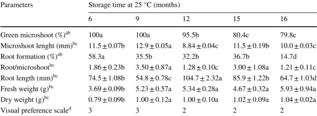
Parameters Storage time at 25 °C (months)
6 9 12 15 16
Green microshoot (%)ab 100a 100a 95.5b 80.4c 79.8c
Microshoot lenght (mm)bc 11.5 ± 0.07b 12.9 ± 0.05a 8.84 ± 0.04c 11.5 ± 0.19b 10.0 ± 0.03c
Root formation (%)ab 58.3a 35.5b 32.2b 36.7b 14.7d
Root/microshootbc 1.86 ± 0.23b 3.50 ± 0.87a 1.28 ± 0.10c 3.00 ± 1.08a 1.21 ± 0.11c Root length (mm)bc 74.5 ± 1.08b 54.8 ± 0.78c 104.7 ± 2.32a 85.9 ± 1.22b 64.7 ± 1.03d Fresh weight (g)bc 3.69 ± 0.09b 5.23 ± 0.57a 5.34 ± 0.28a 4.67 ± 0.32a 5.93 ± 0.94a Dry weight (g)bc 0.79 ± 0.09b 1.00 ± 0.12a 1.00 ± 0.10a 1.02 ± 0.09a 1.04 ± 0.02a
Visual preference scaled 3 3 2 2 2
Fig. 1 Post-storage status of microshoots of fraser photinia contain-ing PGB_invit stored under proliferation conditions (25 °C) together with PCR analysis of genes related with beneficial influence of bacterium. a The status of microshoots after storage for 12 months
(bar = 1.2 cm). b Root formation in fraser photinia microshoots stored for 12 months (bar = 1.2 cm). c Healthy microshoots obtained after 16 months of storage (bar = 1.2 cm)
P.frm<,r
for 16 month]. There is a possibility to obtain about 80% green shoot after storage for 16 months at 25 °C without transferring the plants to fresh medium and not applying low temperature to reduce plant metabolism. After 6 months of conservation in vitro, root development was also occurred on microshoots. The rooted microshoots that were stored for 12 months (were shown in Fig. 1b) and rooted plantlets still could be observed in 16 month-stored microshoots although with a relatively lower percentage (14.7%).
No significant difference was observed in fresh and dry weight of the microshoots that were conserved up to 16 months. However, relatively lower fresh and dry weights (3.69 and 0.79, respectively) were measured in 6 months of conservation. As regard visual preference scale, although relatively lower amount of microshoots with bright green leaves and stems were observed with prolongation of stor-age, they seemed to be healthy with green–brown color (Table 1; Fig. 1a, c).
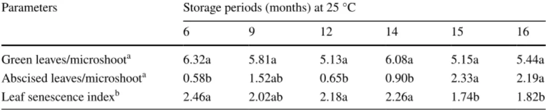
Influences of PGB_invit on number of green/ abscised leaves per microshoot and leaf senescence
There was no significant influence of bacterium on number of green leaves obtained per microshoot (5.13 and 6.32) that conserved up to 16 months (Table 2). However, significantly higher number of abscised leaves and lower leaf senescence index were obtained with conservation of microshoots up to 15 and 16 months.
Influences of PGB_invit on shoot retrieval after in vitro storage
Although the proliferation of shoot apices excised from con-taminated microshoots started to decline after conservation up to 12 months, more than 93% proliferation was obtained after 16 months (Table 3). Moreover, relatively higher num-ber of microshoots was obtained with the prolongation of the storage time as maximum multiple shoot formation was scored with the tested longest storage period. So that the highest SFC index was obtained from shoot apices from microshoots that stored for 9 or 15 months. With increasing the storage time up to 16 months, lower SFC index (4.4) was Table 2 The influences of
PGB_invit on number of green/ abscised leaves per microshoot and leaf senescence after storage of fraser photinia microshoots
Each treatment consisted of at least 50 explants and repeated at least twice
a Means followed by the same letter are not significantly different at P ≤ 0.05 by the ANOVA, followed by the LSD test (i.e., mean significativity is per horizontal lines)
b Leaf senescence index of the microshoots were calculated according to √x/√(y + 0.5) formula
Parameters Storage periods (months) at 25 °C
6 9 12 14 15 16
Green leaves/microshoota 6.32a 5.81a 5.13a 6.08a 5.15a 5.44a
Abscised leaves/microshoota 0.58b 1.52ab 0.65b 0.90b 2.33a 2.19a
Leaf senescence indexb 2.46a 2.02ab 2.18a 2.26a 1.74b 1.82b
Table 3 The influence of PGB_invit on proliferation of shoot apices excised from in vitro-stored fraser photinia microshoots
The data were collected 30 days after culture initiation. Each trial was made with at least 50 explants and the trials were repeated at least 2 times
a The same letters following the percentages show no statistical difference compared to the Post Hoc Multi-ple comparison test (P ≤ 0.05)
b The same letters following the averages show no statistical difference compared to the LSD test following ANOVA (P ≤ 0.05; The average difference was analyzed horizontally)
c Mean ± standard error
d SFC index of microshoots; It is calculated by multiplication the proliferation of the explant by the percent-age of proliferating explants and divided by 100
Parameters Storage time (months)
6 9 12 15 16
Proliferation (%)a 100a 98.9a 95.6b 98.8ab 93.3b
Microshoot/explantbc 4.40 ± 0.17a 3.85 ± 0.18b 3.51 ± 0.14b 4.42 ± 0.18a 4.44 ± 0.13a Microshoot length (cm)bc 3.46 ± 0.14a 3.70 ± 0.15a 3.56 ± 0.01a 4.73 ± 0.14a 4.41 ± 0.14a
obtained due to significantly lower proliferation percentage obtained in that storage period. Also, relatively longer shoots were obtained after 15 and 16 months (4.7 and 4.8 mm, respectively).
Influences of PGB_invit on rooting and acclimatization
Significant differences were obtained on root induction responses between control and contaminated microshoots cultured on different IBA concentration (Table 4). Maximum root formation (70%) was obtained with the tested highest IBA concentration (4.92 µM) in control microshoots whereas 66.6% of rooting was scored with 2.46 µM IBA enriched QL media in contaminated microshoots. Doubling the IBA con-centration resulted in decline as 43.3% of rooting in contami-nated microshoots. Although no significant differences were obtained in number of roots formed per control microshoot in response to different auxin concentrations, the lowest adventitious root formation per contaminated microshoots
(1.2) was obtained with the tested lowest IBA concentration (0.49 µM). On the contrary, the longest roots were obtained in contaminated microshoots cultured on this medium.
Root formation was occurred on control microshoots in between 22 and 27 days while contaminated shoots were rooted in between 27 and 32 days. Except of the roots formed in the QL medium supplemented with the lowest concentration of IBA, no difference was obtained as regard rooting time between contaminated and control microshoots.
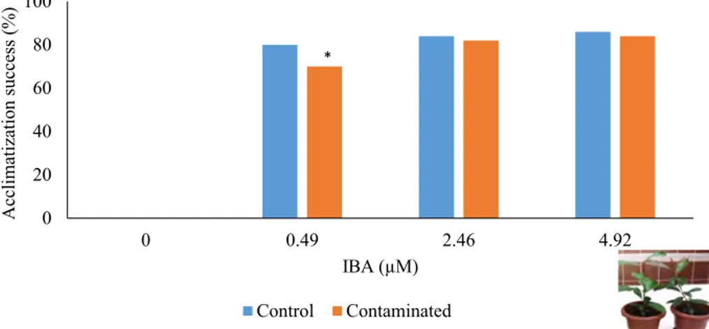
Although spontaneous rooting was observed with the presence of PGB_invit during storage at 25 °C, they were inadequate to support plantlet survival during acclimatiza-tion (Fig. 2). With the inclusion of IBA to the medium, the adventitiously rooted microshoots showed survival during acclimatization to in vivo conditions. Moreover, no statis-tical difference was observed in acclimatization success between control and 12 month in vitro stored contaminated microshoots except the ones that were rooted with the pres-ence of the lowest IBA concentration.
Table 4 Influences of PGB_ invit on root induction
The data were collected 30 days after culture initiation. Each trial was made with at least 50 explants and the trials were repeated at least twice
a The same letters following the percentages show no statistical difference compared to the Post Hoc Multi-ple comparison test (P ≤ 0.05)
b The same letters following the averages show no statistical difference compared to the LSD test following ANOVA (P ≤ 0.05; The average difference was analyzed horizontally)
c Mean ± standard error
d Rooting time of microshoots was calculated according to Σ (N
xTx)/rooting microshoot number. Nx is rooted shoot number; Tx is the number of days between the beginning of the test and the ending of test Parameters IBA concentrations (µM)
0.49 2.46 Control 4.92 0.49 2.46 Contaminated 4.92
Root formation (%)a 40.0b 50.0b 70.0a 40.0b 65.0a 43.0b
Root/microshootbc 2.0 ± 0.6a 1.8 ± 0.2a 1.8 ± 0.3a 1.2 ± 0.2b 1.6 ± 0.1a 2.5 ± 0.5a Root length (cm)bc 1.9 ± 0.6b 1.7 ± 0.4b 1.6 ± 0.4b 3.2 ± 0.6a 2.2 ± 0.3b 2.3 ± 0.3ab
Rooting time(day)d 22.3 26.2 27.6 32.0 27.6 26.5
Fig. 2 Acclimatization results of control and microshoots proliferated from shoot apices excised from 12 month-stored contaminated fraser photinia in vitro cultures on different concentrations of IBA contain-ing medium 0 20 40 60 80 100 0 0.49 2.46 4.92 Acclimatization success (%) IBA (µM) Control Contaminated *
-• •Auxin and cytokinin synthesis
When PCR was performed using aux1 and ipt primers and genomic DNA as a template, no band was obtained. PCR amplification with aux1-FW and aux1-RV primers a sin-gle band of ~ 950 bp for the plasmid DNA of PGB_invit and a single band of 791 bp for the plasmid DNA of R.
rhizogenes ATCC 15834 (Camilleri and Jouanin 1991); with ipt-FW and ipt-RV primers a single band of ~ 800 bp with plasmid DNA of PGB_invit and 427 bp (Haas et al.
1995) with plasmid DNA of R. radiobacter ATCC 15955 were obtained (Fig. 3a). The obtained PCR results show that aux1 and ipt genes in PGB_invit were present in dif-ferent sizes the reference strains. The presence of aux2 genes could not be verified due to the several numbers of non-specific bands obtained from the plasmid DNA as template.
Virulence and nitrogen fixing ability
When PCR with virD1 primers was performed using plasmid DNA of PGB_invit as template, ~ 550 bp sin-gle band (data not shown) was detected. Moreover, expected ~ 750 bp band was obtained with amplification of nifH primers by using bacterial culture (Fig. 3b).
Carbohydrate and alditols production
According to HPLC analysis, no significant difference was obtained between control (MPYE medium) and bacterium, indication that PGB_invit do not secrete tested carbohydrate or alditols into the medium (data not shown).
Discussion
Some endophytic bacteria that are beneficial for host plant can be present naturally in soil and may penetrate the plant and translocate to the above ground organs and, upon colo-nization. Endophytic bacteria can affect the plant growth, health, and productivity positively by enhancing the plant’s capacity for nutrient obtaining, better water management, and/or resistance to abiotic and biotic stresses (some of them may be antagonistic to pathogens) via regulation of hormones and increase expression of defense related genes in plants (Kim et al. 2012). Organisms identified as endo-phytes are usually fungi (Yuan et al. 2016) and bacteria (Fahey et al. 1991; Wilson 1995). Presence of a wide range of common gram positive and negative bacteria including
Enterobacter, Pseudomonas, Staphylococcus, Xanthomonas, Agrobacterium, Methylobacterium spp. have been reported
previously in tissue culture of different plant species (Leif-ert and Cassels 2001; Herman 2004; Kulkarni et al. 2004; Thomas 2004, b, 2007; Thomas et al. 2006). Additionally, Thomas et al. (2008) also identified some uncommon endo-phytic organisms such as Ochrobactrum intermedium,
Alca-ligenes faecalis, Ralstonia mannitolilytica, Oceanobacillus picturae, Bacillus neonatiensis, Brachybacterium, Brevi-bacterium, Kocuria rosea, Tetrasphaera spp. etc. Although
the presence of bacteria in micropropagated plants is gen-erally considered as microbial contamination that must be prevented and eliminated (George et al. 2008; Quambusch et al. 2016), the association of beneficial endophytic bac-teria and micropropagated plants can have positive effects on micropropagation (Dias et al. 2009; Jimtha et al. 2014). For instance, endophytes in tissue cultures of several woody plants showed beneficial influence due to plant growth pro-motion (i.e., Pirttilä et al. 2000; Quambusch et al. 2014; Pham et al. 2017; Perez-Rosales et al. 2018). In accordance with this, PGB_invit exist in in vitro microshoots of fraser photinia did not result any decline in growth and vigor of the cultures.
The culture media used for the proliferation of in vitro plantlets contains minerals, a carbon source, vitamins and generally low concentration of growth regulators. How-ever, in vitro grown plantlets exhausted the nutrients in 2–3 months and therefore they should be transferred frequently (once every 4–6 weeks depending on the species) to fresh media (Ozden-Tokatli et al. 2010). The lengthening of the Fig. 3 PCR analysis of aux1, ipt and nifH genes. a Agarose gel
elec-trophoresis of PCR amplification products obtained with aux1 and
ipt gene primers by using plasmid DNA as template. (1) Size marker,
1 kb DNA ladder (Intron 24074), (2) PCR amplification of
Rhizobac-terium rhizogenes ATCC 15834 plasmid DNA (pRi) with aux1
prim-ers; (3) PCR amplification of endophytic bacterium plasmid DNA with aux1 primers; (4) negative control, (5) PCR amplification of
Rhizobacterium radiobacter ATCC 15955 plasmid DNA (pTi) with ipt primers; (6) PCR amplification of endophytic bacterium plasmid
DNA with ipt primers; and (7) PCR amplification with ipt primers as negative control. b Agarose gel electrophoresis of the nifH gene by using endophytic bacterial culture (1) size Marker, 1 kb and 100 bp DNA Ladders (Thermofisher), (2) PCR amplification of bacterial cul-ture with nifH primers
A
B
subculturing periods through growth rate reduction could be achieved by modification of media components with incor-poration of the culture medium some growth retardants like abscisic acid (ABA) (Kovalchuk et al. 2009) or osmotica like mannitol (Negash et al. 2001; Divakaran et al. 2006) or sucrose (Kovalchuk et al. 2009) together with reduction of the culture temperature usually from 25 to 15 °C (Negash et al. 2001) or 4 °C (Negri et al. 2000; Kovalchuk et al.
2009). In accordance, microshoots of fraser photinia were in vitro-stored at 4 °C up to 15 months on sucrose and man-nitol containing QL medium in both baby food jars and vit-rovents without subculture (Akdemir et al. 2010). However, the presence of PGB_invit enabled the maintenance of the fraser photinia microshoots without any further incorpora-tion of growth retardants to the culture medium or reduc-tion in culture temperature. With the beneficial effect of the bacterium, microshoots could be stored at standard culture medium and conditions for up to 9 months without signifi-cantly losing any viability and vigor. Moreover, all shoot apices excised from 16 months conserved microshoots were capable of resuming or initiating new and organized growth following their transfer to fresh medium.
Alteration of plant growth and development with pro-duction of PGRs (i.e., cytokinins, auxins, etc.) was also reported with the presence of Pseudomonas, Enterobacter,
Staphylococcus, Azotobacter, and Azospirillum (Arshad
and Frankenberger 1991; Leifert et al. 1994; Bashan and Holguin 1997), which some strains of them could also be endophytic. The positive influences of them on plant growth have been attributed to its ability to co-synthesize compounds commonly known as plant products (Zabetakis
1997; Koutsompogeras et al. 2007) and PGRs (Ivanova et al. 2000, 2001; Koenig et al. 2002). Molecular genetic analysis of the PGB_invit reveal that it can produce cyto-kinin and auxin. Thus, the positive influence of endophytic bacteria obtained in in vitro cultures of fraser photinia on growth could also be due to the synthesis of PGRs. The spontaneous rooting observed in microshoots that were cultured on media without any auxin incorpora-tion to medium could also support the presence of opti-mal endogenous levels of PGRs in the original tissues required for rooting (Divakaran et al. 2006). In collabora-tion, it is reported previously that auxin especially IAA synthesis by endophytic bacteria may have not only vari-ous regulatory effects in plant-bacterial interactions but also significant effect on plant growth promotion, i.e., root nodulation (Jasim et al. 2015). For instance, biotization of endophytic plant growth-promoting rhizobacterium (A.
brasilense strain Cd) stimulated in vitro rooting of jojoba
(Perez-Rosales et al. 2018). Moreover, Muromtsev et al. (1987) also reported that the ability of the colonized plants and explants to grow normally on the sucrose-free media and the bright green coloration of the plants infer that; by
producing cytokinins, the methylobacteria promote chloro-plast development and activity. Likewise, this similar posi-tive influence of the isolated bacterium was also observed in contaminated fraser photinia in vitro cultures.
Besides, the beneficial influence of PGB_invit on storage of microshoots at 25 °C in in vitro conditions, it has no nega-tive influence on proliferation of shoot apices excised from stored microshoots. Moreover, there is no statistical differ-ence on rooting and acclimatization results of control and contaminated microshoots, possibly showing the continued beneficial influence of the bacterium. As there is no report on endophytic bacteria that enable to store microshoots in in vitro conditions without subculturing and renewal of the medium such a long-time, endophytes like PGB_invit seemed to be very original and have potential to be used for medium-term storage of plant germplasm.
It should also be noted that diverse species of bacte-ria such as Agrobacterium tumefaciens, Rhizobium sp.,
Sinorhizobium meliloti and Mesorhizobium loti could
transfer genes to plants (Broothaerts et al. 2005). The pres-ence of genes encoding virD1 on the plasmid DNA isolated from PGB_invit may indicate the ability of this bacterium to transfer genes to its host plant as virD1, an endonucle-ase encoded by inducible locus of the virulence (vir) region of the Agrobacterium tumefaciens Ti plasmid, is required for site-specific nicking at T-DNA border sites (Wang et al.
1990).
In our previous study, we showed that the putatively endophytic bacterium may reduce NO3 to NO2 according to its biochemical assays. In this study, ability to fix nitro-gen of PGB_invit was verified by the PCR amplification of nifH gene from this bacterium. Nitrogen is generally a limiting source of plant growth and development. Nif genes encode the enzymes, which are capable of fixing atmos-pheric nitrogen into a form available to plants. Plants only may take nitrogen as ammonia or nitrate forms. Therefore, nitrate reduction by a bacterium is important for the nitrogen availability of the plants (Mbai et al. 2013). In nature there are several nitrogen-fixing bacteria which may benefit the plants (Boddey et al. 1991; Triplett 1996; Malik et al. 1997; Reinhold-Hurek and Hurek 1998; Iniguez et al. 2004).
Although no carbohydrate or alditols production was evident in PGB_invit according to HPLC analysis, the pres-ence of polyhydroxybutyrate (PHB), which was detected in transmission electron microscopy (TEM) analysis (Gul Şeker et al. 2017) could be used as carbon source as it is a carbon reserve of bacteria (Lemoigne 1926) that is syn-thesized when nutrient status is low (Borque et al. 1995; Khosravi-Darani et al. 2013). Thus, PHB accumulation detected in PGB_invit could ensure energy to the bacteria and enabled its survival under metabolic stress. Moreover, it should also be noted that some metabolites (i.e., furanoids and pavettamine) might also be produced by plant bacteria
association (Brader et al. 2014) and this possibility should also be investigated with further analysis.
In conclusion, the presence of PGB_invit in the culture medium and its synergistic effect with its host resulted in a significant improvement in microshoots growth during pro-longed maintenance of fraser photinia shoot cultures in vitro at 25 °C. The positive influence of this bacterium is due to its ability to provide cytokinin and auxin together with its capability to nitrogen fixation. Moreover, the presence of especially virD1 gene is also promising as it may have ability to transfer genes to plant and could be used for future genetic transformation studies. Hence, the isolated bacterium is use-ful for in vitro conservation of fraser photinia germplasm as frequent subculturing can enhance not only the risk of occur-rence of somaclonal variation but also the cost of personnel, energy and materials. It should be noted that PGB_invit will also be inoculated with other plants especially model species (i.e., Arabidopsis thaliana) not only to reveal out its host specificity, but also the molecular mechanism underlying its beneficial influence on plant in our future studies.
Author contributions YÖÇ and ÖA designed the research project; HA and EK carried out the plant storage analyses; IS and ÖA carried out the molecular analysis; IS, ÖA and YÖÇ wrote the paper.
Funding This research was funded by a grant from TUBITAK (#KBAG 114Z579). A partial support was also obtained from Gebze Technical University (2014-A-09).
Compliance with ethical standards
Conflict of interest There is no conflict of interest.
References
Akdemir H, Kaya E, Ozden Y (2010) In vitro proliferation and mini-mum growth storage of fraser photinia: influences of different medium, sugar combinations and culture vessels. Sci Hortic 126:268–275. https ://doi.org/10.1016/j.scien ta.2010.07.005
Akkaya Ö, Gül Şeker M, Özden Çiftçi Y (2019) Plant growth promot-ing microbiome network. In: Dr SJ (ed) Microbes and the environ-ment: plant-soil-microbe-environment interactions on agricultural production, pollution management and waste recycling towards sustainable development (in press)
Aly AH, Debbab A, Kjer J, Proksch P (2010) Fungal endophytes from higher plants: a prolific source of phytochemicals and other bioactive natural products. Fungal Divers 41:1–16. https ://doi. org/10.1007/s1322 5-010-0034-4
Arshad M, Frankenberger WT (1991) Microbial production of plant hormones. In: Keister DL, Cregan PB (eds) The rhizosphere and plant growth, Kluwer Academic Publishers, Dordrecht, pp 327– 334. https ://doi.org/10.1007/BF000 11893
Azevedo JL, Maccheroni Junior W, Pereira JO, Araújo WL (2000) Endophytic microrganisms: a review on insect control and recent advances on tropical plants. Electron J Biotechnol 3:40–65. https ://doi.org/10.2225/vol3-issue 1-fullt ext-4
Bashan Y, Holguin G (1997) Azosprillum-plant relationships: envi-ronmental and physiological advances (1990–1996). Can J Microbiol 43:103–121. https ://doi.org/10.1139/m97-015
Benhamou N, Kloepper JW, Quadth-Hallman A, Tuzun S (1996) Induction of defense-relted ultrastructural modifications in pea root tissues inoculated with endophytic bacteria. Plant Physiol 112:919–929. https ://doi.org/10.1104/pp.112.3.919
Boddey RM, Urquiaga S, Reis V, Döbereiner J (1991) Biologi-cal nitrogen fixation associated with sugar cane. Plant Soil 137:111–117. https ://doi.org/10.1007/BF000 32247
Borque D, Pomerleau Y, Groleau D (1995) High cell density pro-duction of polybhydroxybutyrate (PHB) from methanol by
Methylobacterium extorquens production of high molecular
mass PHB. Appl Microbiol Biotechnol 44:367–376. https :// doi.org/10.1007/BF001 69931
Brader G, Compant S, Mitter B, Trognitz F, Sessitsch A (2014) Met-abolic potential of endophytic bacteria. Curr Opin Biotechnol Sci 27:30–37. https ://doi.org/10.1016/j.copbi o.2013.09.012
Broothaerts W, Mitchell HJ, Weir B, Kaines S, Smith LMA, Yang W, Mayer JE, Roa-Rodriguez C, Jefferson RA (2005) Gene transfer to plants by diverse species of bacteria. Nature 433:629–633.
https ://doi.org/10.1038/natur e0330 9
Cabanas CGL, Schiliro E, Valverde-Corredor A, Mercado-Blanco J (2014) The biocontrol endophytic bacterium Pseudomonas
fluo-rescens PICF7 induces systemic defense responses in aerial
tis-sues upon colonization of olive roots. Front Microbiol 5:1–14.
https ://doi.org/10.3389/fmicb .2014.00427
Camilleri C, Jouanin L (1991) The TR_DNA region carrying the auxin synthesis genes of the Agrobacterium rhizogenes agro-nine-type plasmid pRiA4: nucleotide sequence analysis and introduction into tobacco plants. Mol Plant Microbe Interact 4:155–162. https ://doi.org/10.1094/MPMI-4-155
Card S, Johnson L, Teasdale S, Caradus J (2016) Deciphering endo-phyte behavior: the link between endoendo-phyte biology and effica-cious biological control agents. FEMS Microbiol Ecol 92:1–19.
https ://doi.org/10.1093/femse c/fiw11 4
Dias ACF, Costa FEC, Andreote FD, Lacava PT, Teixeira MA, Assumpçao LC, Araujo WL, Azevedo JL, Melo IS (2009) Iso-lation of micropropagated strawberry endophytic bacteria and assessment of their potential for plant growth promotion. World J Microbiol Biotechnol 25:189–195. https ://doi.org/10.1007/ s1127 4-008-9878-0
Divakaran M, Babu KN, Peter KV (2006) Conservation of
Vanilla species in vitro. Sci Hortic 110:175–180. https ://doi. org/10.1016/j.scien ta.2006.07.003
Fahey JW, Dimock MB, Tomasino SF, Taylor JM, Carlson PS (1991) Genetically engineered endophytes as biocontrol agents: a case study in industry. In: Brock TD (ed) Microbial ecol-ogy of leaves. Springer, New York, pp 402–411. https ://doi. org/10.1007/978-1-4612-3168-4
George EF, Hall MA, De Klerk GJ (2008) Plant propaga-tion by tissue culture. Springer, Dordrecht. https ://doi. org/10.1007/978-1-4020-5005-3_8
Gond SK, Bergen MS, Torres MS, White JF (2015) Endophytic
Bacillus spp. produce antifungal lipopeptides and induce host
defence gene expression in maize. Microbiol Res 172:79–87.
https ://doi.org/10.1016/j.micre s.2014.11.004
Gül-Şeker M, Şah I, Kırdök E, Ekinci H, Özden-Çiftçi Y, Akkaya Ö (2017) A hidden plant growth promoting bacterium iso-lated from in vitro cultures of fraser photinia. Int J Agric Biol 19:1511–1519. https ://doi.org/10.17957 /IJAB/15.0455
Haas JH, Moore LW, Ream W, Manulis S (1995) Universal PCR primers for detection of phytopathogenic Agrobacterium strains. Appl Environ Microbiol 61:2879–2884
Hallman J, Quadt-Hallmann A, Mahaffee WF, Kloepper JW (1997) Bacterial endophytes in agricultural crops. Can J Microbiol 43:895–914. https ://doi.org/10.1139/m97-131
Hardoim PR, van Overbeek LS, van Elsas JD (2008) Properties of bac-terial endophytes and their proposed role in plant growth. Trends Microbiol 16:463–471. https ://doi.org/10.1016/j.tim.2008.07.008
Hardoim PR, van Overbeek LS, Berg G, Pırttila AM, Compant S, Campisano A, Döring M, Sessitsch A (2015) The hidden world within plants: ecological and evolutionary considerations for defining functioning of microbial endophytes. Microbiol Mol Biol Rev 79:293–320. https ://doi.org/10.1128/MMBR.00050 -14
Herman EB (2004) Recent advances in plant tissue culture viii. Micro-bial contaminants in plant tissue cultures: solutions and opportuni-ties 1996–2003. Agritech Consultants, Inc, New York
Iniguez AL, Dong Y, Triplett EW (2004) Nitrogen fixation in wheat provided by Klebsiella pneumonia 342. Mol Plant Microbe Inter-act 17:1078–1085. https ://doi.org/10.1094/MPMI.2004.17. Ivanova EG, Doronina NV, Shepelyakovskaya AO, Laman AG, Brovko
FA, Trotsenko YuA (2000) Facultative and obligate aerobic meth-ylobacteria synthesize cytokinins. Mikrobiologiya 69:764–769.
https ://doi.org/10.1023/A:10266 93805 653
Ivanova EG, Doronina NV, Trotsenko YA (2001) Aerobic methylobac-teria are capable of synthesizing auxins. Microbiology 70:392– 397. https ://doi.org/10.1023/A:10104 69708 107
James EK (2000) Nitrogen fixation in endophytic and associative symbiosis. Field Crops Res 65:197–209. https ://doi.org/10.1016/ S0378 -4290(99)00087 -8
Jasim B, Geethu PR, Mathew J, Radhakrishnan EK (2015) Effect of endophytic Bacillus sp. from selected medicinal plants on growth promotion and diosgenin production in Trigonella foenum grae-cum. Plant Cell Tissue Organ Cult 122:565–572. https ://doi. org/10.1007/s1124 0-015-0788-1
Jimtha JC, Smitha PV, Anisha C, Deepthi T, Meekha G, Radhakrishnan EK, Gayatri GP, Remakanthan A (2014) Isolation of endophytic bacteria from embryogenic suspension culture of banana and assessment of their plant growth promoting properties. Plant Cell Tissue Organ Cult 118:57–66. https ://doi.org/10.1007/s1124 0-014-0461-0
Khosravi-Darani K, Mokhtari Z, Amai T, Tanaka K (2013) Micro-bial production of poly (hydroxybutyrate) from C1 carbon sources. Appl Microbiol Biotechnol 97:1407–1424. https ://doi. org/10.1007/s0025 3-012-4649-0
Kim S, Lowman S, Hou G, Nowak J, Flinn B, Mei C (2012) Growth promotion and colonization of switchgrass (Panicum
vir-gatum) cv. Alamo by bacterial endophyte Burkholderia phyto-firmans strain PsJN. Biotechnol Biofuels 5:37. https ://doi. org/10.1186/1754-6834-5-37
Koenig RL, Morris RO, Polacco JC (2002) tRNA is the source of low-level trans-zeatin production in Methylobacterium spp. J Bacteriol 184:1832–1842. https ://doi.org/10.1128/ JB.184.7.1832-1842.2002
Koutsompogeras P, Kyriacou A, Zabetakis I (2007) The formation of 2,5-dimethyl-4-hydroxy-2H-furan-3-one by cell free extracts of Methylobacterium extorquens and strawberry (Fraga-ria
ananassa cv. Elsanta). Food Chem 104:1654–1661. https ://doi. org/10.1016/j.foodc hem.2007.03.025
Kovalchuk I, Lyudvikova Y, Volgina M, Reed BM (2009) Medium, container and genotype all influence in vitro cold storage of apple germplasm. Plant Cell Tissue Org Cult 96:127–136. https ://doi. org/10.1007/s1124 0-008-9468-8
Kulkarni VM, Ganapathi TR, Bapat VA, Rao PS (2004) Establishment of cell-suspension cultures in banana cv. Gr and Naine and evalu-ation of its sensitivity to gamma-irradievalu-ation. Curr Sci 86:902–904 Lambardi M, Sharma KK, Thorpe TA (1993) Optimization of in vitro
bud induction and plantlet formation from mature embryos of
Aleppo pine (Pinus halepensis Mill.). In Vitro Cell Dev Biol 29:189–199. https ://doi.org/10.1007/BF026 32034
Leifert C, Cassells AC (2001) Microbial hazards in plant tissue and cell cultures. In vitro Cell Dev Biol Plant 37:133–138. https ://doi. org/10.1007/s1162 7-001-0025-y
Leifert C, Morris CE, Waites WM (1994) Ecology of microbial sapro-phytes and pathogens in tissue culture and field grown plants. CRC Crit Rev Plant Sci 13:139–183. https ://doi.org/10.1080/07352 68940 97019 12
Lemoigne M (1926) Produit de d´eshydratation et de polym´erisation de l’acide b-oxybutyrique. Bull Soc Chem Biol 8:770–782 Malboobi MA, Owlia P, Behbahani M, Srakhani E, Moradi S,
Yakh-chali B (2009) Solubilization of organic and inorganic phosphates by three highly efficient soil bacterial isolates. World J Micro-biol Biotechnol 25:1471–1477. https ://doi.org/10.1007/s1127 4-009-0037-z
Malik KA, Bilal R, Mehnaz S, Rasul G, Mirza MS, Ali S (1997) Asso-ciation of nitrogen-fixing, plant-growth-promoting rhizobacteria (PGPR) with Kallar grass and rice. Plant Soil 194:37–44. https :// doi.org/10.1023/A:10042 95714 181
Marascuilo LA, McSweeney M (1977) Nonparametric and distribution-free method for the social sciences. CA Brooks/Cole Publishing Company, Monterey
Mbai FN, Magiri EN, Matiru VN, Ng’ang’a J, Nyambati VCS (2013) Isolation and characterization of bacterial root endophytes with potential to enhance plant growth from Kenyan basmati rice. Am Int J Contemp Res 3:25–40. https ://doi.org/10.30845 /aijcr
Mercado-Blanco J (2015) Life of microbes inside the plant. In: Lugten-berg B (ed) Principles of plant-microbe interactions. Springer, Switzerland, pp 25–32. https ://doi.org/10.1007/978-3-319-08575 -3
Muromtsev GS, Chkanikov DI, Kulaeva ON, Gamburg KZ (1987) Osnovy khimicheskoi regulyatsii rosta I productivnosti rastenii (Basics of chemical regulation of plant growth and productivity). Agropromizdat, Moscow
Nair DN, Padmavathy S (2014) Impact of endophytic microorganisms on plants, environment and humans. Sci World J 2014:1–12. https ://doi.org/10.1155/2014/25069 3
Negash A, Krens F, Schaart J, Vısser B (2001) In vitro conservation of enset under slow-growth conditions. Plant Cell Tissue Organ Cult 66:107–111. https ://doi.org/10.1023/A:10106 47905 508
Negri V, Tosti N, Standardi A (2000) Slow-growth storage of single node shoots of apple genotypes. Plant Cell Tissue Organ Cult 62:159–162. https ://doi.org/10.1023/A:10267 09706 535
Ozden-Tokatli Y, Akdemir H, Tilkat E, Onay A (2010) Current status and conservation of Pistacia germplasm. Biotechnol Adv 28:130– 141. https ://doi.org/10.1016/j.biote chadv .2009.10.006
Patle PN, Navnage NP, Ramteke PR (2018) Endophytes in plant sys-tem: roles in growth promotion, mechanism and their potentiality in achieving agriculture sustainability. Int J Chem Stud 6:270–274 Perez-Rosales E, Alcaraz-Meléndez L, Puente ME, Vázquez-Juárez R, Zenteno-Savín T, Morales-Bojórquez E (2018) Endophytic bacte-ria isolated from wild jojoba (Simmondsia chinensis L. [Schnei-der]) roots improve in vitro propagation. Plant Cell Tissue Organ Cult 135:515–522. https ://doi.org/10.1007/s1124 0-018-1483-9
Pham NT, Meier-Dinkel A, Höltken AM, Quambusch M, Mahnkopp F, Winkelmann T (2017) Endophytic bacterial communities in
in vitro shoot cultures derived from embryonic tissue of hybrid
walnut (Juglans x intermedia). Plant Cell Tissue Organ Cult 130:153–165. https ://doi.org/10.1007/s1124 0-017-1211-x
Pirttilä AM, Laukkanen H, Pospiech H, Myllyla R, Hohtola A (2000) Detection of intracellular bacteria in the buds of Scotch pine (Pinus sylvestris L.) by in situ hybridization. Appl Environ Microbiol 66:3073–3077. https ://doi.org/10.1128/ AEM.66.7.3073-3077.2000
Quambusch M, Pirttila AM, Tejesvi MV, Winkelmann T, Bartsch M (2014) Endophytic bacteria in plant tissue culture: differences between easy- and difficult-to-propagate Prunus avium genotypes. Tree Physiol 34:524–533. https ://doi.org/10.1093/treep hys/tpu02 7
Quambusch M, Brümmer J, Haller K, Winkelmann T, Bartsch M (2016) Dynamics of endophytic bacteria in plant in vitro cul-ture—quantification of three bacterial strains in Prunus avium in different plant organs and in vitro culture phases. Plant Cell Tissue Organ Cult 126:305–317. https ://doi.org/10.1007/s1124 0-016-0999-0
Quoirin M, Lepoivre P (1977) Etude de milieux adaptés aux cultures
in vitro de Prunus sp. Acta Hortic 78:437–442
Reinhold-Hurek B, Hurek (1998) Life in grasses: diazotrophic endo-phytes. Trends Microbiol 139:1–6. https ://doi.org/10.1016/S0966 -842X(98)01229 -3
Remy W, Taylor TN, Haas H, Kerp H (1994) Four hundred-million-year-old vesicular arbuscular mycorrhizae. Proc Natl Acad Sci 91:11841–11843. https ://doi.org/10.1073/pnas.91.25.11841
Rogowsky PM, Powell BS, Shirasu K, Lin TS, Morel P, Zyprian EM, Steck TR, Kado CI (1990) Molecular characterization of the vir regulon of Agrobacterium tumefaciens: complete nucleotide sequence and gene organization of the 28.63-kbp regulon cloned as a single unit. Plasmid 23:85–106. https ://doi.org/10.1016/0147-619x(90)90028 -B
Ryan RP, Germaine K, Franks A, Ryan DJ, Dowling DN (2008) Bacterial endophytes: recent developments and aplica-tions. FEMS Microbiol Lett 278:1–9. https ://doi.org/10.111 1/j.1574-6968.2007.00918 .x
Santoyo G, Moreno-Hagelsieb G, Orozco-Mosqueda C, Glick BR (2016) Plant growth-promoting bacterial endophytes. Microbiol Res 183:92–99. https ://doi.org/10.1016/j.micre s.2015.11.008
Sarkar D, Naik PS (1998) Factors affecting minimal growth conserva-tion of potato microplants in vitro. Euphytica 102:275–280 Sarkar D, Kaushik SK, Naik PS (1999) Minimal growth conservation
of potato microplants silver thiosulfate reduces ethylene-induced growth abnormalities during prolonged storage in vitro. Plant Cell Rep 18: 897–903
Schulz B, Boyle C, Draeger S, Römmert AK, Krohn K (2002) Endo-phytic fungi: a source of novel biologically active secondary metabolites. Mycol Res 106:996–1004. https ://doi.org/10.1017/ S0953 75620 20063 42
Singh M, Kumar A, Singh R, Pandey KD (2017) Endophytic bacteria: a new source of bioactive compounds. 3Biotech 7:1–14. https :// doi.org/10.1007/s1320 5-017-0942-z
Thomas P (2004a) In vitro decline in plant cultures: detection of a legion of covert bacteria as the cause for degeneration of long-term micropropagated triploid walong-termelon cultures. Plant Cell Tissue Org Cult 77:173–179. https ://doi.org/10.1023/ B:TICU.00000 16824 .09108 .c8
Thomas P (2004b) A three-step screening procedure for detection of covert and endophytic bacteria in plant tissue cultures. Curr Sci 87:67–72
Thomas P (2007) Isolation and identification of five alcohol defying
Bacillus spp. covertly associated with in vitro culture of seedless
watermelon. Curr Sci 92:983–987
Thomas P, Prabhakara BS, Pitchaimuthu M (2006) Cleansing the long-term micropropagated triploid walong-termelon cultures from covert bacteria and field testing the plants for clonal fidelity and fertility during the 7–10 year period in vitro. Plant Cell Tissue Org Cult 85:317–329. https ://doi.org/10.1007/s1124 0-006-9083-5
Thomas P, Swarna GK, Roy PK, Patil P (2008) Identification of cul-turable and originally non-culcul-turable endophytic bacteria iso-lated from shoot tip cultures of banana cv. Grand Naine. Plant Cell Tissue Organ Cult 93:55–63. https ://doi.org/10.1007/s1124 0-008-9341-9
Triplett EW (1996) Diazotrophic endophytes: progress and prospects for nitrogen fixation in monocots. Plant Soil 186:29–38. https :// doi.org/10.1007/BF000 35052
Vigani G, Rolli E, Marasco R, Dell’Orto M, Michoud G, Soussi A, Raddadi N, Borin S, Sorlini C, Zocchi G, Daffonchio D (2018) Root bacterial endophytes confer drought resistance and enhance expression and activity of a vacuolar H+- pumping pyroph-osphatease in pepper plants. Environ Microbiol. https ://doi. org/10.1111/1462-2920.14272
Wang K, Herrera-Estrella A, Van Montagu M (1990) Overexpression of virD1 and virD2 genes in Agrobacterium tumefaciens enhances T-complex formation and plant transformation. J Bacteriol 172:4432–4440. https ://doi.org/10.1128/jb.172.8.4432-4440.1990
Wilson D (1995) Endophyte-the evolution of a term and clarifica-tion of its use and definiclarifica-tion. Oikos 73:274–276. https ://doi. org/10.2307/35459 19
Yuan J, Zhou JY, Li X, Dai CC (2016) The primary mechanism of endophytic fungus Gilmaniella sp. AL12 promotion of plant growth and sesquiterpenoid accumulation in Atractylodes
lan-cea. Plant Cell Tissue Organ Cult 125:571–584. https ://doi. org/10.1007/s1124 0-016-0971-z
Zabetakis I (1997) Enhancement of flavour biosynthesis from straw-berry (Fragaria ananassa) callus cultures by Methylobacterium species. Plant Cell Tissue Org Cult 50:179–183. https ://doi. org/10.1023/A:10059 68913 237
Publisher’s Note Springer Nature remains neutral with regard to jurisdictional claims in published maps and institutional affiliations.