Idiopathic Hypereosinophilic Syndrome Presenting With Acute
Heart Failure and Mesenteric Thromboembolism: Review of the
Literature
*
Akut Kalp Yetmezliği ve Mezenterik Tromboembolizm ile Ortaya Çıkan İdiyopatik Hipereozinofilik Sendrom: Literatürün Gözden Geçirilmesi
Hüseyin Göksülük
1, Demet Menekșe Gerede
1, İrem Müge Akbulut
1, Mustafa Fırat Coșkun
1,
Cebrail Yarlıoğlueș
1, İrem Dinçer
11 Ankara University, Cardiology Department, Ankara, Turkey * Case report was accepted as poster presentation on 32. Turkish
Society Of Cardiology Congreess (2016).
İdiyopatik hipereozinofilik sendromun kardiyak tutulumuna bağlı gelișen akut kalp yetmezliği kötü prognoza ve yüksek mortalite hızına sahiptir. Burada, akut kalp yetmezliği ve mezenterik tromboembolizme sebep olan hipereozinofilik sendromlu 52 yașında bir erkek hastayı sunuyoruz. Akut kalp yetmezliğinin ayırıcı tanısında ST yükselmeli olmayan myokart infarktüsü (troponin yüksekliği) düșünüldü. Ancak akut koroner sendrom tanısı koroner anjiyografi ile dıșlandı. Laboratuvar bulgularında hipereozinofili vardı. Kardiyak manyetik rezonans görüntülemede (MR) difüz sol ventriküler genișleme, duvar hareket bozukluğu ve subendokardiyal fibrozis olmaksızın apikal trombüs izlendi. Hasta kalp yetmezliği kılavuzuna göre tedavi edildi ve hastanın kliniği düzeldi. Kortikosteroid tedavisi ile eozinofil sayısı ve CRP seviyesi normale sınırlara geriledi. Buna rağmen masif mezenterik tromboembolizm nedeniyle hastanın klinik durumu kötülești ve kardiyojenik șok ve sepsis nedeniyle hasta kaybedildi. Hastalığın patogenezinde, eozinofil kaynaklı granüller, kardiyak hasardan ve akut kalp yetmezliğinden endomyokardiyal fibrozise kadar değișen klinik çeșitlilikten sorumlu olabilir. Erken tanı ve tedavi ile morbidite ve mortalite önlenebilir.
Anahtar Sözcükler: Hipereozinofilik sendrom, eozinofili, kalp yetmezliği, tromboembolizm.
Acute heart failure due to cardiac involvement of idiopathic hypereosinophilic syndrome has poor prognosis and high mortality rate. We present a 52 year old male patient with hypereosinophilic syndrome resulting in acute heart failure and mesenteric thromboembolism. Non ST elevation myocardial infarction (high troponin levels) was considered in the differential diagnosis of acute heart failure. However acute coronary syndrome was excluded with diagnostic coronary angiography. The laboratory results showed hypereosinophilia on the blood count. Cardiac magnetic resonance imaging (CMR) showed diffuse left ventricular dilatation, wall motion abnormalities and left ventricular apical thrombus without subendocardial fibrosis. He was treated according to heart failure guidelines and the patient status improved clinically. With the corticosteroid treatment, eosinophil count, C-reactive protein levels returned to normal range. Nevertheless, his clinical condition deteriorated due to massive mesenteric thromboembolism and died after cardiogenic shock and sepsis. In the pathogenesis of the disease, eosinophil derived granüls may be responsible for the cardiac damage and clinical variability that was ranging from cardiogenic shock to endomyocardial fibrosis. Early diagnosis and treatment can prevent morbidity and mortality.
Key Words: Hypereosinophilic syndrome, eosinophilia, heart failure, thromboembolism.
Hypereosinophilic syndrome (HES) is a myeloproliferative disorder characterized by primary eosinophilia (persist for longer than 6 months) without secondary causes and multiple organ damage (1). HES has multiple clinical manifestations. Cardiac system is the most frequently affected system and cardiac complications are a leading cause of morbidity and mortality (2). The first presentation of the disease can be mural thrombus with endomyocardial fibrosis. Mitral and tricuspid valve involvement can
contribute heart failure and symptomatology (3). However, acute heart failure due to eosinophil-mediated myocytes damage is a rare presentation of HES. Acute heart failure with thromboembolism are associated with high mortality rate (4). Glucocorticoids are the first-line therapy for disease. Antineoplastic agents (e.g., vincristine, etoposide, imatinib) and immunomodulators (e.g. peginterferon alfa 2a, interferon alfa 2b) may be used in patients whose are refractory to steroid treatment (5).
AnkaraÜniversitesiTıpFakültesiMecmuası 2018, 71 (1) DOI: 10.1501/Tıpfak_000000999 DAHİLİ TIP BİLİMLERİ/ MEDICAL SCIENCES
Olgu Sunumu / Case Report
Received: Oct. 30, 2017 Accepted: Nov. 08, 2017 Corresponding Author: Huseyin Goksuluk E-mail: [email protected] GSM: +90 (532) 548 61 23 Phone: +90 (312) 508 25 27 Fax: +90 (312) 312 52 51
Ankara University, İbni Sina Hospital, Cardiology Department Sıhhiye, 06410 Ankara/Turkey.
Ankara Üniversitesi Tıp Fakültesi Mecmuası 2018, 71 (1)
Idiopathic Hypereosinophilic Syndrome Presenting With Acute Heart Failure and Mesenteric Thromboembolism: Review of the Literature
66
In this case report, we present a patient with HES, presenting with acute heart failure and mesenteric thromboembolism.
Case Presentation
A 52 year-old man admitted to our emergency department with dyspnea, tachypnea. His previous medical history was insignificant. He was hemodynamically unstable (blood pressure 85/55 mmHg; heart rate 112/ bpm, saturation 80% with 5 liters per minute of oxygen inhalation) on admission. His physical examination revealed a S3 and fine inspiratory crackles in both lung zones up to mid segments. His electrocardiogram (ECG) was consistent with sinus tachycardia and 1.5-2 mm ST segment depressions in precordial leads V1-V4. The chest X-ray revealed alveolar edema and bilateral pleural effusion. His cardiac biomarkers were elevated (troponin I=4.02 ng/ml, normal range: 0-0.06 ng/ml). Routine blood tests revealed an elevated C-reactive protein level (150 mg/L, normal range 0-5 mg/L), erythrocyte sedimentation rate (48 mg/dl, normal range 0-20 mm/hour), eosinophil count (2.68x10⁹ cells/L, normal range 0-0.7x
109 cells/L) with mild leukocytosis. The
eosinophil count measured in another center 4 months ago was normal. Consecutive eosinophil levels were detected elevated in clinical course before the corticosteroid treatment (3.1x10⁹-3.4x10⁹-2.9x10⁹). The
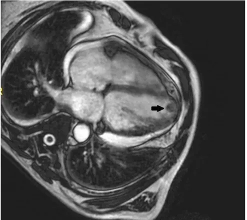
transthoracic echocardiography showed diffuse hypokinesia, mild to moderate pericardial effusion and left ventricular apical thrombus with the low ejection fraction (EF: %20) (Figure 1). Non-ST elevation myocardial infarction was considered in the differential diagnosis of acute heart failure. However acute coronary syndrome was excluded with diagnostic coronary angiography that showed normal coronary anatomy. Cardiac magnetic resonance imaging (MRI) showed diffuse left ventricular dilatation, wall motion abnormality and left ventricular apical thrombus
Figure 1: The transthoracic echocardiography with diffuse ventricular dilatation and large apical
thrombi (arrow) (performed at the admission)
Figure 2: The left ventricle are shown in long axis planes in cardiac magnetic resonance
imaging which was performed 30 days later from the admission: left ventricular dilatation and apical trombi (black arrow).
Journal of Ankara University Faculty of Medicine 2018, 71 (1)
Hüseyin Göksülük, Demet Menekșe Gerede, İrem Müge Akbulut, Mustafa Fırat Coșkun, Cebrail Yarlıoğlueș, İrem Dinçer 67
(Figure 2). Other causes to be
considered in the differential diagnosis
of hypereosinophilic syndrome were excluded: e.g., allergic diseases, collagen vascular diseases, drug reactions, eosinophilic pneumonia, malignancy with secondary eosinophilia (Hodgkin disease, acute myeloid leukemia M4 with bone marrow eosinophilia), parasitic infections. Bone marrow aspiration and biopsy was performed for the detection of an atypical presentation of chronic
eosinophilic leukemia. Biopsy showed
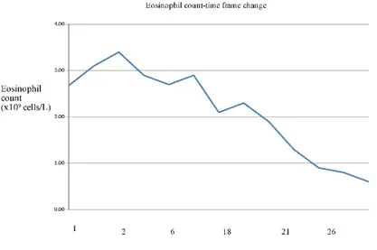
eosinophilic granulocytes rich material (21% eosinophils) without FIP1L1/ PDGFRA fusion gene and JAK2 V617F mutation. Diuretics, low dose beta-blockers, angiotensin converting enzyme (ACE) inhibitors, mineralocorticoid receptor antagonists were started stepwise for heart failure based on the current guidelines. Anticoagulant treatment with warfarin initiated immediately. When the patient began to improve clinically, methylprednisolone (1000 mg a day for three days) was started. The eosinophil count (Figure 3) and C-reactive protein levels returned to normal range with the corticosteroid treatment. However, no improvement was observed in left ventricular function. Apical thrombus increased in size despite the anticoagulant therapy. The patient's status suddenly deteriorated with abdominal pain and dyspnea. Massive mesenteric thromboemboli (Figure 4) was occurred 35 days later from the admission, while the INR and PT levels were effective (2.1 and 22 second). The patient died because of bacterial translocation, sepsis and shock.
Discussion
Idiopatic hypereosinophilic syndrome (IHES) is a heterogeneous group of conditions defined by Chusid, with the criteria of: (a) a sustained elevated eosinophil count of >1.5×10⁹/L (HE) in peripheral blood (b)
no secondary causes of hypereosinophilia, (c) signs and symptoms of multiple organ involvement (6). There is a male predominance in hypereosinophilic
syndrome which is most commonly
diagnosed in patients aged 20-50 years (7).
Figure 3: Changing of eosinophil count with time frame (Corticosteroid treatment was started
15 days later after hospitalization)
Figure 4: Computed tomography showing mesenteric emboli (arrow).
Table 1: Case series of hypereosinophilic syndrome patients with heart failure and thrombus
formation.
References Gender Age Eosinophil
Count Modality Imaging Failure Heart Thrombi Location Outcome Yu IT (12) F 2 150,000/μl Echo,
autopsy + Biventricular mural, inflow tract thrombi
LV and RV Fatal
Tanino M (13) F 68 - Autopsy + Posterior mitral
valve LV Fatal
Christen R (14) F 32 - Autopsy + None LV Fatal Rasche RFH (15) F 5 83 % Autopsy + Extensive mural
trombi LV and RV Fatal Niemeijer ND
(16) M 37 87% Echo, MRI + Posterior leaflet of the mitral valve
LV Surgical replacement of mitral valve, good
Echo: Echocardiography, F:Female, LV: Leftventricle, MRI: Magnetic resonance imaging, RV: Right ventricle
Ankara Üniversitesi Tıp Fakültesi Mecmuası 2018, 71 (1)
Idiopathic Hypereosinophilic Syndrome Presenting With Acute Heart Failure and Mesenteric Thromboembolism: Review of the Literature
68
The cardiac system is one of the most frequently involved systems (75% of patients with HES) and cardiac
complications are the major cause of
morbidity and mortality (8, 9).Heart
failure, intracardiac thrombus, myocardial ischemia, arrhythmia, pericarditis and syncope are the clinical manifestations of cardiac involvement (10). Cardiac damage typically occurs in 3 stages: acute necrosis, thrombotic stage and fibrotic stage (2). The acute necrosis stage is characterised by asymptomatic eosinophilic infiltration of the myocardium. On the second stage, thrombus formation occurs along with the damaged endocardium typically at the apex of the ventricles (11). Thromboembolic complications may develop up to 25% of patients. Mortality rates can be as high as 10% (10). In the last stage, hyaline fibrosis replaces the thrombus and restrictive cardiomyopathy occurs. Common symptoms in these phases include chest pain, dyspnea, or orthopnea.
Nearly 75% of the patients present with signs and symptoms of heart failure.
We searched the literature in “PubMed” ("hypereosinophilic syndrome" [MESH] OR eosinophilia [MESH] OR loeffler) AND ("Heart failure" OR "cardiac failure") AND (thromboembolism). Five case report of hypereosinophilic syndrome associated with heart failure and thromboembolism were detected. Table 1 shows the results: majority of patients had poor outcomes with 80% mortality rates despite the treatment. In one patient, surgical replacement was performed to the mitral valve which was embedded by large thrombus. All patients were admitted to the hospital with acute heart failure. Initiating steroid treatment, which may
worsen heart failure, can create dilemma while waiting specific tests for the diagnosis of HES in the patient who was treated for acute heart failure. In such
cases, detection of diffuse subendocardial fibrosis on the late gadolinium enhancement with MRI may help to make the diagnosis (17). Endomyocardial biopsy (EMB) is the gold standart for the definite diagnosis, altough it has complications such as bleeding, pneumothorax, infection, ventricular perforation, arrhythmia, conduction abnormality. Also, biopsy material can be inadequate for the diagnosis, because of patchy distributed myocarditis (18). Treatment responce is usually more important than the laboratory results. Glucocorticoids decrease the eosinophil counts and prevent end-organ damage by inhibiting the production of the inflammatory mediators.
In conclusion, idiopathic hypereosinophilic syndrome is a rare condition. As a result, HES and associated eosinophilic myocarditis should be included in the differential diagnoses of a patient with acute heart failure. Patients should be monitored carefully for the potential complications of the disease.
REFERENCES
1. Simon HU, Rothenberg ME, Bochner BS et al. Refining the definition of hypereosinophilic syndrome. J Allergy Clin Immunol. 2010;126:45-49.
2. Mankad R, Bonnichsen C, Mankad S. Hypereosinophilic syndrome: cardiac diagnosis and management. Heart 2016;102(2):100-6. 3. Ommen SR, Seward JB, Tajik AJ. Clinical and
echocardiographic features of hypereosinophilic syndromes. Am J Cardiol 2000;86(1):110-3.
4. Kleinfeldt T, Nienaber CA, Kische S et al. Cardiac manifestation of the hypereosinophilic syndrome: newinsights. Clin Res Cardiol 2010;99(7):419-27.
5. Gotlib J. World Health Organization-defined eosinophilic disorders: 2014 update on diagnosis, risk stratification, and management. Am J Hematol 2014 Mar. 89(3):325-37. 6. Chusid MJ, Dale DC, West BC et al. The
hypereosinophilic syndrome: analysis of fourteen cases with review of the literature. Medicine (Baltimore). 1975;54(1): 1-27. 7. Weller PF, Bubley GJ. The idiopathic
hypereosinophilic syndrome. Blood. 1994;83(10):2759-79.
8. Cincin AA, Ozben B, Tanrikulu MA et al. Large apical thrombus in a patient with persistent heart failure and hypereosinophilia: Löffler endocarditis. J Gen Intern Med 2008; 23:1713-8.
9. Fuzellier JF, Chapoutot L, Torossian PF et al. Mitral valve replacement in idiopathic eosinophilic endocarditis without peripheral eosinophilia. J Card Surg 2005; 20(5):472-4. 10. Ogbogu PU, Rosing DR, Horne MK.
Cardiovascular manifestations of hypereosinophilic syndromes. Immunol Allergy Clin North Am 2007;27(3):457-75. 11. Gottdiener JS, Maron BJ, Schooley RT et al.
Two-dimensional echocardiographic assessment of the idiopathic hypereosinophilic syndrome. Anatomic basis of mitral regurgitation and peripheral embolization. Circulation 1983;67(3):572-8.
12. Yu IT, Chiang H, Wang JH. Hypereosinophilic syndrome with Löffler's endomyocarditis: report of an autopsy case. Taiwan Yi Xue Hui Za Zhi. 1989;88(7):740-3.
13. Tanino M, Kitamura K, Ohta G et al. Hypereosinophilic syndrome with extensive myocardial involvement and mitral valve
thrombus instead of mural thrombi. Acta Pathol Jpn. 1983;33(6):1233-42.
14. Christen R, Morant R, Schneider J et al. Progressive dilated cardiomyopathy in a patient with longstanding and complete prednisone-induced hematological remission of idiopathic hypereosinophilic syndrome. Klin Wochenschr. 1989;67(6):358-65.
15. Rasche RFH, Kelsch RD, Weaver DK. Loffler's endocarditis in childhood. British Heart Journal, I973, 35, 774-6.
16. Niemeijer ND, van Daele PL, Caliskan K et al. Löffler endocarditis: a rare cause of acute cardiac failure. J Cardiothorac Surg. 2012;7:109.
17. Syed IS, Martinez MW, Feng DL et al. Cardiac magnetic resonance imaging of eosinophilic endomyocardial disease. Int J Cardiol. 2008; 126(3):e50–2.
18. Holzmann M, Nicko A, Kuhl U et al. Complication rate of right ventricular endomyocardial biopsy via the femoral approach: a retrospective and prospective study analyzing 3048 diagnostic procedures over an 11-year period. Circulation 2008; 118(17):1722-8