DOI:10.2478/jvetres-2020-0052
Characterisation
and antibiotic susceptibility profile
of
Clostridioides (Clostridium) difficile
isolated from chicken carcasses
Enver Baris Bingol
1, Hamparsun Hampikyan
2, Karlo Muratoglu
1,
Esra Akkaya
1, Omer Cetin
1, Hilal Colak
11Department of Food Hygiene and Technology, Faculty of Veterinary Medicine, Istanbul University-Cerrahpasa, 34500, Istanbul, Turkey
2Faculty of Fine Arts, Department of Gastronomy and Culinary Arts, Beykent University, 34500, Istanbul, Turkey
Received: January 16, 2020 Accepted: July 27, 2020
Abstract
Introduction: Clostridioides (Clostridium) difficile is a Gram+, anaerobic, spore-forming, rod-shaped bacterium that can
produce toxins, and it is mainly because its virulence is attributed. The objective of this study was to evaluate the presence of
C. difficile and hyper virulent ribotypes in chicken carcasses and the antibiotic susceptibility of isolated strains. Material and
Methods: C. difficile was isolated from chicken carcasses by microbiological methods, its ribotypes were identified by means of
PCR, the toxin production ability was defined by ELISA, and the susceptibility of the isolates to selected antibiotics was determined by minimum inhibitory concentration evaluator strips. Results: The bacterium was isolated from 69 out of 185 (37.3%) examined chicken carcass samples, and six out of the 69 (8.7%) isolates were identified as ribotype 027. All isolates were susceptible to amoxicillin-clavulanic acid (100.0%), vancomycin (97.1%), metronidazole (88.4%), and tetracycline (95.7%), whereas they were resistant to cefotaxime (97.1%) and imipenem (89.9%). Conclusion: The results of this study demonstrate the presence of toxigenic
C. difficile isolates such as ribotype 027 (one of the most common causes of C. difficile infection in humans) in chicken carcasses.
Although there is no case for stating that C. difficile is a food-borne pathogen, the presence of C. difficile in chicken may be considered to be a potential risk to consumers.
Keywords: chicken, C. difficile, ribotype, antibiotic susceptibility, toxin.
Introduction
Clostridioides (Clostridium) difficile is a Gram+, anaerobic, spore-forming, rod-shaped bacterium which can colonise the entire intestinal tract of humans and various animal species (19, 29). The most frequent predisposing risk factor for C. difficile infection (CDI) in humans and animals is long-duration antibiotic usage that results in the destruction of regular intestinal microflora. As a result, C. difficile can multiply throughout the intestines and lead to gastrointestinal symptoms that vary but usually include mild to serious diarrhoea. Deaths can even be seen in some critical cases (6, 22, 28).
The tcdA and tcdB genes of C. difficile encode the production of its toxins, which are A (enterotoxin) and B (cytotoxin), and some strains have cdtA/B genes which
encode binary toxin production (adenosine diphosphate-ribosyltransferase). The virulence of this bacterium is mostly related to the existence of these toxins. Certain C. difficile ribotypes have increased toxin generation and efficient sporulation characteristics, which makes them hypervirulent, and in this subset, human pathogenic ribotypes like RT027 and RT078 are at the forefront and known as the cause of human CDI (10, 20, 24).
C. difficile can be found in the environment (soil and water), poultry, slaughter animals, seafoods, meat products, vegetables, and ready-to-eat food varieties. Recently, prevalence studies on C. difficile and its human pathogenic ribotypes in chicken carcasses have drawn attention to these animals as a presumptive source of contamination with this bacterium for humans (10, 17, 20, 21).
The objective of this study conducted in the Marmara Region of Turkey was to quantify the presence
© 2020 E.B. Bingol et al. This is an open access article distributed under the Creative Commons Attribution-NonCommercial-NoDerivs license (http://creativecommons.org/licenses/by-nc-nd/3.0/)
of C. difficile in whole chicken carcasses, to identify C. difficile strains, to analyse ribotype diversity including RT027 and RT078 by PCR, to gauge the toxin production ability by ELISA, and to determine the susceptibility of the isolates to the antibiotics that are most widely used for the treatment of C. difficile infection.
Material and Methods
Samples and sampling technique. A total of 185
whole chicken carcasses were obtained from butchers (at least 15 different establishments in each city) located in nine different cities in the Marmara Region of Turkey (population: over 25,000,000; surface area: 67,000 km2). The samples were collected once a month from each of the different cities and were promptly taken to the laboratories of Istanbul University-Cerrahpasa in an insulated icebox, and the analyses were started on the same day (in less than 24 h).
The sampling technique was performed by using the whole carcass rinsing method as per the US Department of Agriculture Food Safety and Inspection Service directive (30). For this purpose, the chicken carcass samples were picked up by the legs and placed in a sterile sample bag, 400 mL of buffered peptone water was added, and the carcass was rinsed for approximately 1 min and inverted about 30 times in the process.
Isolation of C. difficile from samples. A 50 mL
volume of rinsate was collected immediately and mixed with 50 mL of C. difficile moxalactam norfloxacin (CDMN) broth with 0.1% taurocholate (Biological Reference Preparation, European Pharmacopoeia (EP) Reference Standard, Sigma-Aldrich, St Louis, MO, USA) (15). Then, the mixture was incubated at 37°C for 10 days under anaerobic conditions using an Anaerogen Kit (SR0173, Oxoid, Basingstoke, UK). After alcohol shocking, the sediment was spread on C. difficile selective agar (CM0601+ CDMN supplement SR 0173 + 5% defibrinated horse blood, Oxoid), and then, the Petri dishes with the agar were incubated for 48–72 h at 37°C under anaerobic conditions (10). Colonies with a greyish ground glass appearance and a horse manure odour were classified as suspected colonies, and further analyses were carried out on them such as Gram staining and
a latex agglutination test according to the manufacturer’s manual (DR1107A C. difficile test kit, Oxoid). Before PCR analysis, a pure culture of C. difficile was isolated on tryptic soy agar (CM0131, Oxoid) including 5.0% defibrinated horse blood and incubated anaerobically at 37°C for 48–72 h.
Genomic DNA preparation. For amplification
process, a loopful of colony which had been cultivated in blood agar was diluted in 1 mL of sterile saline solution (0.85%) and boiled for 10 min. Then, extracted DNA was stored at −20°C.
Molecular confirmation of isolates and detection of toxin-producing genes. The C. difficile-specific
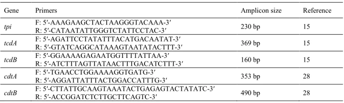
triose phosphate isomerase (tpi) gene and tcdA and tcdB toxin-producing genes were detected by PCR. For this purpose, the primers listed in Table 1 were used according to Lemee et al. (14) with minor modifications. The PCRs were performed on a CG 1-96 Palm-Cycler (Genetix Biotech Asia, New Delhi, India) in a final volume of 25 µL containing: 5 µL of DNA template, 10% (v/v) glycerol, 1 µM of each primer (except for tpi-F and tpi-R, of which there was 0.5 µM), 200 µM of each deoxynucleoside triphosphate, and 0.5 U of Taq DNA polymerase in a 1× amplification buffer (10 mM Tris-HCl, pH 8.3) (Thermo Fisher Scientific, Waltham, MA, USA).
The PCR mixtures were denatured at 95°C for 3 min and then, a touchdown step was applied at 95°C for 30 s. An annealing step for 30 s at temperatures decreasing from 65°C to 55°C in the first 11 cycles and a final extension step at 72°C for 30 s were performed (in total 40 cycles). Binary toxin genes (cdtA and cdtB) were determined by means of a multiplex PCR as described by Stubbs et al. (27) (Table 1). The PCRs were performed in a final volume of 50 µL containing: 10 µL of DNA template, 0.15 µM of each primer, 1.5 mM of MgCl2, 1U of Taq polymerase, and 200 µM of each deoxynucleoside triphosphate in a 1× amplification buffer (10 mM Tris-HCl (pH 8.3) and 50 mM KCl) (Thermo Fisher Scientific). The mixtures were put through 30 cycles of a denaturation step at 94°C for 45 s, an annealing step at 52°C for 1 min, and a final extension step at 72°C for 80 s. For the electrophoresis process, 1.5% agarose gel with the addition of ethidium bromide was used and for gel screening, a UV transilluminator provided imaging with the Dolphin-Doc analysing system (Wealtec, Sparks, NV, USA).
Table 1. Primer sequence list used in the study
Gene Primers Amplicon size Reference
tpi F: 5′-AAAGAAGCTACTAAGGGTACAAA-3′ R: 5′-CATAATATTGGGTCTATTCCTAC-3′ 230 bp 15
tcdA F: 5′-AGATTCCTATATTTACATGACAATAT-3′ R: 5′-GTATCAGGCATAAAGTAATATACTTT-3′ 369 bp 15
tcdB F: 5′-GGAAAAGAGAATGGTTTTATTAA-3′ R: 5′-ATCTTTAGTTATAACTTTGACATCTTT-3′ 160 bp 15
cdtA F: 5′-TGAACCTGGAAAAGGTGATG-3′ R: 5′-AGGATTATTTACTGGACCATTTG-3′ 353 bp 28
In this research, the ATCC 9689 C. difficile strain was used as the positive control for the tcdA and tcdB genes, the BAA 1870 strain was the equivalent for the cdtA, and cdtB genes, both of these references could serve as the positive control for the tpi gene, and Milli-Q water served as the negative control (Merck, Darmstadt, Germany).
PCR – ribotyping. The 16S–23S intergenic spacer
regions of C. difficile isolates were amplified according to Bidet et al. (3), and capillary electrophoresis was carried out by means of an ABI 310 Genetic Analyser using performance-optimised polymer 4 and GeneScan 1200 LIZ size standard (all Applied Biosystems, Carlsbad, CA, USA), with a 36 cm array length and provision of default fragment analysis. The WEBRIBO database was used for ribotype determination after Gene Mapper v4.9 (Applied Biosystems) software processing (12).
Detection of C. difficile toxin A and B production.
A Ridascreen ELISA kit (C0801, R-Biopharm AG, Darmstadt, Germany) was used for the detection of toxin production. A loopful of colony cultured on blood agar and confirmed as C. difficile was diluted in 1 mL of sample dilution buffer and centrifuged at 2,500 × g for 5 min. After centrifugation, the supernatant was used for the detection of toxin presence according to the manufacturer’s protocol.
Antibiotic susceptibility test. The antibiotic
susceptibility of C. difficile isolates was examined by minimum inhibitory concentration evaluator strips (Oxoid) according to the supplied instructions. The breakpoint values for imipenem, cefotaxime, amoxicillin-clavulanic acid, tetracycline, clindamycin, ampicillin, and metronidazole were taken from the CLSI (5), and for vancomycin the values derived from EUCAST (8).
Results
A total of 185 chicken carcasses were analysed for the presence of the tpi gene, which is specific for C. difficile by PCR, and the gene was found in 69 (37.3%) isolates. According to the PCR ribotyping results, 6/69 (8.7%) isolates were determined as RT027, whereas the other hypervirulent human pathogenic strain, RT078, could not be detected in any chicken carcass samples.
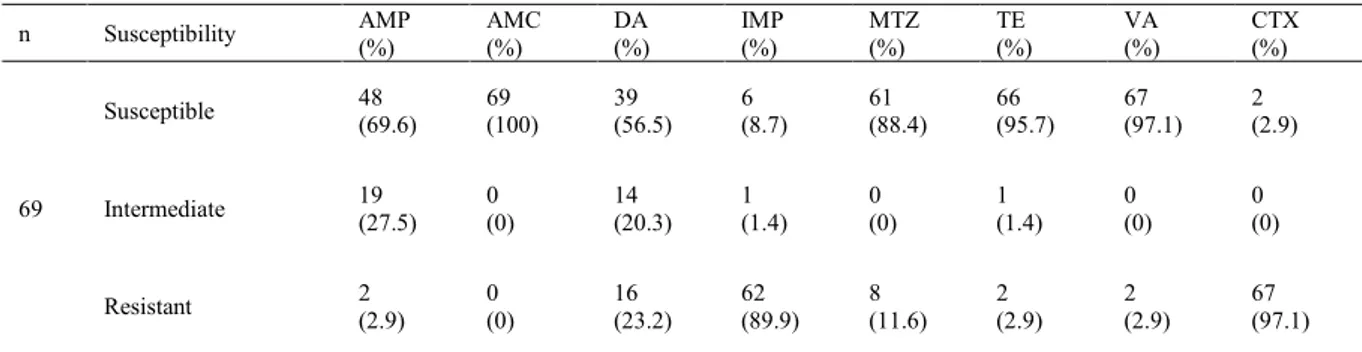
When the antibiotic sensitivity of isolates was evaluated, it was determined that 69 (100%) isolates were susceptible to amoxicillin-clavulanic acid, 61 (88.4%) to metronidazole, 66 (95.7%) to tetracycline, and 67 (97.1%) to vancomycin. On the other hand, 62 (89.9%) and 67 (97.1%) out of 69 chicken carcass isolates were resistant to imipenem and cefotaxime, respectively (Table 2). The susceptibility profiles of C. difficile isolates obtained from chicken carcasses are shown in Table 3.
Table 2. Susceptibility profiles of C. difficile isolates from chicken carcasses
n Susceptibility AMP (%) AMC (%) DA (%) (%) IMP MTZ (%) TE (%) VA (%) CTX (%)
69
Susceptible 48 (69.6) 69 (100) 39 (56.5) (8.7) 6 61 (88.4) 66 (95.7) 67 (97.1) 2 (2.9)
Intermediate 19 (27.5) 0 (0) 14 (20.3) (1.4) 1 0 (0) 1 (1.4) 0 (0) 0 (0)
Resistant 2 (2.9) 0 (0) 16 (23.2) (89.9) 62 8 (11.6) 2 (2.9) 2 (2.9) 67 (97.1) AMP – ampicillin; AMC – amoxicillin-clavulanic acid; DA – clindamycin; IMP – imipenem; MTZ – metronidazole; TE – tetracycline; VA – vancomycin; CTX – cefotaxime; n –number of samples
Table 3. The distribution of determined ribotypes in terms of antibiotic susceptibility
RTs n AMP S I R AMC S I R DA S I R IMP S I R S MTZ I R TE S I R VA S I R CTX S I R
RT027 6 4 2 - 6 - - 4 1 1 - - 6 6 - - 6 - - 6 - - - - 6 RT087 4 3 1 - 4 - - 3 1 - - - 4 4 - - 4 - - 4 - - - - 4 RT470 4 2 2 - 4 - - 4 - - - - 4 4 - - 4 - - 4 - - - - 4 RT085 4 4 - - 4 - - 3 1 - - - 4 4 - - 4 - - 4 - - - - 4 RT456 2 2 - - 2 - - 2 - - - - 2 2 - - 2 - - 2 - - - - 2 RT020 2 2 - - 2 - - 1 1 - - - 2 2 - - 2 - - 2 - - - - 2 RT010 1 1 - - 1 - - 1 - - - - 1 1 - - 1 - - 1 - - - - 1 RT003 1 1 - - 1 - - 1 - - - - 1 1 - - 1 - - 1 - - - - 1 ML027 6 3 3 - 6 - - 3 2 1 - - 6 6 - - 6 - - 6 - - - - 6 NR 39 26 11 2 39 - - 17 8 14 6 1 32 31 - 8 36 1 2 37 - 2 2 - 37 TOTAL 69 48 19 2 69 - - 39 14 16 6 1 62 61 - 8 66 1 2 67 - 2 2 - 67 AMP – ampicillin; AMC – amoxicillin-clavulanic acid; DA – clindamycin; IMP – imipenem; MTZ – metronidazole; TE – tetracycline; VA – vancomycin; CTX – cefotaxim; n – ribotype number; ML – most likely; S – susceptible; I – intermediate; R – resistant; RTs – ribotypes
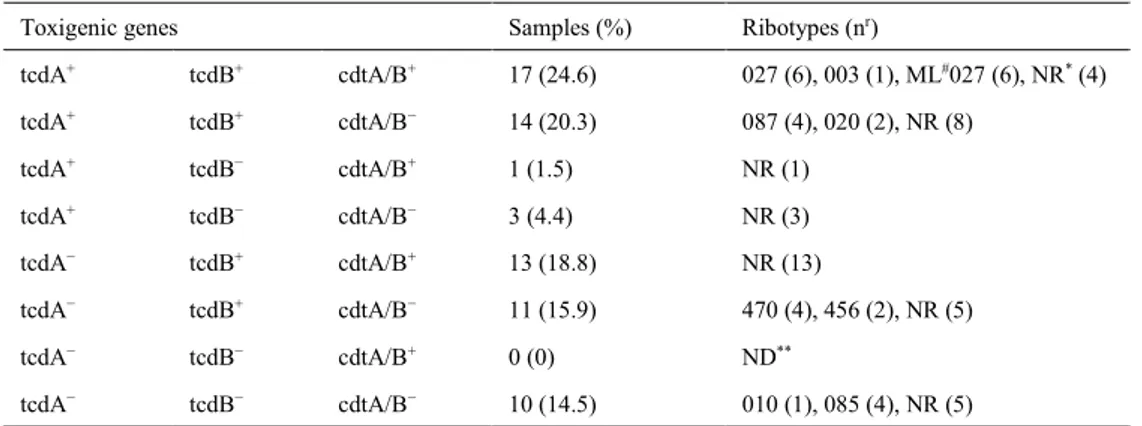
Table 4. The distribution of the virulence genes and ribotypes of C. difficile isolates (n = 69)
Toxigenic genes Samples (%) Ribotypes (nr)
tcdA+ tcdB+ cdtA/B+ 17 (24.6) 027 (6), 003 (1), ML#027 (6), NR* (4) tcdA+ tcdB+ cdtA/B− 14 (20.3) 087 (4), 020 (2), NR (8) tcdA+ tcdB− cdtA/B+ 1 (1.5) NR (1) tcdA+ tcdB− cdtA/B− 3 (4.4) NR (3) tcdA− tcdB+ cdtA/B+ 13 (18.8) NR (13) tcdA− tcdB+ cdtA/B− 11 (15.9) 470 (4), 456 (2), NR (5) tcdA− tcdB− cdtA/B+ 0 (0) ND** tcdA− tcdB− cdtA/B− 10 (14.5) 010 (1), 085 (4), NR (5) NR* – new ribotype; ND** – not detected; ML# – most likely; nr – number of ribotypes
In this study, the toxin genes of C. difficile isolates were detected by PCR and these, the tcdA, tcdB, and cdtA/B genes, were determined in 35 (50.7%), 55 (79.7%), and 31 (44.9%) out of 69 chicken isolates, respectively.
The distribution of the toxin genes and the number of ribotypes which were detected in chicken carcass isolates are shown in Table 4. Seventeen (24.6%) isolates had all three toxin genes (six were RT027 and one was RT003). In contrast, 10 (14.5%) isolates did not include any tcdA, tcdB, or cdtA/B genes.
ELISA was used for the detection of C. difficile toxins A and B. A total of 47 out of 69 (68.1%) chicken isolates had the toxin production attribute, whereas no toxin production was observed in 22 out of 69 (31.9%) isolates.
Discussion
The results of this research are further proof of the presence of C. difficile in chicken carcasses. There are also a number of studies from different countries confirming the detection of this organism in poultry and poultry-originated products. In a study performed by de Boer et al. (6), C. difficile was found in 7 out of 257 (2.7%) chicken carcass samples. In another piece of research conducted in Canada, Weese et al. (32) isolated the organism from 12.8% (26/203) of chicken carcasses (thigh, wing, and leg). Guran and Ilhak (9) conducted similar research in which they obtained 310 chicken samples from supermarkets and butcher’s shops located in the eastern part of Turkey and found that 25 (8.1%) of them were contaminated with the bacterium. From Indra et al. (13), in Austria came research results in which C. difficile was noted in three out of 59 (5.1%) broiler chicken samples. Our results are higher than those seen in these studies. Contrary to this trend, in the USA, Mooyottu et al. (18) reported that they could not detect any C. difficile strains in 100 chicken wing samples. Limbago et al. (15) found similar findings: the researchers could not determine the bacterium in 614 minced turkey or 259 chicken breast samples obtained
from retail markets. Likewise, Ersöz and Coşansu (7) from Turkey could not ascertain any C. difficile presence in 27 chicken breast samples.
The existence of the bacterium not only in chicken carcasses but also in chicken faeces was detected and reported by other researchers (25, 26, 33). The persistence of C. difficile and its spores in the environment (in soil and water), ineffective hygiene in rearing operations, and deficient manufacturing practices such as unhygienic slaughterhouse conditions (insufficient cleaning, sub-structural deficiencies, etc.), an unsuitable plucking process, the contamination of carcasses with faeces because of careless evisceration or contact with the floor, improper chilling processes, unhygienic storage conditions, poor personnel and equipment hygiene (contaminated hands, clothes, knives, etc.), and inattentive and improper disposal of animal remnants and extraneous matter are some important factors fostering C. difficile presence in poultry carcasses (10, 21, 31).
Recently, C. difficile isolates detected in poultry carcasses have shown similarities with some strains like RT027 and RT078, which are related to CDI outbreaks in humans. In this context, Varshney et al. (31) examined 76 minced turkey meat samples and found C. difficile in 11 (14.5%) of them (including one RT027 and two RT078 isolates). In another study, Weese et al. (32) found C. difficile in 26 out of 203 (12.8%) chicken samples, and all isolates were R078. In contrast to this, RT078 could not be detected in the present study; however, RT027 was found in 6 out of 69 (8.7%) examined chicken samples. As a counterpoint, neither RT027 nor RT078 could be isolated in chicken meat samples by a number of researchers (1, 6, 31). In various studies, C. difficile and its hypervirulent ribotypes were reported in poultry carcasses with different rates of prevalence. Guran and Ilhak (9) and Rodriguez-Palacios et al. (23) reported that the prevalence of C. difficile was generally higher in winter than in other seasons. In another study, Lund and Peck (16) reported that the isolation rates of C. difficile were relatively low (4.3%) in Europe, whereas they were higher (44.0%) in North America, and they indicated that one of the reasons for
this difference may be the different natures of each continent’s enrichment, isolation, and identification methods. Blanco et al. (4) stated that the procedure used to isolate C. difficile can have a significant impact on prevalence data for this organism. Zidaric et al. (33) reported that C. difficile colonisation in chickens was probably established within the first two weeks post-hatching and subsequently decreases with age. In the light of this data, dissimilar characteristics of the sampled animals (age, breed, etc.), geographical and seasonal differences, the use of distinct sampling techniques (material, sampling amount, etc.), and the adoption of different isolation methods can be the explanation of the differences in prevalence rates of C. difficile in chickens (7, 10, 31).
Concerns about the use of antibiotics in poultry have gradually increased in recent years. Although a number of countries have prohibited their use, still different antibiotics are used in the poultry industry to promote growth, to treat sick animals, and to prevent diseases. Therefore, concern about antibiotic resistance developing in C. difficile in poultry would seem to be founded. On the other hand, some antibiotics such as vancomycin and metronidazole are the medicines of first resort for the treatment of CDI and CDI-related diarrhoea in humans. Several research publications demonstrated that the majority of the isolated C. difficile strains from various foods are resistant to imipenem and cefotaxime but susceptible to amoxicillin, ampicillin, tetracycline, metronidazole, and vancomycin (2, 10, 11, 20, 28, 29). Simango and Mwakurudza (26) reported that all isolated C. difficile strains from chicken samples were found susceptible to vancomycin, metronidazole and tetracycline, despite them all being resistant to cefotaxime. As shown in Table 2, our findings parallel these results. Our investigation demonstrated that the isolates were susceptible to amoxicillin, tetracycline, vancomycin, and metronidazole at rates of 100.0%, 95.7%, 97.1%, and 88.4%, respectively. The cefotaxime and imipenem resistance rates in chicken carcass samples were found to be 97.1% and 89.9%, respectively.
Toxin production from the genes with this purpose can be regarded as the primary virulence factor of C. difficile; however, the presence of toxin genes does not mean that they have toxin production capacity. In this research, toxin production was detected in 47 out of 69 (68.1%) chicken carcass isolates, whereas toxin production could not be detected in the other 22 (31.9%) examined samples. In the study performed by Guran and Ilhak (9), it was reported that five out of 25 C. difficile strains isolated from chicken parts had toxin A, and eight of 25 isolates had toxin B, whereas the isolates did not contain any binary toxin. In a similar study, Simango and Mwakurudza (26) determined C. difficile in 29 out of 100 (29.0%) chickens, and they reported that 26 (89.7%) of these isolates had the toxin production attribute. In Iran, Rahimi et al. (20) analysed 368 ready-to-eat food products, among which the organism was
found in only five samples, and they detected that three out of five (60.0%) strains produced toxins A and B.
In conclusion, the results of this study conducted in Turkey reveal the presence of C. difficile isolates in chicken carcasses. Although the significance of foods contaminated with C. difficile in human infection is still unclear, chicken carcasses can be a presumptive C. difficile contamination route for humans, and in consequence, chicken and chicken products can be considered one of the probable transmission pathways for humans and a potential risk for consumers.
Conflict of Interests Statement: The authors declare
that there is no conflict of interests regarding the publication of this article.
Financial Disclosure Statement: This work was
financially supported by the Scientific and Technological Research Council of Turkey (TÜBİTAK), Project Number: 114O860.
Animal Rights Statement: None required.
References
1. Abdel-Glil M.Y., Thomas P., Schmoock G., Abou-El-Azm K., Wieler L.H., Neubauer H., Seyboldt C.: Presence of Clostridium
difficile in poultry and poultry meat in Egypt. Anaerobe 2018, 51,
21–25, doi: 10.1016/j.anaerobe.2018.03.009.
2. Bakri M.: Prevalence of Clostridium difficile in raw cow, sheep, and goat meat in Jazan, Saudi Arabia. Saudi J Biol Sci 2018, 25, 783–785, doi: 10.1016/j.sjbs.2016.07.002.
3. Bidet P., Barbut F., Lalande V., Burghoffer B., Petit J.C.: Development of a new PCR-ribotyping method for Clostridium
difficile based on ribosomal RNA gene sequencing. FEMS
Microbiol Lett 1999, 175, 261–266, doi: 10.1111/j.1574-6968.1999.tb13629.x.
4. Blanco J.L., Álvarez-Pérez S., García M.E.: Is the prevalence of
Clostridium difficile in animals underestimated? Vet J 2013, 197,
694–698, doi: 10.1016/j.tvjl.2013.03.053.
5. Clinical and Laboratory Standards Institute: Performance Standards for Antimicrobial Susceptibility Testing, CLSI Document M100. Clinical and Laboratory Standards Institute, Wayne, 2018.
6. De Boer E., Zwartkruis-Nahuis A., Heuvelink A.E., Hurmanus C., Kuijper E.J.: Prevalance of Clostridium difficile in retailed meat in the Netherlands. Int J Food Microbiol 2011, 144, 561–564, doi: 10.1016/j.ijfoodmicro.2010.11.007.
7. Ersöz Ş.Ş., Coşansu S.: Prevalence of Clostridium difficile isolated from beef and chicken meat products in Turkey. Korean J Food Sci Anim Resour 2018, 38, 759–767, doi: 10.5851/kosfa.2018.e14.
8. European Committee for Antimicrobial Susceptibility Testing (EUCAST): EUCAST Clinical Breakpoint Table v. 9.0, 2019, p. 72. https://www.eucast.org/fileadmin/src/media/PDFs/EUCAST_ files/Breakpoint_tables/v_9.0_Breakpoint_Tables.pdf.
9. Guran H.S., Ilhak O.I.: Clostridium difficile in retail chicken meat parts and liver in the Eastern Region of Turkey. J Verbr Lebensm 2015, 10, 359–364, doi: 10.1007/s00003-015-0950-z.
10. Hampikyan H., Bingol E.B., Muratoglu K., Akkaya E., Cetin O., Colak H.: The prevalence of Clostridium difficile in cattle and sheep carcasses and the antibiotic susceptibility of isolates. Meat Sci 2018, 139, 120–124, doi: 10.1016/j.meatsci.2018.01.020.
11. Harvey R.B., Norman K.N., Andrews K., Hume M.E., Scanlan C.M., Hardin M.D., Scott H.M.: Clostridium difficile in retail meat and processing plants in Texas. J Vet Diagn Invest 2011, 23, 807–811, doi: 10.1177/1040638711407893.
12. Indra A., Huhulescu S., Schneeweis M., Hasenberger P., Kernbichler S., Fiedler A., Wewalka A., Allerberger F., Kuijper E.J.: Characterization of Clostridium difficile isolates using capillary gel electrophoresis-based PCR ribotyping. J Med Microbiol 2008, 57, 1377–1382, doi: 10.1099/jmm.0.47714-0. 13. Indra A., Lassnig H., Baliko N., Much P., Fiedler A., Huhulescu S.,
Allerberger F.: C. difficile: a new zoonotic agent? Wien Klin Wochenschr 2009, 121, 91–95, doi: 10.1007/s00508-008-1127-x. 14. Lemee L., Dhalliun A., Testelin S., Mattrat M.A., Maillard K., Lemeland J.F., Pons J.L.: Multiplex PCR targeting tpi (triose phosphate isomerase), tcdA (toxin A), and tcdB (toxin B) genes for toxigenic culture of Clostridium difficile. J Clin Microbiol 2004, 42, 5710–5714, doi: 10.1128/jcm.42.12.5710-5714.2004. 15. Limbago B., Thompson A.D., Greene S.A., MacCannell D.,
MacGowan C.E., Jolbitado B., Hardin H.D., Estes S.R., Weese J.S., Songer J.G., Gould L.H.: Development of a consensus method for culture of Clostridium difficile from meat and its use in a survey of U.S. retail meats. Food Microbiol 2012, 32, 448–451, doi: 10.1016/j.fm.2012.08.005.
16. Lund B.M., Peck M.W.: A possible route for foodborne transmission of Clostridium difficile? Foodborne Pathog Dis 2015, 12, 177–182.
17. Metcalf D.S., Costa M.C., Dew W.M.V., Weese J.S.: Clostridium
difficile in vegetables. Canada Lett Appl Microbiol 2010, 51, 600–
602, doi: 10.1111/j.1472-765X.2010.02933.x.
18. Mooyottu S., Flock G., Kollanoor-Johny A., Upadhyaya I., Jayarao B., Venkitanarayanan K.: Characterization of a multidrug resistant C. difficile meat isolate. Int J Food Microbiol 2015, 192, 111–116, doi: 10.1016/j.ijfoodmicro.2014.10.002.
19. Pelaez T., Alcalá L., Blanco J.L., Álvarez-Pérez S., Marín M., Martín-López A., Catalán P., Reigadas E., García M.E., Bouza E.: Characterization of swine isolates of Clostridium difficile in Spain: A potential source of epidemic multidrug resistant strains? Anaerobe 2013, 22, 45–49, doi: 10.1016/j.anaerobe.2013.05.009. 20. Rahimi E., Afzali Z.S., Baghbadorani Z.T.: Clostridium difficile in ready-to-eat foods in Isfahan and Shahrekord, Iran. Asian Pac J Trop Biomed 2015, 5, 128–131, doi: 10.1016/S2221-1691(15) 30156-8.
21. Rodriguez C., Avesani V., Van Broeck J., Taminiau B., Delmée M., Daube G.: Presence of Clostridium difficile in pigs and cattle intestinal contents and carcass contamination at the slaughterhouse in Belgium. Int J Food Microbiol 2013, 166, 256–262, doi: 10.1016/j.ijfoodmicro.2013.07.017.
22. Rodriguez C., Taminiau B., Van Broeck J., Avesani V., Delmée M., Daube G.: Clostridium difficile in young farm animals and slaughter animals in Belgium. Anaerobe 2012, 18, 621–625, doi: 10.1016/j.anaerobe.2012.09.008.
23. Rodriguez-Palacios A., Reid-Smith R.J., Staempfli H.R., Daignault D., Janecko N., Avery B.P., Martin H., Thompson A.D., McDonald L.C., Limbago B., Weese J.C.: Possible seasonality of
Clostridium difficile in retail meat, Canada. Emerg Infect Dis
2009, 15, 802–805.
24. Romano V., Albanese F., Dumontet S., Krovacek K., Petrini O., Pasquale V.: Prevalence and genotypic characterization of
Clostridium difficile from ruminants in Switzerland. Zoonoses
Public Hlth 2012, 59, 545–548, doi: 10.1111/j.1863-2378.2012.01540.x.
25. Simango C.: Prevalence of Clostridium difficile in the environment in a rural community in Zimbabwe. Trans R Soc Trop Med Hyg 2006, 100, 1146–1150, doi: 10.1016/j.trstmh.2006.01.009.
26. Simango C., Mwakurudza S.: Clostridium difficile in broiler chickens sold at market places in Zimbabwe and their antimicrobial susceptibility. Int J Food Microbiol 2008, 124, 268–270, doi: 10.1016/j.ijfoodmicro.2008.03.020.
27. Stubbs S., Rupnik M., Gibert M., Brazier J., Duerden B., Popoff M.: Production of actin-specific ADP-ribosyltransferase (binary toxin) by strains of Clostridium difficile. FEMS Microbiol Lett 2000, 186, 307–312, doi: 10.1111/j.1574-6968.2000.tb09122.x. 28. Thitaram S.N., Frank J.F., Siragusa G.R., Bailey J.S., Dargatz D.A.,
Lombart J.E., Haley C.A., Lyon S.A., Fedorka-Cray P.J.: Antimicrobial susceptibility of Clostridium difficile isolated from food animals on farms. Int J Food Microbiol 2016, 227, 1–5. doi: 10.1016/j.ijfoodmicro.2016.03.017.
29. Troiano T., Harmanus C., Sanders I.M.J.G., Pasquale V., Dumontet S., Capuano F., Romano V., Kuijper E.J.: Toxigenic
Clostridium difficile PCR ribotypes in edible marine bivalve
molluscs in Italy. Int J Food Microbiol 2015, 208, 30–34, doi: 10.1016/j.ijfoodmicro.2015.05.002.
30. United States Department of Agriculture Food Safety and Inspection Service: Salmonella and Campylobacter verification program for raw meat and poultry products, Directive 10,250.1. U.S. Department of Agriculture Food Safety and Inspection Service, Washington, 2013.
31. Varshney J.B., Very K.J., Williams J.L., Hegarty J.P., Stewart D.B., Lumadue J., Venkitanarayanan K., Jayarao B.M.: Characterization of Clostridium difficile isolates from human fecal samples and retail meat from Pennsylvania. Foodborne Pathog Dis 2014, 11, 822–829, doi: 10.1089/fpd.2014.1790.
32. Weese J.S., Reid-Smith R.J., Avery B.P., Rousseau J.: Detection and characterization of Clostridium difficile in retail chicken. Lett Appl Microbiol 2010, 50, 362–365, doi: 10.1111/j.1472-765X.2010.02802.x.
33. Zidaric V., Zemljic M., Janezic S., Kocuvan A., Rupnik M.: High diversity of Clostridium difficile genotypes isolated from a single poultry farm producing replacement laying hens. Anaerobe 2008, 14, 325–327, doi: 10.1016/j.anaerobe.2008.10.001.