http://journals.tubitak.gov.tr/biology/ © TÜBİTAK
doi:10.3906/biy-1705-66
Determining expression of miRNAs that potentially regulate
STAT5A and 5B in dasatinib-sensitive K562 cells
Asu Fergün YILMAZ1,*, Burçin KAYMAZ2, Çağdaş AKTAN3, Nur SOYER4, Buket KOSOVA2, Ajda GÜNEŞ5, Fahri ŞAHİN4, Melda CÖMERT6, Güray SAYDAM4, Filiz VURAL4
1Department of Hematology, İzmir Kâtip Çelebi University Atatürk Training and Research Hospital, İzmir, Turkey 2Department of Medical Biology, Ege University Hospital, İzmir, Turkey
3Department of Medical Biology, School of Medicine, Beykent University, İstanbul, Turkey 4Department of Hematology, Internal Medicine, Ege University Hospital, İzmir, Turkey
5Department of Hematology, Sivas Numune Hospital, Sivas, Turkey
6Department of Hematology, Internal Medicine, İnönü University Hospital, Malatya, Turkey
1. Introduction
Chronic myeloid leukemia (CML) is a hematologic malignancy characterized by the presence of a reciprocal chromosomal translocation of the Abelson (ABL1) oncogene on the 9th chromosome and a breakpoint cluster region (BCR) on the 22nd chromosome, resulting in t(9,22). This results in chimeric fusion, producing oncoprotein BCR–ABL with tyrosine kinase activity (Groffen et al., 1984). The subsequent uncontrolled tyrosine kinase activity results in excess activation of multiple signaling pathways such as RAS/RAF/MAPK, PI3K/Akt, JUN, MYC, and Janus kinase/signal transducers and activators of transcription (JAK/STAT), leading to persistent cell proliferation, reduced apoptosis, and malignant expansion of pluripotent stem cells in bone marrow (Steelman et al., 2004).
Since CML is due to a well-recognized translocation, it is possible to inhibit the aberrant BCR–ABL tyrosine kinase (TK) activity using molecularly targeted therapies.
Imatinib was the first BCR–ABL TK inhibitor (TKI) introduced for the treatment of CML (Kantarjian et al, 2002). Although imatinib and other second-generation (including dasatinib and nilotinib) and third-generation TKIs are generally well tolerated, ensuing resistance remains a major clinical challenge (Apperley, 2007).
Cytokines and growth factors can activate the JAK/ STAT signaling pathway, which has been well investigated in CML. Thus, the cascade transmits information from extracellular chemical signals to the nucleus, resulting in DNA transcription and expression of genes involved in immunity, proliferation, cellular migration, angiogenesis, differentiation, apoptosis, and oncogenesis (Rawlings et al., 2004). Therefore, overexpression of STATs, especially STAT5, is associated with leukemogenesis and carcinogenesis (Bowman et al., 2000; Rawlings et al., 2004). BCR/ABL chimeric protein constitutively activates the JAK/STAT cascade and causes antiapoptotic activity and uncontrolled proliferation of the malignant clone
Abstract: In the era of tyrosine kinase inhibitors, resistance still constitutes a problem in chronic myeloid leukemia (CML) patients;
thus, new pathway-specific inhibitors like miRNAs have become important in the treatment of refractory patients. There are no satisfying data regarding the miRNAs and anti-miRNA treatment targeting STAT5A and 5B. In this study, we first researched the effect of dasatinib on apoptosis in the CML cell line K562. The expressions of miRNAs possibly targeting both STAT5A and 5B were then determined. The down- and upregulation of the miRNAs were compared using the ΔΔCT method. At the last stage of the study, we used a new primer probe in order to validate the results. The level of hsa-miR-940 was decreased 4.4 times and the levels of hsa-miR-527 and hsa-miR-518a-5p were increased 12.1 and 8 times, respectively, in the dasatinib-treated group when compared to the control group. We detected similar results in the validation step. As a conclusion, we determined the expression profiles of miRNAs targeting STAT5A and 5B that had an important role in the pathogenesis of CML. The data obtained could lead to determining new therapeutic targets for CML patients.
Key words: miRNA expressions, STAT5A, STAT5B, chronic myeloid leukemia, dasatinib, K562 cells
Received: 20.05.2017 Accepted/Published Online: 28.09.2017 Final Version: 18.12.2017 Research Article
(Benekli et al., 2009). Accordingly, suppression of STATs results in inhibition of leukemogenesis and apoptosis of tumor cells including CML, which makes STAT proteins ideal targets for emerging cancer therapies (Steelman et al., 2004; Yu and Have, 2004; Kosova et al., 2010). In this regard, it is imperative to target the uncontrolled STAT proteins without affecting normal cells. In this setting, new treatment strategies such as microRNAs (miRNAs) attract a great deal of attention.
miRNAs are small endogenously synthesized noncoding RNA sequences with 19–24 nucleotides. Over 2000 miRNAs reported in humans regulate gene expression by binding to a 3’-untranslated region (3’UTR) within target messenger RNAs (mRNAs) and inducing translational repression or RNA destabilization (Bartel, 2004). Through this mechanism, they regulate many cellular processes such as cellular proliferation, differentiation, apoptosis, metabolic activities, and immunity (Cheng et al., 2005; Spizzo et al., 2009; Farazi et al., 2011). More than 50% of miRNAs reported in humans are located at specific chromosomes that are associated with several cancers (Calin and Croce, 2006), including hematologic malignancies (Calin et al., 2002); thus, dysregulation of miRNAs seems to play a key role in carcinogenesis (Spizzo et al., 2009). In light of these observations, it appears that these small molecules may potentially be exploited for prognostic, diagnostic, or therapeutic purposes in patients with neoplastic disorders, including hematologic malignancies and solid tumors such as lymphoma and leukemia, as well as breast, lung, gastric, and pancreatic cancers (Avery-Kiejda et al., 2014; Miao et al., 2014; Troppan et al., 2014; Zheng et al., 2014). In CML patients, several miRNAs such as miR-29b, miR-138, and miR-130a/b, which were up- or downregulated aberrantly,
were reported as either oncogene or tumor suppressors (Suresh et al., 2011; Li et al., 2013; Xu et al., 2014).
In this study, we analyzed the miRNAs that are potential targets of STAT5A and STAT5B and varying expression profiles of these determined miRNAs in CML cell line model K562 cells treated with dasatinib or not. Furthermore, apoptosis associated with dasatinib treatment in leukemic cells was explored.
2. Materials and methods
2.1. Culturing conditions of cells and dasatinib treatment
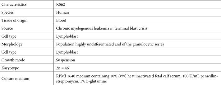
Cell line K562 “human CML” (the characteristics of the K562 cell line are provided in Table 1) was purchased from ECACC (European Collection of Cell Cultures) and was cultured in RPMI 1640 medium containing 10% (v/v) heat-inactivated fetal calf serum, 100 U/mL penicillin-streptomycin, and 1% L-glutamine at 37 °C in humidified air containing 5% CO2. Cells with 95% survival rates and 80% confluency were used in experimental analyses.
Dasatinib (BMS-35482) was provided by Bristol-Myers-Squibb (Princeton, NJ, USA) and dissolved in dimethyl sulfoxide (DMSO). Experiments were generated with 3.3 nM dasatinib treatment, indicated as the IC50 value of the K562 cell line, as well as with untreated control group cells. After 48 h of the IC50 dose of dasatinib treatment, cells were collected for apoptotic analyses and miRNA profiling. Experiments were done in triplicate with 2 separate biological replicas, and the average of the results was taken.
2.2. Apoptotic analysis assays
The amounts of apoptotic and live cells were detected by measuring caspase-3 activity in both untreated control cells and cells treated with the IC50 dose of dasatinib via the Caspase-3 Colorimetric Assay Kit (BioVision Research
Table 1. Characteristics of the K562 cell line.
Characteristics K562
Species Human
Tissue of origin Blood
Source Chronic myelogenous leukemia in terminal blast crisis
Cell type Lymphoblast
Morphology Population highly undifferentiated and of the granulocytic series
Cell type Lymphoblast
Growth mode Suspension
Karyotype 2n = 46
Products, Mountain View, CA, USA) according to the kit’s manual. Each sample’s absorbance was measured spectrophotometrically at 405 nm with an ELISA reader. Finally, apoptotic cell rates were calculated after taking the average values of triplicated absorbance measurements for each sample.
2.3. miRNA profiling
All 23 miRNAs chosen for the qPCR array were picked for their potential roles in the JAK/STAT pathway and targeted to either STAT5A or STAT5B genes. The miRTar database (http://mirtar.mbc.nctu.edu.tw) was used for determining appropriate miRNAs. The 3’UTR regions of STAT5A and STAT5B gene transcripts were selected. The analyzing parameters were in 3’UTR: MFE ≤ –14 kcal/mol and Score ≥ 140.
Total RNA containing miRNA was isolated from cells using the miRVana miRNA Isolation Kit (Life Technologies, Carlsbad, CA, USA) according to the manufacturer’s protocol. The isolated RNAs were transcribed into cDNA
by using an All-in-One First-Strand cDNA Synthesis Kit (GeneCopoeia, Inc., Rockville, MD, USA). qPCR array analyses were performed with the All-in-One qPCR Mix using GeneCopoeia custom miRNA qPCR arrays on an ABI 7500 Fast Real-Time PCR system. The miRNA qPCR array also included RNU-2, SNORD 44, SNORD 48, SNORD 47, SNORD 49a, and SNORD 6 endogenous controls for normalization. The list of miRNAs is given in Table 2. All raw threshold cycle (Ct) values of the qPCR machine-setting baseline and threshold were calculated using SDS software v.2.1. The fold change of each miRNA was calculated from the expression levels between dasatinib-treated and untreated cells using the 2–ΔΔCT
method.
2.4. Validation of miRNA expression patterns
The evaluated expression profiles of miRNAs (hsa-miR-940, hsa-miR-527, and hsa-miR-518a-5p) were further validated using the All-in-One miRNA qRT-PCR Detection Kit (GeneCopoeia). Validated primers for
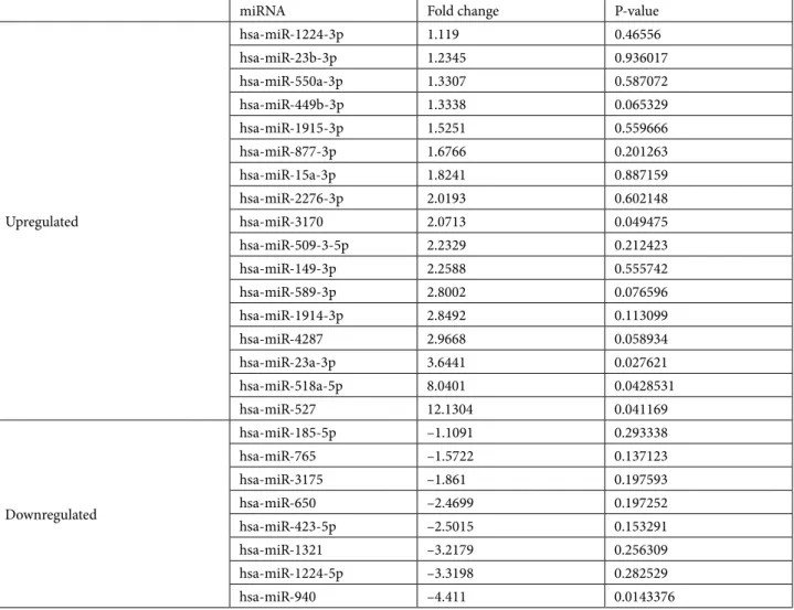
Table 2. miRNA alterations in response to dasatinib treatment.
miRNA Fold change P-value
Upregulated hsa-miR-1224-3p 1.119 0.46556 hsa-miR-23b-3p 1.2345 0.936017 hsa-miR-550a-3p 1.3307 0.587072 hsa-miR-449b-3p 1.3338 0.065329 hsa-miR-1915-3p 1.5251 0.559666 hsa-miR-877-3p 1.6766 0.201263 hsa-miR-15a-3p 1.8241 0.887159 hsa-miR-2276-3p 2.0193 0.602148 hsa-miR-3170 2.0713 0.049475 hsa-miR-509-3-5p 2.2329 0.212423 hsa-miR-149-3p 2.2588 0.555742 hsa-miR-589-3p 2.8002 0.076596 hsa-miR-1914-3p 2.8492 0.113099 hsa-miR-4287 2.9668 0.058934 hsa-miR-23a-3p 3.6441 0.027621 hsa-miR-518a-5p 8.0401 0.0428531 hsa-miR-527 12.1304 0.041169 Downregulated hsa-miR-185-5p –1.1091 0.293338 hsa-miR-765 –1.5722 0.137123 hsa-miR-3175 –1.861 0.197593 hsa-miR-650 –2.4699 0.197252 hsa-miR-423-5p –2.5015 0.153291 hsa-miR-1321 –3.2179 0.256309 hsa-miR-1224-5p –3.3198 0.282529 hsa-miR-940 –4.411 0.0143376
hsa-miR-940, hsa-miR-527, and hsa-miR-518a-5p were purchased from GeneCopoeia; their catalog numbers are HmiRQP0845, HmiRQP0579, and HmiRQP0579, respectively. First-strand cDNA reaction was performed using Techne prime thermal cyclers (Techne, Staffordshire, UK) with samples incubated at 37 °C for 60 min and 85 °C for 5 min to inactivate the enzyme and stored at –20 °C until ready for use. cDNA was diluted 1:10 prior to use for qPCR. The qPCR reactions were performed using 2X All-in-One qPCR Mix (GeneCopoeia) and an ABI 7500 Fast Real-Time PCR system. All reactions were performed in triplicate and analyzed using the 2–ΔΔCT method.
2.5. Statistical analyses
Statistical analyses were performed with SPSS 15.0 (SPSS Inc., Chicago, IL, USA). Student’s t-test was used to analyze the differences between treatment and control groups in miRNA expression levels. P ≤ 0.05 was considered statistically significant.
3. Results
3.1. Dasatinib triggered apoptotic cell death of leukemic cells
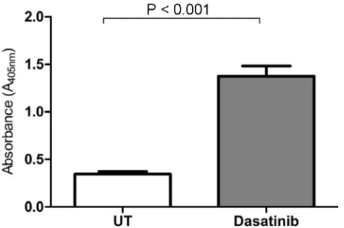
The number of apoptotic cells significantly increased in dasatinib-treated cells compared to untreated (UT) control cells at 3.58-fold (P < 0.001) (Figure 1). This suggested that dasatinib was very effective in triggering leukemic cell apoptosis.
3.2. Alterations in miRNA profiling that potentially regulate STAT5A and STAT5B
Expression profiles of selected miRNAs that potentially regulate STAT5A and STAT5B mRNAs were evaluated following an IC50 dose of dasatinib treatment for the study group and untreated control cells for a duration of 48 h. For this purpose, we detected 23 miRNAs’ expression profiles that were selected after in silico analyses from the
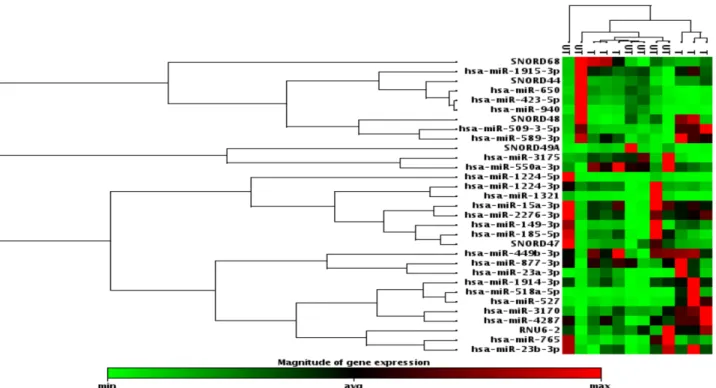
miRTar web site (http://mirtar.mbc.nctu.edu.tw). Among these 23 miRNAs, while 8 miRNA expressions were downregulated, ranging between –1.109-fold and –4.411-fold inhibitions, 15 were upregulated, ranging between 1.119-fold and 12.13-fold regulations. All obtained data are summarized in Table 1 and hierarchical clustering is shown in Figure 2.
Accordingly, while there was a –4.411-fold significant decrease in hsa-miR-940 expression (P = 0.0143) in the dasatinib-treated group compared to untreated control cells, an 8.0401-fold (P = 0.0428) increase in hsa-miR-518a-5p expression and a 12.1304-fold increase in has-miR-527 expression (P = 0.0411) were found. The subsequent scatter plot and volcano plot graphics with log2 transformations are presented in Figure 3.
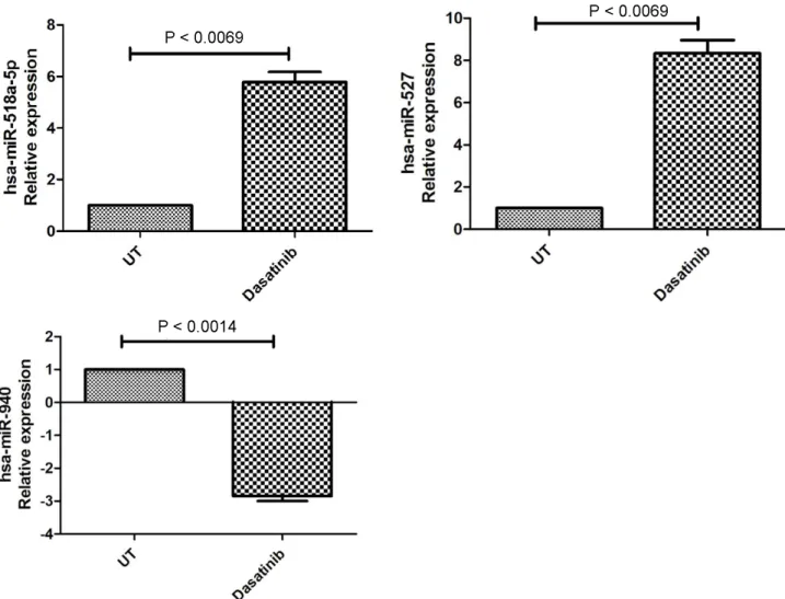
In the last phase of the study, we used new primer probe sets for hsa-miR-518a-5p, hsa-miR-940, and hsa-miR-527 in order to validate the initial results regarding miRNA expression. We detected similar results and determined that target miRNA expressions were again significantly regulated in dasatinib-treated cells. In detail, while hsa-miR-518a-5p and hsa-miR-527 expressions were upregulated 5.78 ± 0.692-fold (P = 0.0069) and 8.337 ± 1.077-fold (P = 0.0071), respectively, hsa-miR-940 expression was 2.85 ± 0.2516-fold (P < 0.0014) downregulated (Figure 4).
4. Discussion
The JAK/STAT pathway and STAT5 in particular have become novel molecular targets in refractory CML patients. Permanent activation of STATs is responsible for the development of many neoplasms, including hematologic malignancies and solid tumors (Bowman et al., 2000). In the case of CML, STAT5 comes into the focus of investigation since BCR/ABL activates STAT5 proteins and causes permanent proliferation of leukemic cells (Kosova et al., 2010; Dalgiç et al., 2015). Therefore, molecules with an inhibitory effect on the JAK/STAT cascade as well as STAT5A and STAT5B have attracted significant attention with regard to their therapeutic utility in relapsed and refractory CML patients.
miRNAs are small noncoding RNAs that control many cellular activities by influencing cellular cascades including JAK/STAT. In this study, we attempted to determine the miRNAs that potentially regulate STAT5A and STAT5B before and after dasatinib treatment. Sensitivity of K562 cells to dasatinib was evaluated by measuring caspase-3 activity. According to our results, while hsa-miR-940 expression was significantly decreased (–4.411-fold, P = 0.0143) in the dasatinib-treated group compared to untreated control cells, hsa-miR-518a-5p (8.0401-fold, P = 0.0428) and hsa-miR-527 (12.1304-fold, P = 0.0411) expressions were upregulated. These observations were confirmed during the validation steps. We also detected
Figure 1. Measurement of caspase-3 activity. Apoptosis was
significantly induced due to dasatinib treatment in leukemic cells. UT: Untreated.
that apoptosis was 3.58-fold upregulated following dasatinib treatment in leukemia cells. Dasatinib-induced apoptosis of leukemic cells has also been reported by many authors previously (Simara et al., 2013; Heo et al., 2014; Dalgiç et al., 2015; Ferreira et al., 2015).
These small molecules also have roles in the pathogenesis of several hematopoietic malignancies (Ramkissoon et al., 2006; Garzon et al., 2007; Dixon-McIver et al., 2008; Troppan et al., 2014), which makes them promising targets for treatment (Garzon et al., 2008a; Saumet et al., 2009).
Figure 2. Hierarchical cluster analyses of miRNA profiling. This figure was constructed by hierarchical clustering analysis. This analysis
was used to organize the miRNAs based on similarities in their expression profiles. Green indicates low expression, red indicates high expression, and black indicates intermediate expression. UT: Untreated, T: treated.
Figure 3. miRNA profiling results in scatter plot and volcano plot graphics in log2 transformation. While red plots indicate upregulated
miRNAs are not only a promising treatment approach; they are also candidates for being prognostic indicators (Garzon et al., 2008b) or being found to be involved in the pathogenesis (Han et al., 2010) of acute myeloid leukemia. They can also be target molecules in differential diagnosis in leukemias (Mi et al., 2007). However, further research is required to approve a specific miRNA as a diagnostic indicator.
Since CML is a genetically well-documented hematologic malignancy, the role of miRNA is also being studied in CML cell lines. It was reported previously that certain miRNAs were either downregulated or upregulated in CML and associated with prognosis and refractory disease. It was reported that miR150 and miR146a were downregulated in CML and were restored after imatinib treatment (Flamant et al., 2010). Agirre (2008) demonstrated that miR10a expression was decreased in bone marrow-derived CD34+ CML cells when compared
to healthy controls. miR17-5p, miR18a, and miR106a also exhibited high expressions in chronic-phase CML, with a change in their expression profile during the blastic phase (Venturini et al., 2007).
Other studies in the literature demonstrated different miRNA expressions in CML pathogenesis. Some miRNAs’ (miR203, miR328, and miR181a) expressions were repressed, especially during the blastic phase of CML. Among these miRNAs, miR203 was located at the 14q32.33 chromosome; this miRNA has potential to be a promising molecule in the treatment of CML patients, since its genetic and epigenetic silencing is associated with ABL1 and BCR–ABL1 oncogene expression (Bueno et al., 2008; Eiring et al., 2010; Zimmerman et al., 2010).
One of the most important problems in CML patients is gaining resistance to TKIs. Liu (2012) found that c-Myc expression was upregulated; in turn, miR144/miR451 expressions were repressed in CML cell lines that were
Figure 4. Validation results by qRT-PCR for hsa-miR-518a-5p, hsa-miR-940, and hsa-miR-527 expressional changes due to dasatinib
resistant to imatinib. This finding suggested that the pathways and molecules inhibiting Myc expressions or restoring miR144/miR451 expression would bypass imatinib resistance (Liu et al., 2012). Similarly, miR424, which directly targeted BCR–ABL1, exhibited markedly low expression in CML lines and in patients at diagnosis. The restoration of this miRNA suppressed proliferation and induced apoptosis, which suggested that it could be a potential therapeutic target in CML patients (Hershkovitz-Rokah et al., 2015).
In the literature, the expression profiles of miRNAs have been studied, but the precise molecular pathways could not be very well documented. In this study, we determined differing miRNA profiles in CML cell lines that were potentially regulated by STAT5A and STAT5B, which could exhibit a pivotal outcome to discover more about CML pathogenesis.
In the literature, miR518a-5p, 527, and miR-940 were not directly reported with leukemia-related malignancies, especially in CML; however, in a very recent study, miR-518a-5p was studied in imatinib mesylate-resistant gastrointestinal stromal tumors. It was determined that its expression was downregulated, and PIK3C2A was reported as a gene-specific target of miRNA-518a-5p (Shi et al., 2016). Since we have detected that miR-518a-5p expression was upregulated in our dasatinib-treated CML cells, we can conclude that miR-518a-5p can be a promising molecular target for CML therapy. Additionally, Rubie et al. (2014) reported that chemokine receptor CCR6 expression was also regulated by miR-518a-5p in colorectal cancer cells. In another study, Port et al. (2011) reported that miR518 expression was associated with cisplatin-resistant germ cell tumors.
According to our results, miR527 expression increased as a result of dasatinib treatment. In a recent study, miR-527 was reported to be a member of the network involved in the regulation of physiological wound-healing responses and was suppressed by tumor cells to promote metastatic dissemination (Rodriguez Calleja et al., 2016).
Finally, we detected that miR-940 expression was decreased in dasatinib-treated leukemic cells. Although this study marks the first time that it has been put forward that miR-940 might take place in CML pathogenesis, Liu et al. (2015) reported that downregulation of plasma miR-940 might serve as a novel biomarker for detection of gastric cancer.
In some other recent studies, researchers claimed that low miR-940 expressions were correlated with reduced survival in hepatocellular carcinoma and pancreatic ductal adenocarcinoma (Song et al., 2015; Yuan et al., 2015). Although these reported studies seem to conflict with our results, it is known that miRNAs can exhibit different
effects in different cancer types; thus, it will be necessary to study miR-940 in CML in order to clarify the exact impact. When we look at the studies including miRNAs in leukemia, Rokah et al. (2012) reported that miR31, miR155, and miR564 expressions were downregulated in CML cell line model K562 cells. They added that this expressional downregulation was dependent on BCR– ABL activity.
There are some other miRNAs reported to also display therapeutic effects on CML cells by inhibiting STATs. As an example of this phenomenon, it was reported that miR-520a-5p targeted STAT3 and enhanced the antitumoral effects of capsaicin (Kaymaz et al., 2014); in addition, miR-2278 was reported as a tumor suppressor and thus a player in the regaining of chemotherapeutic imatinib response by targeting STAT5A in imatinib-resistant K562 leukemia cells (Kaymaz et al., 2015).
There are also limitations to our study. This present study reported preliminary data concerning miRNAs regulating both STAT5A and STAT5B. The data were obtained by a study conducted on the K562 cell line. A study conducted with samples from CML patients should be the next step in evaluating the role of reported miRNAs in CML. Additionally, a second study with mimics and inhibitors of reported miRNAs is needed to confirm the results. Overall, our study seems to be unique since we have evaluated the expression profiles of miRNAs that potentially regulate STAT5A and 5B, which have a major role in the development and progression of CML.
In conclusion, studies about the miRNAs targeting STAT molecules and the JAK/STAT pathway are appearing more often in the literature. We collected preliminary data about miRNAs regulating both STAT5A and STAT5B, and miRNAs 518a-5p, hsa-miR-527, and hsa-miR-940 were highlighted in this study for exhibiting significant expressional changes after dasatinib treatment. We believe these miRNAs could be promising therapeutic targets in CML. Further studies with the inhibitors of hsa-miR-940 and the mimics of hsa-miR-527 and hsa-miR-518a-5p should be conducted on resistant CML cells. Since it is accepted that miRNAs exhibit very important roles in the pathogenesis of cancers including CML, the precise mechanism and cascades of the specific miRNAs should be identified with new studies for more curative clinical outcomes.
Acknowledgment
This study was financially supported by the Scientific Research Projects Branch Directorate of Ege University, Turkey (2013 – TIP-019) and the Hematology Specialist Association.
References
Agirre X, Jiménez-Velasco A, San José-Enériz E, Garate L, Bandrés E, Cordeu L, Aparicio O, Saez B, Navarro G, Vilas-Zornoza A et al. (2008). Down-regulation of hsa-miR-10a in chronic myeloid leukemia CD34+ cells increases USF2-mediated cell growth. Mol Cancer Res 6: 1830-1840.
Apperley JF (2007). Part I: Mechanisms of resistance to imatinib in chronic myeloid leukaemia. Lancet Oncol 8: 1018-1029.
Avery-Kiejda KA, Braye SG, Mathe A, Forbes JF, Scott RJ (2014). Decreased expression of key tumour suppressor microRNAs is associated with lymph node metastases in triple negative breast cancer. BMC Cancer 31: 14-51.
Bartel DP (2004). MicroRNAs: genomics, biogenesis, mechanism, and function. Cell 116: 281-297.
Benekli M, Baumann H, Wetzler M (2009). Targeting signal transducer and activator of transcription signaling pathway in leukemias. J Clin Oncol 27: 4422-4432.
Bowman T, Garcia R, Turkson J, Jove R (2000). STATs in oncogenesis. Oncogene 19: 2474-2488.
Bueno MJ, Pérez de Castro I, Gómez de Cedrón M, Santos J, Calin GA, Cigudosa JC, Croce CM, Fernández-Piqueras J, Malumbres M (2008). Genetic and epigenetic silencing of microRNA-203 enhances ABL1 and BCR-ABL1 oncogene expression. Cancer Cell 13: 496-506.
Calin GA, Croce CM (2006). MicroRNA signatures in human cancers. Nat Rev Cancer 6: 857-866.
Calin GA, Dumitru CD, Shimizu M, Bichi R, Zupo S, Noch E, Aldler H, Rattan S, Keating M, Rai K et al. (2002), Frequent deletions and down-regulation of micro- RNA genes miR15 and miR16 at 13q14 in chronic lymphocytic leukemia. P Natl Acad Sci USA 99: 15524-15529.
Cheng AM, Byrom MW, Shelton J, Ford LP (2005). Antisense inhibition of human miRNAs and indications for an involvement of miRNA in cell growth and apoptosis. Nucleic Acids Res 33: 1290-1297.
Dalgiç CT, Kaymaz BT, Dalmizrak A, Cömert Özkan M, Kosova B, Şahin F, Saydam G (2015). Investigating the role of JAK/STAT pathway on dasatinib-induced apoptosis for CML cell model K562. Clin Lymphoma Myeloma Leuk 15: 161-166.
Dixon-McIver A, East P, Mein CA, Cazier JB, Molloy G, Chaplin T, Lister TA, Young BD, Debernardi S (2008). Distinctive patterns of microRNA expression associated with karyotype in acute myeloid leukaemia. PLoS One 3: e2141.
Eiring AM, Harb JG, Neviani P, Garton C, Oaks JJ, Spizzo R, Liu S, Schwind S, Santhanam R, Hickey CJ et al. (2010). miR-328 functions as an RNA decoy to modulate hnRNP E2 regulation of mRNA translation in leukemic blasts. Cell 140: 652-665.
Farazi TA, Spitzer JI, Morozov P, Tuschl T (2011). miRNAs in human cancer. J Pathol 223: 102-115.
Ferreira AF, de Oliveira GL, Tognon R, Collassanti MD, Zanichelli MA, Hamerschlak N, de Souza AM, Covas DT, Kashima S, de Castro FA (2015). Apoptosis-related gene expression profile in chronic myeloid leukemia patients after imatinib mesylate and dasatinib therapy. Acta Haematol 133: 354-364.
Flamant S, Ritchie W, Guilhot J, Holst J, Bonnet ML, Chomel JC, Guilhot F, Turhan AG, Rasko JE (2010). Micro-RNA response to imatinib mesylate in patients with chronic myeloid leukemia. Haematologica 95: 1325-1333.
Garzon R, Garofalo M, Martelli MP, Briesewitz R, Wang L, Fernandez-Cymering C, Volinia S, Liu CG, Schnittger S, Haferlach T et al. (2008a). Distinctive microRNA signature of acute myeloid leukemia bearing cytoplasmic mutated nucleophosmin. P Natl Acad Sci USA 105: 3945-3950.
Garzon R, Pichiorri F, Palumbo T, Visentini M, Aqeilan R, Cimmino A, Wang H, Sun H, Volinia S, Alder H et al. (2007). MicroRNA gene expression during retinoic acid-induced differentiation of human acute promyelocytic leukemia. Oncogene 26: 4148-4157.
Garzon R, Volinia S, Liu CG, Fernandez-Cymering C, Palumbo T, Pichiorri F, Fabbri M, Coombes K, Alder H, Nakamura T (2008b). MicroRNA signatures associated with cytogenetics and prognosis in acute myeloid leukemia. Blood 111: 3183-3189.
Groffen J, Stephenson JR, Heisterkamp N, Groffen J, Stephenson JR, Heisterkamp N, de Klein A, Bartram CR, Grosveld G (1984). Philadelphia chromosomal breakpoints are clustered within a limited region, bcr, on chromosome 22. Cell 36: 93-99. Han YC, Park CY, Bhagat G, Zhang J, Wang Y, Fan JB, Liu M, Zou
Y, Weissman IL, Gu H (2010). microRNA-29a induces aberrant self-renewal capacity in hematopoietic progenitors, biased myeloid development, and acute myeloid leukemia. J Exp Med 207: 475-489.
Heo SK, Noh EK, Yoon DJ, Jo JC, Park JH, Kim H (2014). Dasatinib accelerates valproic acid-induced acute myeloid leukemia cell death by regulation of differentiation capacity. PLoS One 9: e98859.
Hershkovitz-Rokah O, Modai S, Pasmanik-Chor M, Toren A, Shomron N, Raanani P, Shpilberg O, Granot G (2015). Restoration of miR-424 suppresses BCR-ABL activity and sensitizes CM cells to imatinib treatment. Cancer Lett 360: 245-256.
Kantarjian H, Sawyers C, Hochhaus A, Guilhot F, Schiffer C, Gambacorti-Passerini C, Niederwieser D, Resta D, Capdeville R, Zoellner U et al. (2002). Hematologic and cytogenetic responses to imatinib mesylate in chronic myelogenous leukemia. N Engl J Med. 346: 645-652.
Kaymaz BT, Çetintaş VB, Aktan Ç, Kosova B (2014). MicroRNA-520a-5p displays a therapeutic effect upon chronic myelogenous leukemia cells by targeting STAT3 and enhances the anticarcinogenic role of capsaicin. Tumour Biol 35: 8733-8742.
Kaymaz BT, Günel NS, Ceyhan M, Çetintaş VB, Özel B, Yandım MK, Kıpçak S, Aktan Ç, Gökbulut AA, Baran Y et al. (2015). Revealing genome-wide mRNA and microRNA expression patterns in leukemic cells highlighted “hsa-miR-2278” as a tumor suppressor for regain of chemotherapeutic imatinib response due to targeting STAT5A. Tumour Biol 36: 7915-7927.
Kosova B, Tezcanli B, Ekiz HA, Cakir Z, Selvi N, Dalmizrak A, Kartal M, Gunduz U, Baran Y (2010). Suppression of STAT5A increases chemotherapeutic sensitivity in imatinib-resistant and imatinib-sensitive K562 cells. Leuk Lymphoma 51: 1895-1901.
Li Y, Wang H, Tao K, Xiao Q, Huang Z, Zhong L, Cao W, Wen J, Feng W (2013). miR-29b suppresses CML cell proliferation and induces apoptosis via regulation of BCR/ABL1 protein. Exp Cell Res 319: 109-101.
Liu L, Wang S, Chen R, Wu Y, Zhang B, Huang S, Zhang J, Xiao F, Wang M, Liang Y (2012). Myc induced miR-144/451 contributes to the acquired imatinib resistance in chronic myelogenous leukemia cell K562. Biochem Biophys Res Commun 425: 368-373.
Liu X, Ge X, Zhang Z, Zhang X, Chang J, Wu Z, Tang W, Gan L, Sun M, Li J (2015). MicroRNA-940 promotes tumor cell invasion and metastasis by downregulating ZNF24 in gastric cancer. Oncotarget 6: 25418-25428.
Mi S, Lu J, Sun M, Li Z, Zhang H, Neilly MB, Wang Y, Qian Z, Jin J, Zhang Y et al. (2007). MicroRNA expression signatures accurately discriminate acute lymphoblastic leukemia from acute myeloid leukemia. P Natl Acad Sci USA 104: 19971-19976.
Miao L, Xiong X, Lin Y, Cheng Y, Lu J, Zhang J, Cheng N (2014). miR-203 inhibits tumor cell migration and invasion via caveolin-1 in pancreatic cancer cells. Oncol Lett 7: 658-662.
Port M, Glaesener S, Ruf C, Riecke A, Bokemeyer C, Meineke V, Honecker F, Abend M (2011). Micro-RNA expression in cisplatin resistant germ cell tumor cell lines. Mol Cancer 10: 52.
Ramkissoon SH, Mainwaring LA, Ogasawara Y, Keyvanfar K, McCoy JP Jr, Sloand EM, Kajigaya S, Young NS (2006). Hematopoietic-specific microRNA expression in human cells. Leuk Res 30: 643-647.
Rawlings JS, Rosler KM, Harrison DA (2004). The JAK/STAT signaling pathway. J Cell Sci 117: 1281-1283.
Rodriguez Calleja L, Jacques C, Lamoureux F, Baud’huin M, Tellez Gabriel M, Quillard T, Sahay D, Perrot P, Amiaud J, Charrier C et al. (2016). ΔNp63α silences a microRNA program to aberrantly initiate a wound healing program that promotes TGFβ-induced metastasis. Cancer Res 76: 3236-3251. Rokah OH, Granot G, Ovcharenko A, Modai S, Pasmanik-Chor
M, Toren A, Shomron N, Shpilberg O (2012). Downregulation of miR-31, miR-155 and miR-564 in chronic myeloid leukemia cells. PLoS One 7: 35501.
Rubie C, Kruse B, Frick VO, Kölsch K, Ghadjar P, Wagner M, Grässer F, Wagenpfeil S, Glanemann M (2014). Chemokine receptor CCR6 expression is regulated by miR-518a-5p in colorectal cancer cells. J Transl Med 12: 48.
Saumet A, Vetter G, Bouttier M, Portales-Casamar E, Wasserman WW, Maurin T, Mari B, Barbry P, Vallar L, Friederich E et al. (2009). Transcriptional repression of microRNA genes by PML-RARA increases expression of key cancer proteins in acute promyelocytic leukemia. Blood 113: 412-421.
Shi Y, Gao X, Hu Q, Li X, Xu J, Lu S, Liu Y, Xu C, Jiang D, Lin J et al. (2016). PIK3C2A is a gene-specific target of microRNA-518a-5p in imatinib mesylate-resistant gastrointestinal stromal tumor. Lab Invest 96: 652-660.
Simara P, Stejskal S, Koutna I, Potesil D, Tesarova L, Potesilova M, Zdrahal Z, Mayer J (2013). Apoptosis in chronic myeloid leukemia cells transiently treated with imatinib or dasatinib is caused by residual BCR–ABL kinase inhibition. Am J Hematol 88: 385-393.
Song B, Zhang C, Li G, Jin G, Liu C (2015). MiR-940 inhibited pancreatic ductal adenocarcinoma growth by targeting MyD88. Cell Physiol Biochem 35: 1167-1177.
Spizzo R, Nicoloso MS, Croce CM, Calin GA (2009). SnapShot: MicroRNAs in cancer. Cell 137: 586-586.
Steelman LS, Pohnert SC, Shelton JG, Franklin RA, Bertrand FE, McCubrey JA (2004). JAK/STAT, Raf/MEK/ERK, PI3K/Akt and BCR-ABL in cell cycle progression and leukemogenesis. Leukemia 18: 189-218.
Suresh S, McCallum L, Lu W, Lazar N, Perbal B, Irvine AE (2011). MicroRNAs 130a/b are regulated by BCR-ABL and downregulate expression of CCN3 in CML. J Cell Commun Signal 5: 183-191.
Troppan K, Wenzl K, Deutsch A, Ling H, Neumeister P, Pichler M (2014). MicroRNAs in diffuse large b-cell lymphoma: implications for pathogenesis, diagnosis, prognosis and therapy. Anticancer Res 34: 557-564.
Venturini L, Battmer K, Castoldi M, Schultheis B, Hochhaus A, Muckenthaler MU, Ganser A, Eder M, Scherr M (2007). Expression of the miR-17-92 polycistron in chronic myeloid leukemia (CML) CD34+ cells. Blood 109: 4399-4405.
Xu C, Fu H, Gao L, Wang L, Wang W, Li J, Li Y, Dou L, Gao X, Luo X et al. (2014). BCR-ABL/GATA1/miR-138 mini circuitry contributes to the leukemogenesis of chronic myeloid leukemia. Oncogene 33: 44-54.
Yu H, Hove R (2004). The stats of cancer: new molecular targets come of age. Nat Rev Cancer 4: 97-105.
Yuan B, Liang Y, Wang D, Luo F (2015). MiR-940 inhibits hepatocellular carcinoma growth and correlates with prognosis of hepatocellular carcinoma patients. Cancer Sci 106: 819-824.
Zheng G, Xiong Y, Xu W, Wang Y, Chen F, Wang Z, Yan Z (2014). A two-microRNA signature as a potential biomarker for early gastric cancer. Oncol Lett 7: 679-684.
Zimmerman EI, Dollins CM, Crawford M, Grant S, Nana-Sinkam SP, Richards KL, Hammond SM, Graves LM (2010). Lyn kinase-dependent regulation of miR181 and myeloid cell leukemia-1 expression: implications for drug resistance in myelogenous leukemia. Mol Pharmacol 78: 811-817.