Protective effect of CDP-choline on hypotension and tissue injury in
septic shock model
Çiğdem SEVİM
1, Burçin ALTINBAŞ
2, Murat YALÇIN
2, Sevda İNAN
3, Musa Özgür ÖZYİĞİT
3,
İlker ARICAN
4, Mustafa Sertaç YILMAZ
11Uludag University, Faculty of Medicine, Department of Medical Pharmacology; 2Department of Physiology; 3Department of Pathology; 4Faculty of Veterinary Medicine,Department of Anatomy, Bursa, Turkey.
Summary: CDP-choline is an endogen molecule and also a drug that is used in several trauma and ischemic conditions. It has
been demonstrated that it improves the hemodynamic parameters in different shock models and prevents tissue damage in rats. The current study tested the effect of CDP-choline on hypotension, inflammation and tissue injury induced by septic shock model in rats. Twenty-four adult, male Spraque-Dawley rats, weighing 250-300 g were used. Septic shock was induced by cecal ligation-incision (CLI). CDP-choline (100 mg/kg) injected intravenously (i.v.) at the 180th minute of the experiment. The animals were observed for 180 minutes after the injection, then blood and tissue samples were obtained for cytokine measurements and histological examinations, respectively. The cecal ligation-incision decreased arterial pressure and increased heart rate. Intravenous injection of CDP-choline reversed hypotension and increased arterial pressure up to control levels within the first 60 minutes without changing the increase in heart rate. The effect lasted for 3 hours. CDP-choline attenuated the increases in TNF-α, IL-1β and IL-6 levels in septic shock. Moreover, the drug exerted protective effects for the injury induced by septic shock in lungs, liver and kidney tissues; whereas this effect was not present on spleen. In conclusion, the present data suggested that intravenous CDP-choline administration can improve the deteriorations in hemodynamic and inflammatory parameters and can prevent the tissue injury in septic shock-induced by CLI in rats.
Keywords: CDP-choline, cytokine, multiple organ failure, septic shock.
CDP-kolin’in septik şok modelinde hipotansiyon ve doku hasarı üzerine koruyucu etkileri
Özet: CDP-kolin, endojen bir molekül olmasının yanısıra travma ve iskemik koşullarda da kullanılan bir ilaçtır. Değişik şok
modellerinde, sıçanlarda hemodinamik parametreleri iyileştirdiği ve doku hasarına karşı koruyucu etki gösterdiği ortaya konmuştur. Bu çalışmada, CDP-kolin’in sıçanlarda septik şokun neden olduğu hipotansiyon, inflamasyon ve doku hasarı üzerine olan etkileri incelendi. Deneylerde, ağırlıkları 250-300 g arasında değiş en 24 adet Spraque-Dawley ırkı erkek sıçan kullanıldı. Septik şok modeli oluşturmak için çekal bağlama-kesme yöntemi uygulandı. CDP-kolin (100 mg/kg), deneyin 180. dakikasında intravenöz (i.v.) olarak uygulandı. Hayvanlar enjeksiyon sonrası 180 dakika daha gözlendikten sonra sitokin ölçümleri ve histolojik incelemeler için sırasıyla kan ve doku örnekleri toplandı. Çekal bağlama-kesme kan basıncını azaltırken kalp atım sayısını artırdı. İntravenöz CDP-kolin uygulaması hipotansiyonu düzeltti ve arteryel basıncı ilk 60 dakika içerisinde, kalp hızındaki yüksekliği değiştirmeden, kontrol düzeylerine kadar artırdı. Etki 3 saat kadar sürdü. CDP-kolin, septik şoktaki TNF-α, IL-1β ve IL-6 düzeylerindeki yükselmeleri de azalttı. Bu etkilere ek olarak ilaç; akciğer, karaciğer ve böbreklerde (ancak dalakta değil) septik şokun sebep olduğu hasara karşı koruyucu etki gösterdi. Sonuç olarak mevcut veriler intravenöz CDP-kolin uygulamasının; çekal bağlama-kesme ile oluşturulan septik şokun neden olduğu hemodinamik ve inflamatuar parametrelerdeki bozukluklarda iyileşmeye ve meydana gelen doku hasarına karşı koruyucu olduğunu ortaya koymaktadır.
Anahtar sözcükler: CDP-kolin, çoklu organ yetmezliği, septik şok, sitokin.
Introduction
Sepsis is a complicated syndrome ensuing from a systemic inflammatory reaction to infection and is the primary reason of death in critically ill patients (19). Despite the importance of early antibiotic therapy, several additional therapeutic strategies are still under investigation in order to alleviate some of the serious effects of septic shock (42). Cholinergic modalities are
one of the recently investigated approaches based on the importance of vagal anti-inflammatory cholinergic signaling in endotoxemia and sepsis (26, 40).
CDP-choline (cytidine-5-diphosphate choline; citicoline) is an endogenous molecule and a drug which has many physiological and pharmacological effects in several conditions (1, 2, 36). Clinical studies mainly focused on drug’s anti-ischemic and tissue protective
effects, because CDP-choline protects the membrane from ischemic insult through the preclusion of fatty acids release (14), stimulation of phosphatidylcholine synthesis (28), prevention of cardiolipin and sphingomyelin levels (27). The drug also decreases oxidative stress by increasing glutathione synthesis (3). CDP-choline can also have the antiapoptotic activity by reducing the expression of all procaspases involved in apoptosis, particularly by inhibiting the caspase-3 activation (17). When administered exogenously, i.e., orally or intravenously (i.v.), CDP-choline quickly hydrolyzed to choline and cytidine (18, 35). These final metabolites increase in the circulation, cross blood–brain barrier, are taken up by the cells/neurons and mediate re-synthesis of the molecule in the cell (18). We have been investigating its cholinergic nature and usefulness in several shock conditions for almost a decade. Our studies have shown that CDP-choline can exert significant hemodynamic and endocrine effects in normal and stimulated situations as osmotic stimulation and hemorrhage (9, 10, 31, 32, 33). It increases blood pressure in normal circumstances (31), restores hypotension and increases survival in hemorrhagic shock (32). The activation of central cholinergic receptors through the increase in brain choline levels mediates these effects (32). We also reported that CDP-choline is able to decrease neuronal injury in spinal cord transected rats by limiting oxidative injury (11). Additionally, we demonstrated that intravenously given CDP-choline exposes intense coverage versus arrhythmias and increase survival rates in short-term ischemia-reperfusion of myocardium by activating efferent vagal pathways followed by increased brainstem cholinergic transmission throughout the initiation of central muscarinic receptors in rats (45). More recently we reported that CDP-choline has protective effects on myocardium from long-term coronary occlusion–reperfusion induced injury in rats (12). Furthermore, there were reports that demonstrated that CDP-choline mediated some protective effect in endotoxic shock. It was reported that CDP-choline amends serum lipid responses to endotoxin and precludes hepatic and renal damage during endotoxemia via a mechanism which is mediated by nicotinic acetylcholine receptor (21). CDP-choline alleviates the anomalies in the hemostasis and avoids the development of dissemine intravascular coagulopathy during experimental endotoxemia in dogs (46). These reports also showed that CDP-choline enhanced survival rate in lethal endotoxin shock (21). Despite these reports, there is still not enough information on the possible protective effects of CDP-choline on septic shock induced negative hemodynamics and tissue injury. Therefore we hypothesized that CDP-choline might exert beneficial effect in septic shock by improving hemodynamic parameters and protect tissue injury due to its anti-inflammatory and antioxidant effect.
Materials and Methods
Animal preparation and experimental design: 24
adult (3-4 months old) male Sprague-Dawley rats weighing 250-300 g (Uludag University Experimental Animals Breeding and Research Center, Bursa, Turkey) were used in the current study. Rats were housed three per cage (Euro Type 3 rat cage, dimensions 425 x 265 mm x 185 mm, floor area: 800 cm2) under a 12-h light/dark cycle
in a temperature-controlled environment (20 oC to 22 oC)
with food pellets (standard diet of rat chow) and tap water available ad libitum. Experiments were performed on animals after an acclimation period of one week. Uludag University Animal Care and Use Ethical Committee approved both surgical and experimental protocols of the current study (Approval number: 2010-08/07 and date: 26.11.2010). Animal experiments were performed in accordance with the Guide for the Care and Use of Laboratory Animals which is issued by the National Institutes of Health.
Twenty-four rats were randomly assigned to 3 groups (8 rats/group): group 1 (sham), group 2 (CLI + saline 1 ml/kg), group 3 (CLI + CDP-choline 100 mg/kg). All animals’ blood pressure and heart rate recorded during the experiment as described below. Animals in the second and third group treated by saline or CDP-choline 3 hours after undergoing CLI. The rats in each group were killed at 6 hours after undergoing CLI, blood and tissue specimens were collected for histological analysis and cytokine measurements.
Concisely, in sevoflurane anesthetized rats (inducted by 4% and maintained by 1.5% sevoflurane); left arteria femoralis and vena jugularis were cannulated for recording blood pressure and drug injections. Blood pressure and heart rate were monitored and recorded using Power Lab data acquisition system and Lab Chart software (AD Instruments, New Zealand) at 1 min intervals for 6 h. Mean arterial pressure (MAP) was reported as mmHg and heart rate (HR) was expressed as beats per minute (bpm).
Cecal ligation-incision procedure: For CLI, midline
laparotomy was accomplished in animals. The cecum was exteriorized in these animals by cotton sticks, which had been located in saline (0.9% NaCl) solution. In sham operated animals, the cecum was slotted back into the abdomen following a kindly operation. In the CLI-groups, for avoiding the bowel obstruction; the cecum and the mesenteric artery and vein were ligated just below the ileocecal valve. Consequently, the cecum was opened through a 1.5 cm incision. The cecum was then placed back into the abdomen. In all groups, saline (2 ml/kg) was administered intraperitoneally for fluid resuscitation just before the abdomen was closed. CDP-choline (100 mg/kg) or saline (1 ml/kg) was intravenously injected 3 hours after CLI induction. Sham group did not receive any injection.
CDP-choline dose was chosen from dose-response studies that published previously (45).
Cytokine measurements: For the measurement of
plasma cytokine (IL-1β, IL-6,TNF-) levels, blood samples (200 µl) were obtained from rats into EDTA containing cold tubes and centrifuged at +4 °C, 1800 rpm, for 20 min. All the samples stored at −80 °C until the measurement. The plasma levels of cytokines were determined by enzyme immunoassay (ELISA) as per company's guide (Signosis Inc., CA, USA) using a microplate reader (BioTek Instruments, Inc., VT, USA) where the absorbances were read at 450 nm.
Histopathological staining: Animals were perfused
with a fixative that contains paraformaldehyde (4%) in 0.1 mol/L phosphate buffer (pH 7.6) under profound anaesthesia, at the end of the study. Following the perfusion, tissue samples (liver, lung, kidney, spleen) were removed. They fixed for 24–48 h at 37 °C in the same solution. After consecutive dehydration in 70% ethanol, 80% ethanol, 96% ethanol, and absolute ethanol, the specimens were fixed in paraffin, and sections were cut 5 µm thick. Finally, they stained with hematoxylin-eosin. Each slide was examined for congestion, hyperemia, hemorrhage, degeneration, necrosis, infiltration of polymorphonuclear leukocytes, necrosis on x40 power magnification.
Drugs: CDP-choline (Sigma Aldrich Co. Deisenhofen,
Germany) was dissolved in saline (100 mg CDP-choline/1 ml saline).
Statistical analysis: All data are expressed as mean ±
standard error of the mean (S.E.M.) and statistical analyses were performed by repeated measure one- and two-way analysis of variance (ANOVA) followed by Bonferroni’s multiple-comparisons test using SigmaStat 3.0 (Chicago, IL, USA). P<0.05 was considered statistically significant.
Results
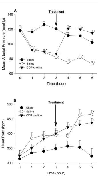
Effect of CDP-choline on blood pressure and heart rate in septic shock-induced by CLI: Baseline mean
arterial pressure (MAP) values of rats were 119 ± 2 mm Hg (n=24). These values did not change significantly in sham group (p>0.05) (Figure 1A). Cecal ligation incision (CLI) procedure caused the decrease in MAP within 60 min (p<0.05) (Figure 1A). Saline injection 3 h after CLI did not change the arterial blood pressure in CLI-induced rats and arterial blood pressure continued to decrease throughout 3 hours (p>0.05) (Figure 1A). Intravenous injection of CDP-choline at 3 hours after CLI ameliorated hypotension in 1 hour (p<0.05) (Figure 1A). Arterial blood pressure of rats maintained at these levels until the end of the study in CDP-choline group (Figure 1A).
Heart rate of rats in CLI + saline and CLI + CDP-choline groups increased significantly (p<0.05) (Figure 1B). CDP-choline administration did not affect the rise in heart rate significantly in septic shock conditions (p>0.05) (Figure 1B). Measured MAP and HR values during the experimental protocol are presented in Table 1.
Effect of CDP-choline on IL-1β, IL-6 and TNF-α levels in septic shock-induced by CLI: Plasma IL-1β, IL-6
and TNF-α levels were 266 ± 54, 1201 ± 125 and 27 ± 9 pg/ml respectively at the end of experiments in sham group (Figure 2). Cecal ligation incision procedure caused significant increases in the levels of IL-1β (p<0.05) (Figure 2A), IL-6 (p<0.05) (Figure 2B) and TNF-α (p<0.05) (Figure 2C) as observed in saline injected control group. CDP-choline injection abolished the increases in IL-1β (p<0.05) and TNF-α (p<0.05), attenuated the increase in IL-6 levels (p<0.05) (Figure 2) induced by CLI procedure.
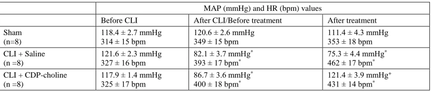
Table 1. Comparison of the values of mean arterial pressure (MAP) and heart rate (HR) in sham, saline and CDP-choline groups before and after cecal ligation-incision (CLI). Data are means ± S.E.M. (n = 8 rats in each group). *p<0.05, vs sham group. +p < 0.05 vs CLI + saline group.
Tablo 1. Çekal ligasyon-insizyon öncesi/sonrasında sham, tuzlu su ve CDP-kolin gruplarında ortalama arter basıncı (MAP) ve kalp hızı (HR) değerlerinin karşılaştırılması. Veriler ortalama ± ortalamanın standart hatası olarak verilmiştir. *p<0.05, sham grubuna göre. +p < 0.05 tuzlu su tedavisi uygulanan gruba göre.
MAP (mmHg) and HR (bpm) values
Before CLI After CLI/Before treatment After treatment Sham (n=8) 118.4 ± 2.7 mmHg 314 ± 15 bpm 120.6 ± 2.6 mmHg 349 ± 15 bpm 111.4 ± 4.3 mmHg 353 ± 18 bpm CLI + Saline (n =8) 121.6 ± 2.3 mmHg 327 ± 16 bpm 82.1 ± 3.7 mmHg * 393 ± 17 bpm* 75.3 ± 4.4 mmHg* 462 ± 17 bpm* CLI + CDP-choline (n =8) 117.9 ± 1.4 mmHg 325 ± 17 bpm 86.7 ± 3.6 mmHg * 400 ± 18 bpm* 121.4 ± 3.9 mmHg+ 431 ± 14 bpm*
Figure 1. Cardiovascular effect of intravenously injected CDP-choline in CLI-induced septic shock. 3 hours after the CLI induction, rats were treated with CDP-choline (100 mg/kg; i.v.) or saline (1 ml/kg; i.v.) and then mean arterial pressure (A) and heart rate (B) were recorded for the next 3 hours. ‘‘0’’ shows CLI induction time point. Data are means ± S.E.M. (n = 8 rats in each group). *p<0.05, vs sham group. +p < 0.05 vs CLI + saline group. Şekil 1. İntravenöz olarak uygulanan CDP-kolin’in çekal bağlama-kesme yöntemi ile oluşturulan septik şoktaki kardiyovasküler etkileri. Çekal bağlama ve kesme işleminin başlangıcından 3 saat sonra, sıçanların CDP-kolin (100 mg/kg, i.v.) veya tuzlu su (1 ml/kg; i.v.) ile tedavi edilmelerini takiben ortalama arter basıncı (A) ve kalp hızı (B) sonraki 3 saat boyunca kaydedildi. ‘‘0’’ zaman noktası çekal bağlama ve kesme işleminin uygulandığı anın verisini göstermektedir. Veriler ortalama ± ortalamanın standart hatası olarak verilmiştir. *p<0.05, sham grubuna göre. +p < 0.05 tuzlu su tedavisi uygulanan gruba göre.
Figure 2. The effect of CDP-choline on CLI induced alterations in plasma cytokine levels. Rats were treated with CDP-choline (100 mg/kg; i.v.) or saline (1 ml/kg; i.v.) 3 hours after the CLI induction. At the end of the study (3h after treatment), blood samples (200 µl) were collected for IL-1β (A), IL-6 (B), and TNF (C) levels. Data are means ± S.E.M. (n = 8 rats in each group). *p<0.05, vs sham group. +p < 0.05 vs CLI + saline group. Şekil 2. CDP-kolin’in çekal bağlama-kesmenin neden olduğu plazma sitokin seviyeleri değişiklikleri üzerine etkileri. Çekal bağlama ve kesme işleminin başlangıcından 3 saat sonra, sıçanlar CDP-kolin (100 mg/kg, i.v.) veya tuzlu su (1 ml/kg; i.v.) ile tedavi edildi. Çalışmanın sonunda (Tedaviden sonra 3 saat) IL-1β (A), IL-6 (B) ve TNF (C) seviyelerinin ölçümü için kan örnekleri (200 µl) toplandı. Veriler ortalama ± ortalamanın standart hatası olarak verilmiştir. *p<0.05, sham grubuna göre. +p < 0.05 tuzlu su tedavisi uygulanan gruba göre.
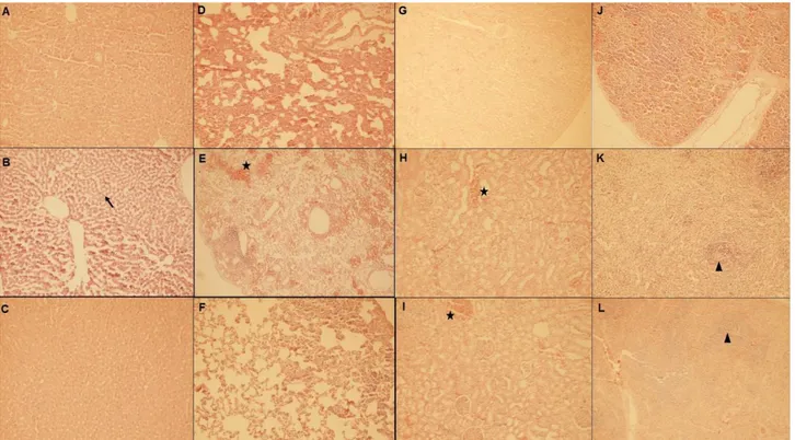
Figure 3. The effect of CDP-choline on CLI induced organ failure. Rats were treated with CDP-choline (100 mg/kg; i.v.) or saline (1 ml/kg; i.v.) 3 hours after the CLI induction. Histopathological comparison of liver [sham (A), saline (B), CDP-choline (C) groups], lung [sham (D), saline (E), CDP-choline (F) groups], kidney [sham (G), saline (H), CDP-choline (I) groups] and spleen [sham (J), saline (K), CDP-choline (L) groups] tissues. “arrow” shows vacuolar degeneration; “star” shows hemorrhage; “triangle” shows necrosis.
Şekil 3. CDP-kolin’in çekal bağlama-kesmenin neden olduğu doku hasarı üzerine etkileri. Çekal bağlama ve kesme işleminin başlangıcından 3 saat sonra, sıçanlar CDP-kolin (100 mg/kg, i.v.) veya tuzlu su (1 ml/kg; i.v.) ile tedavi edildi. Karaciğer, [sham (A), tuzlu su (B), kolin (C) grupları], akciğer [sham (D), tuzlu su (E), kolin (F) grupları], böbrek [sham (G), tuzlu su (H), CDP-kolin (I) grupları] ve dalak [sham (J), tuzlu su (K), CDP-CDP-kolin (L) grupları] dokularının histopatolojik karşılaştırılması. “ok”, vasküler dejenerasyonu; “yıldız”, hemorajiyi; “üçgen”, nekrozu göstermektedir.
Effect of CDP-choline on tissue injury induced by septic shock model in rats: Since septic shock leads to the
multiple organ failure due to the tissue injury, we investigated whether CDP-choline can exert tissue protective effect in septic shock condition. Therefore histopathological examination was performed on lung, liver, kidney and spleen tissue. In CLI + saline group, vacuolar degeneration, necrosis and inflammatory reaction, increasing inflammatory cells in portal area were seen in liver widely to moderate; while these changes were observed in CLI + CDP- choline group slightly to moderate. In lung; severe inflammatory response (including bronchopneumonia and increased BALT activity) and severe hemorrhage were observed in CLI + saline group, while inflammatory response and hemorrhage were just moderate in CLI + CDP- choline group. In kidney, degeneration in tubules and necrosis were seen slightly in CLI + saline group. The severity of lesions in CLI + CDP- choline group was moderate extent. In spleen, there were remarkable siderosis, necrosis and depletion of lymphoid tissue in saline treated CLI-induced sepsis group. However this changes decreased in CDP- choline treated CLI sepsis (Figure 3).
Discussion and Conclusion
The present data shows that intravenously injected CDP-choline restores blood pressure, attenuates the increase in plasma IL-6, IL-1, TNF-a levels and prevents lung, liver and kidney injury in septic shock induced by cecal ligation and incision model.
Cecal ligation and incision model decreased arterial blood pressure and caused hypotension within 3 hours (Figure 1). CDP-choline that was injected at 3rd hours after
CLI increased blood pressure and reversed hypotension without affecting the heart rate changes. The dose of CDP-choline was chosen from our earlier experiments in which the drug was shown to exert both cardiovascular and tissue protective effects in several conditions (11, 31, 32, 45). Our previous reports repeatedly demonstrated that CDP-choline, administered either intracerebroventricularly or intravenously, can affect blood pressure and exert pressor response in normal and hypotensive conditions (31, 32). Therefore the present finding showing that CDP-choline restores hypotension induced by CLI is in aggreement with those previous reports. Besides, the observation that CDP-choline did not alter the increase in heart rate caused by CLI is in good accordance with the previous data
demonstrating that CDP-choline did not influence heart rate changes while producing pressor effect in normal and haemorrhaged conditions (31, 32).
Although the mechanism of the CDP-choline’s pressor effect was not investigated in this study, we may imply that the activation of central cholinergic nicotinic and/or muscarinic receptors followed by the enhancement of central cholinergic transmission is the leading part of its pressor effect. Because we have previously shown that intravenously injected CDP-choline increases choline levels in the plasma and the brain, improves neuronal acetylcholine synthesis and release; the increase in neuronal Ach release activates central nicotinic receptors which results in the pressor response through the stimulation of the peripheral catecholamine and vasopressin release (32, 33). On the other hand, it has been shown that, choline is a selective agonist for 7 nicotinic acetylcholine receptors (6). It has also been reported that choline exerts pressor effect by directly activating 7-nicotinic acetylcholine receptors (23). Hence, we may also suggest that choline metabolite may mediate the pressor effect of CDP-choline observed in this study.
Septic shock induced by cecal ligation insicion model caused an increase in plasma TNF-, 6 and IL-1 levels. CDP-choline administration significantly attenuated the increase of these cytokine levels (Figure 2). The excessive increase of these proinflammatory molecules indicates the progressive and deleterious inflammatory response to infection in septic shock. Therefore, treatment approaches aimed at reducing the release of these molecules in endotoxemia or sepsis are very important (29). One of those treatment strategies is to activate vagal anti-inflammatory cholinergic signaling because the stimulation of efferent vagus nerve inhibits the release of proinflammatory molecules and attenuates the progression of shock in endotoxemia (8). CDP-choline can be considered one of those cholinergic modalities since it has been shown to activate efferent vagal cholinergic pathway by stimulating central muscarinic receptors throughout the enhanced cholinergic transmission in short term myocardial ischemia-reperfusion injury (45). Moreover, 7-nicotinic acetylcholine receptors have a unique role in mediating vagal anti-inflammatory pathway (25, 39). Several reports revealed that the activation of 7-nicotinic acetylcholine receptors by nicotine (a nonselective nicotinic agonist) or GTS-21 (a selective 7 nicotinic acetylcholine receptor agonist) blocked the TNF- release and improved survival in experimental septic conditions (26, 40). Considering that choline is the main functional mediator of CDP-choline and is a selective agonist for 7 nicotinic acetylcholine receptors together with the above information, we may suggest that the stimulation of vagal cholinergic anti-inflammatory
signaling through the initiation of central muscarinic cholinergic receptors or peripheral 7-nicotinic acetylcholine receptors may mediate the CDP-choline induced inhibiton of the proinflammatory cytokine release in septic shock conditions.
In the present study, histological data demonstrated that CLI model caused multiple organ injury including liver, kidney, lung and spleen. Reduced tissue perfusion during the hypotensive phase, inflammation through the excessive release of proinflammatory cytokins and oxidative stress are the main reasons of these tissue damages in septic shock conditions (5). CDP-choline administration attenuated tissue injuries and decreased vacuolar degeneration, necrosis and hemorrhage in liver, kidney and lung tissue. CDP-choline-induced tissue protection may have been through i) the improved tissue perfusion due to drug’s pressor effect, ii) the alleviation of inflammatory conditions due to the drug-induced decrease in plasma proinflammatory cytokins. CDP-choline also can reduce oxidative stress in several pathological conditions (3, 11). Therefore the antioxidant effect of CDP-choline may have another explanation of its tissue protective effect.
In summary, the outcomes of the current study show that CDP-choline is able to restore hypotension, to decrease the plasma levels of proinflammatory cytokins and to protect tissue injury induced by septic shock model.
Acknowledgements
This work was supported by a grant from the Uludag University Commission of Scientific Research Projects (UAP (T) – 2011/13) awarded to Dr. M. Sertaç Yilmaz. This study was presented in FASEB meeting in 2013 where abstract only is published in the meeting abstract supplement (47).
References
1. Adibhatla RM, Hatcher JF (2002): Citicoline mechanisms and clinical efficacy in cerebral ischemia. J Neurosci Res,
70, 133-139.
2. Adibhatla RM, Hatcher JF (2005): Cytidine 5’-diphosphocholine (CDP-choline) in stroke and other CNS disorders. Neurochem Res, 30, 15-23.
3. Adibhatla RM, Hatcher JF, Dempsey RJ (2001): Effects of citicoline on phospholipid and glutathione levels in transient cerebral ischemia. Stroke, 32, 2376-2381. 4. Adibhatla RM, Hatcher JF, Dempsey RJ (2004):
Cytidine-5'-diphosphocholine (CDP-choline) affects CTP: Phosphocholine cytidylyltransferase and lyso-phosphatidylcholine after transient brain ischemia. J Neurosci Res, 76, 390-396.
5. Ahmad A, Druzhyna N, Szabo C (2016): Delayed treatment with sodium hydrosulfide improves regional blood flow and alleviates cecal ligation and puncture (CLP)-induced septic shock. Shock, Epub ahead of print (doi: 10.1097/SHK.0000000000000589)
6. Alkondon M, Pereira EF, Barbosa CT, et al. (1997): Neuronal nicotinic acetylcholine receptor activation modulates gamma-aminobutyric acid release from CA1 neurons of rat hippocampal slices. J Pharmacol Exp Ther,
283, 1396-1411.
7. Blusztajn JK, Wurtman RJ (1983): Choline and cholinergic neurons. Science, 22, 614-620.
8. Borovikova LV, Ivanova S, Zhang M, et al. (2000): Vagus nerve stimulation attenuates the systemic inflammatory response to endotoxin. Nature, 405, 458-462.
9. Cavun S, Savci V (2004): CDP-choline increases plasma ACTH and potentiates the stimulated release of GH, TSH and LH: The cholinergic involvement. Fundam Clin Pharmacol, 18, 513-523.
10. Cavun S, Savci V, Ulus IH (2004): Centrally injected CDP-choline increases plasma vasopressin levels by central cholinergic activation. Fundam Clin Pharmacol, 18, 71-77.
11. Coskun C, Avci B, Ocak N, et al. (2010): Effect of repeatedly given CDP-choline on cardiovascular and tissue injury in spinal shock conditions: Investigation of the acute phase. J Pharm Pharmacol, 62, 497-506.
12. Coskun C, Avci B, Yalcin M, et al. (2014): Protective effect of CDP-choline on ischemia-reperfusion-induced myocardial tissue injury in rats. Ir J Med Sci, 183, 539-548. 13. Deitch EA (1998): Animal models of sepsis and shock: A
review and lessons learned. Shock, 9, 1-11.
14. Dorman RV, Dabrowiecki Z, Horrocks LA (1983): Effects of CDP-choline and CDP-ethanolamine on the alterations in rat brain lipid metabolism induced by global ischemia. J Neurochem, 40, 276-279.
15. Downing JEG, Miyan JA (2000): Neural immunoregulation: Emerging roles for nerves in immune homeostasis and disease. Immunol Today, 21, 281-289. 16. Esmon CT (2004): Why do animal models (sometimes) fail
to mimic human sepsis? Crit Care Med, 32, 219-222. 17. Fiedorowicz M, Makarewicz D, Stan´czak-Mrozek KI,
et al. (2008): CDP-choline (citicoline) attenuates brain
damage in a rat model of birth asphyxia. Acta Neurobiol Exp, 68, 389-397.
18. G-Coviella IL, Wurtman RJ (1992): Enhancement by cytidine of membrane phospholipids synthesis. J Neurochem, 59, 338-343.
19. Hotchkiss RS, Karl IE (2003): The pathophysiology and treatment of sepsis. N Engl J Med, 348, 138.
20. Ilcol YO, Gurun MS, Taga Y, et al. (2003): Choline increases serum insulin in rat when injected intraperitoneally and augments basal and stimulated acetylcholine release from the rat minced pancreas in vitro. Eur J Biochem, 270, 991-999.
21. Ilcol YO, Yilmaz Z, Cansev M, et al. (2009): Choline or CDP-choline alters serum lipid responses to endotoxin in dogs and rats: Involvement of the peripheral nicotinic acetylcholine receptors. Shock, 32, 286-294.
22. Klein J (2000): Membrane breakdown in acute and chronic neurodegeneration: Focus on choline-containing phospholipids. J Neural Transm, 107, 1027-1063.
23. Li XD, Buccafusco JJ (2004): Role of alpha7 nicotinic acetylcholine receptors in the pressor response to intracerebroventricular injection of choline: Blockade by
amyloid peptide Abeta1-42. J Pharmacol Exp Ther, 309, 1206-1212.
24. Marshall JC, Vincent JL, Fink MP, et al. (2003): Measures, markers, and mediators: toward a staging system for clinical sepsis. A report of the Fifth Toronto Sepsis Roundtable, Toronto, Ontario, Canada, October 25-26, 2000. Crit Care Med, 31, 1560-1567.
25. Parrish WR, Rosas-Ballina M, Gallowitsch-Puerta M, et al. (2008): Modulation of TNF release by choline requires
alpha7 subunit nicotinic acetylcholine receptor-mediated signaling. Mol Med, 14, 567-574.
26. Pavlov VA, Ochani M, Yang LH, et al. (2007): Selective alpha7-nicotinic acetylcholine receptor agonist GTS-21 improves survival in murine endotoxemia and severe sepsis. Crit Care Med, 35, 1139-1144.
27. Rao AM, Hatcher JF, Dempsey RJ (2000): Lipid alterations in transient forebrain ischemia: Possible new mechanisms of CDP-choline neuroprotection. J Neurochem, 75, 2528-2535.
28. Rao AM, Hatcher JF, Dempsey RJ (2001): Does CDP-choline modulate phospholipase activities after transient forebrain ischemia? Brain Res, 893, 268-272.
29. Riedemann NC, Guo RF, Ward PA (2003): Novel strategies for the treatment of sepsis. Nat Med, 9, 517-524. 30. Rivera CA, Wheeler MD, Enomoto N, et al. (1998): A choline rich diet improves survival in a rat model of endotoxin shock. Am J Physiol, 275, 862-867.
31. Savci V, Cavun S, Goktalay G, et al. (2002): Cardiovascular effects of intracerebroventricularly injected CDP-choline in normotensive and hypotensive animals: The involvement of cholinergic system. Naunyn Schmiedebergs Arch Pharmacol, 365, 388-398.
32. Savci V, Goktalay G, Cansev M, et al. (2003): Intravenously injected CDP-choline increases blood pressure and reverses hypotension in haemorrhagic shock: Effect is mediated by central cholinergic activation. Eur J Pharmacol, 468, 129-139.
33. Savci V, Goktalay G, Ulus IH (2002): Intracerebroventricular choline increases plasma vasopressin and augments plasma vasopressin response to osmotic stimulation and hemorrhage. Brain Res, 942, 58-70.
34. Savci V, Ulus IH (1997): Cardiovascular effects of central choline during endotoxin shock in the rat. J Cardiovasc Pharmacol, 30, 667-675.
35. Savci V, Wurtman RJ (1995): Effect of cytidine on membrane phospholipid synthesis in rat striatal slices. J Neurochem, 64, 378-384.
36. Secades JJ (2011): Citicoline: Pharmacological and clinical review, 2010 update. Rev Neurol, 52, 1-62. 37. Stoll AL, Renshaw PF, DeMicheli E, et al. (1995):
Choline ingestion increases the resonance of choline containing compound in human brain: An in vivo proton magnetic resonance study. Biol Psych, 37, 170-174. 38. Ulus IH, Wurtman RJ, Mauron C, et al. (1995): Choline
increases acetylcholine release and protects against the stimulation-induced decrease in phosphatide levels within membranes of rat corpus striatum. Brain Res, 484, 217-227. 39. Wang H, Yu M, Ochani M, et al. (2003): Nicotinic acetylcholine receptor alpha7 subunit is an essential regulator of inflammation. Nature, 421, 384-388.
40. Wang H, Liao H, Ochani M, et al. (2004): Cholinergic agonists inhibit HMGB1 release and improve survival in experimental sepsis. Nat Med, 10, 1216-1221.
41. Weiss GB (1995): Metabolism and actions of CDP-choline as an endogenous compound and administered exogenously as citicoline. Life Sci, 56, 637-660.
42. Wenzel RP, Edmond MB (2012): Septic shock devaluating another failed treatment. N Engl J Med, 366, 2122. 43. Wijdicks EF, Stevens M (1992): The role of hypotension
in septic encephalopathy following surgical procedures. Arch Neurol, 49, 653.
44. Wurtman RJ (1992): Choline metabolism as a basis for the selective vulnerability of cholinergic neurons. Trends Neurosci, 15, 117-122.
45. Yilmaz MS, Coskun C, Yalcın M, et al. (2008): CDP-choline prevents cardiac arrhythmias and lethality induced by short-term myocardial ischemia-reperfusion injury in the rat: involvement of central muscarinic cholinergic mechanisms. Naunyn Schmiedebergs Arch Pharmacol, 378, 293-301.
46. Yilmaz Z, Ilcol YO, Torun S, et al. (2006): Intravenous administration of choline or cdp-choline improves platelet count and platelet closure times in endotoxin-treated dogs. Shock, 25, 73-79.
47. Yilmaz MS, Sevim C, Altinbas B, et al. (2013): CDP-choline protects against sepsis-induced acute tissue injury in rats. The FASEB J, 27, Supplement 888.8.
Geliş tarihi: 25.12.2015 / Kabul tarihi: 20.07.2016
Address for correspondence:
Mustafa Sertaç Yılmaz, MD, PhD Uludağ Universitesi Tıp Fakultesi, Tıbbi Farmakoloji Anabilim Dalı, Gorukle, 16059, Bursa, Turkey.
Tel: + 90 224 295 3566 Fax: + 90 224 442 8102 e-mail: [email protected]