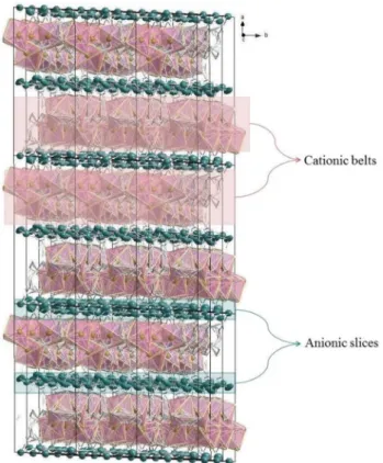
Metalo components exhibiting significant anticancer and antibacterial properties: a novel sandwich-type like polymeric structure
Tam metin
Şekil

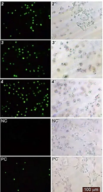
![Figure 6. Effects of 2, 3, 4 and [Ag(CN) 2 ]-on the proliferation of Vero cells, HeLa, C6, and HT-29](https://thumb-eu.123doks.com/thumbv2/9libnet/4367022.73297/9.892.235.715.81.429/figure-effects-ag-cn-proliferation-vero-cells-hela.webp)
Benzer Belgeler
Bunun yanında; temel ücret yapısına yönelik adaletin gözetilmesi, performans ücret ilişkisinin kurularak işgörenlerin katkılarının ücretlerine yansıtılması,
SEM experiments were conducted to in- vestigate the morphology of the product (Fig. A large number of structures with spherical shapes are observed; whose average particle size is..
Our trust record system provides a natural infrastructure that can he used for evaluating the service level of the peers: At the time of a download, the priority of
Using the entire training data for learning, we give the parameter analysis performed on the test set in Figure 4.4 for the window size and the number of states and in Figure 4.5
Asllnda bu kawam, Tiirkge iedmdeki gibi zaman zaman bir oy veya oylana belirgin bir dalraruS, hatta bir tepki leklini almalla birlikte, daha gok kamuca ijziimsenmil
Zira Hâzım’ın ve Mu- ammer’in tiyatroculuğa başladığı 1920’li yıllarda Direklerarası’nda büyük komedyen Naşit Bey (o günlerin) deyimiyle ‘Komik-i Şehir Naşit
Kendisini bir alkış şelâlesi içinde kar şılayan halkı, bir elini havaya kaldı rarak, ötekini de kalbinin üstüne ba sarak selâmlaması ne
Tablo 6.5’te elektriksel veriler sonucunda hesaplanan kirlilik etkisi (KE) ve görüntü işleme işlemi sonucunda elde edilen kirlilik oranı (KO) verilmiştir.. Kirlilik etkisi
