ORIGINAL ARTICLE
Neonatal calf meningitis associated with
Streptococcus gallolyticus
subsp. gallolyticus
Fuat Aydın1&Vehbi Güneş2&Latife Çakır Bayram3&Seçil Abay1&Emre Karakaya1&Kemal Varol4&Gencay Ekinci2& Kadir Semih Gümüşsoy1&Hamit Kaan Müştak5&Kadir Serdar Diker5
Received: 29 May 2018 / Accepted: 12 September 2018 / Published online: 19 September 2018 # Institute of Microbiology, Academy of Sciences of the Czech Republic, v.v.i. 2018
Abstract
Here, we report a case of neonatal calf meningitis due to Streptococcus gallolyticus subsp. gallolyticus (SGG). Clinical, patho-logical and microbiopatho-logical findings were evaluated. API Strep, 16S rRNA gene sequencing, rpoB gene sequencing and sodA gene sequencing were used for the complete identification of SGG. This is the first documented report of neonatal calf meningitis due to SGG in veterinary medicine.
Introduction
Until 2003, Streptococcus bovis (S. bovis) was divided into three different biotypes (biotype I, biotype II/1 and biotype II/ 2). Thereafter, in view of their phenotypic and genotypic char-acteristics, S. bovis biotypes were reclassified as Streptococcus gallolyticus subsp. gallolyticus (SGG) (biotype I), S. infantarius subsp. infantarius (biotype II/1), S. lutetiensis (biotype II/1) and S. gallolyticus subsp. pasteurianus (biotype II/2). Also, S. gallolyticus subsp. macedonicus was placed as a new subspecies of S. gallolyticus. (Beck et al.2008; Osawa et al.1995; Poyart et al.2002; Schlegel et al.2003; Jans et al. 2015; Dekker and Lau2016). SGG is a Gram-positive inhab-itant bacterium usually found in the gastrointestinal tract of healthy humans (Dumke et al.2017) and animals (Sasaki et al. 2004; Schulz et al.2015). Also known to be an opportunistic pathogen (Dumke et al. 2017; Kambarev et al. 2017), this
bacterium has been isolated from cases of meningitis (Beneteau et al. 2015), colonic malignant diseases (Boleij and Tjalsma2013; Butt et al.2016), septic arthritis and oste-omyelitis (García-País et al.2016), bacteraemia (Corredoira-Sánchez et al.2012) endocarditis (Vollmer et al.2010) and biliary tract infections (Corredoira et al. 2014) in humans. Furthermore, this microorganism has also been reported to be isolated from animals in cases of endocarditis in chickens (Sekizaki et al.2008), mastitis in cows (Sasaki et al. 2004), mortality in turkey poults (Droual et al. 1997), infection in ducklings (Hogg and Pearson2009), purulent lesions in vari-ous organs in calves (Seimiya et al.1992) and haemorrhages in the pulmonary artery and aortic valve in a roe deer (Velarde et al.2009). Recent research has shown that SGG is also of zoonotic nature (Dumke et al.2014; Dumke et al.2015).
In this study, it was aimed to report the isolation of SGG from a neonatal calf with meningitis. In addition, advanced molecular analysis of the isolated bacterium was performed and the results were evaluated.
Materials and methods
Patient
’s description and clinical data
A 5-day-old male Simmental calf, weighing 40 kg with central nervous system (CNS) signs, was admitted to the Internal Medicine Clinic of the Education, Research and Practice Hospital of Faculty of Veterinary Medicine, Erciyes University. The patient’s anamnesis revealed that the calf, which was born to a dam with dystocia, had
* Seçil Abay [email protected]
1
Department of Microbiology, Faculty of Veterinary Medicine, Erciyes University, Kayseri, Turkey
2 Department of Internal Medicine, Faculty of Veterinary Medicine,
Erciyes University, Kayseri, Turkey
3
Department of Pathology, Faculty of Veterinary Medicine, Erciyes University, Kayseri, Turkey
4
School of Burdur Vocational Education, Department of Veterinary Science, Mehmet Akif Ersoy University, Burdur, Turkey
5 Department of Microbiology, Faculty of Veterinary Medicine,
received neither enough colostrum nor the required care of the navel region, and had been administered antibacte-rial, vitamin and mineral treatment after being examined by a veterinarian. But we could not get more information from the owner about this treatment including dose and name of drugs used. At clinical examination, the animal showed a general deterioration of the body condition, lethargy, stiffness of the neck muscles, painful paraspinal muscle spasm, hyperaesthesia, anorexia, dyspnoea, opisthotonus, ataxia, tension of the extremities, slight de-hydration, mild cough, increased vesicular sounds on the auscultation of the thorax, exophthalmos and excitation. Measurements demonstrated that while the body temper-ature of the animal was 38.4 °C, its pulsation and respi-ration rate were 140/min and 35/min, respectively. Five-millilitre blood samples were collected into EDTA-coated tubes for haematological analyses, and 9-ml blood sam-ples were collected into dry tubes for biochemical analy-ses. The patient was hospitalised so as to be monitored, and was regularly checked and treated in the following days. The calf underwent regular daily examinations and was sampled for blood. Furthermore, in view of the CNS signs that the animal showed, 4-ml sterile cerebrospinal fluid (CSF) samples were taken from the lumbosacral re-gion into EDTA-coated and dry tubes using a spinal can-nula (Spinocan® 0.90 × 88 mm/20 G × 3.5″). It was ob-served that the CSF was yellowish grey in colour, opaque and cloudy. Treatment was started with the parenteral ad-ministration of enrofloxacin (Baytril® 10%, 5 mg/kg, in-travenous, twice daily (IV BID), metronidazole (Flagyl®, ampoule 0.5% inj. 100 ml solution, 20 mg/kg IV BID), sulfamethoxazole/trimethoprim (Animar®, 5 mg/kg IV BID), vitamins B1 and B6 (Nervit®, 10 ml IV, once daily) and dexamethasone (Deksavet®, 0.1 ml/kg intramuscular (IM) once daily). Also, balanced electrolyte solutions (0.9% NaCl Polifleks®, Polifarma, Turkey) and lactated Ringer’s solution (Medifleks®, Eczacıbaşı, Turkey) were given intravenously via the auricular vein. Furthermore, 5% O2therapy was administered by nasal route.
Haematological analyses
Blood with EDTA samples were analysed by the haemocytometer (Mindray BC-2800Vet, China) during hospitalisation. In addition, CSF taken from first day of the hospitalisation was evaluated in the same way.
Biochemical analyses of serum and CSF
Biochemical analyses of serum and CSF samples obtained only on the first day of the hospitalization were performed by using an autoanalyzer (BT-3000 Plus; Italy).
Glutaraldehyde coagulation test
We performed glutaraldehyde coagulation test in the detection of failure of passive transfer according to Turgut et al. (1998) and Weaver et al. (2000).
Cytological analysis
Cytospin smears from CSF were prepared for morphological e v a l u a t i o n b y c y t o c e n t r i f u g a t i o n ( C y t o s p i n 3 , ThermoShandon, Pittsburgh, PA, USA) for 5 min at 1000 rpm using disposable plastic chambers (Cytofunnel, T h e r m o S h a n d o n ) a n d g l a s s s l i d e s ( C y t o s l i d e , ThermoShandon) (Scott and Penny 1993). Then, cytology smears were stained with Diff-Quik (Richard Allan Scientific, Thermo Electron Corporation, 9990700, USA).
Bacteriological analysis
Gram staining was used for slides prepared from the CSF. The CSF was inoculated onto 7% sheep blood agar (blood agar base no. 2, CM0271, Oxoid), McConkey (105,465, Merck, Germany) and eosin methylene blue (EMB, CM0069B, Oxoid, UK) agar; the plates were incubated at 37 °C under aerobic, anaerobic (Anaerocult A, Merck) and microaerobic (Anaerocult C, Merck) conditions, and were examined daily. Three colonies were selected at random to obtain pure cultures of the bacteria. Gram staining, and the oxidase, catalase, oxi-dation fermentation and motility tests were used for the phe-notypic identification of the colonies grown (Quinn et al. 2011). API 20 Strep system (bioMerieux SA, Marcy-l’Etoile, France) was performed for the identification at the species and subspecies levels of the bacteria. In addition, an-timicrobial susceptibility testing was done for the bacteria iso-lated (Bauer et al. 1966). Metronidazole (MET, 5 μg), trimethoprim-sulfamethoxazole (SXT, 25 μg), gentamicin (CN, 10μg), ampicillin (AM, 10 μg), amoxicillin-clavulanic acid (AMC, 30μg), cephazolin (KZ, 30 μg) and enrofloxacin (ENR, 5μg) discs were used in the test. The disc diffusion test results were interpreted using the criteria published by Clinical and Laboratory Standards Institute (CLSI 2008a). After the necropsy, samples (lungs, liver, spleen, kidneys and brain) taken from calf were used for microbiological examinations. The isolation and identification procedure followed for the CSF was also used for the organ samples.
DNA extraction
DNA extraction for the molecular analyses was performed by using UltraClean® Microbial DNA Isolation Kit (12224-50, Mo Bio Laboratories, Carlsbad, CA, USA) following the manufacturer’s instructions.
Molecular analysis
16S rRNA gene sequencing, rpoB gene sequencing and sodA gene sequencing were used for the molecular analysis of the recovered isolate. The universal primers 27F and 1492R were used to amplify the 16S rRNA gene (Lane1991). The primers 31F and 830R were used to amplify the rpoB gene, which encodes the beta subunit of RNA polymerase (Drancourt et al.2004). In addition, sodA partial sequencing has been performed by using the degenerated primers d1 (5-CCI TAY ICI TAY GAY G CI YTI GAR CC-3) and d2 (5-ARR TAR TAI GCR TGY TCC CAI ACR TC-3) reported by the Poyart et al. (1998). The amplified products were purified using the QIA-quick PCR Purification Kit (Qiagen, USA), and se-quence analysis was performed using the Big Dye Direct Cycle Sequencing Kit (Applied Biosystems, USA) according to the manufacturer’s instructions. After cycle sequencing, the amplicons were purified with Sephadex G-50 (Sigma-Aldrich, USA) by using spin columns and sequenced on the Applied Biosystems 3500 Genetic Analyser (Applied Biosystems, USA). All sequences were analysed with the CLC Main Workbench 6 and compared with reference se-quences available on the website of the National Centre for Biotechnology Information using the Basic Local Alignment Search Tool for Nucleotides (BLASTn) programme according to the criteria of the CLSI, MM18-A guideline (CLSI,2008b). The genotyping of the isolates obtained from the CSF and the organ samples was performed with Enterobacterial repet-itive intergenic consensus (ERIC)-PCR (Aydin et al.2007).
Histopathological analysis
After the animal died (on fifth day after the administered to the clinic), a necropsy was performed. Samples were taken from several organs (lungs, liver, spleen, kidneys, small intestine and brain) for histopathological examinations. Tissue samples were fixed in 10% formalin, routinely processed and stained with haematoxylin and eosin for histopathological evaluation. Tissue sections were also stained with Brown-Brenn Gram’s stain to investigate the presence of bacteria.
Results
Clinical data
On day 3, treatment with metronidazole and sulfamethoxa-zole/trimethoprim, to which the bacteria were found to be resistant, was ceased and therapy was continued with enrofloxacin and amoxicillin-clavulanic acid. Treatment was continued until day 5, when the animal died.
Haematological analyses
Haematological analysis results pointed out to leucocytosis (18.9 × 10⁶/μl), monocytosis (1.1 × 10⁶/μl), granulocytosis (13.4 × 10⁶/μl) and low-packed cell volume (PCV) level (24.0%), and no significant alteration was observed in these values until day 4 (Table1). CSF analysis revealed the pres-ence of leucocytosis (0.1 × 10⁶/μl). While the platelet level was within the normal range on day 1 (572 × 106/μl), thrombocytosis was detected in the measurements on days 2 (2524 × 106/μl), 3 (2993 × 106/μl) and 4 (1814 × 106/μl) (Table 1). The other haematological parameters measured were within the normal range (Klinkon and Ježek2012).
Biochemical analyses
Biochemical analyses of serum and CSF
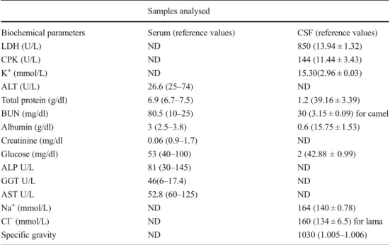
BUN (blood urea nitrogen) (80.5 mg/dl) and GGT (gamma-glutamyl transferase) (46 U/L) levels were higher than those of references values reported previously for bovine (10– 25 mg/dl and 6–17.4 U/L respectively) (Table2). Results of serum biochemistry were evaluated according to Russell and Roussel (2007) and Kaneko et al. (2008). While levels of LDH (lactate dehydrogenase), CPK (creatine phosphokinase), K+, BUN, Na+and Cl−were found to be high, albumin and glucose levels were low in CSF of the calf (Table 2). Biochemistry results of CSF were evaluated according to pub-lications conducted by various researchers (St Jean et al.1997; Welles et al.1992; Al-Sagair et al.2005).
Glutaraldehyde coagulation test
Negative result was detected in the glutaraldehyde coagula-tion test, so it was confirmed to the failure of colostrum uptake of the calf.
Cytological analyses
Cytology smears of the CSF stained with Diff-Quik showed abundant neutrophils and a few aggregates of degenerated neutrophils and numerous Gram-positive cocci (Fig.1A–C).
Bacteriological analyses
The Gram staining of CSF and organs revealed the presence of Gram-positive cocci. Following 24 h of incubation, bacterial growth was not observed in the EMB and McConkey plates, whereas in the blood agar plates incubated under all three atmospheric conditions, grey-white coloured non-haemolytic smooth colonies of a diameter of 2–3 mm were observed. On the basis of the results of phenotypic tests, the bacteria were identified as Streptococcus spp. Results of the Api 20 Strep
test were analysed by the API 20 Strep V7.0 software avail-able at http://210.242.211.31/servlet/Identify?action= prepareNew&stripId=6. Three isolates recovered from CSF were identified as S. bovis I (S. gallolyticus subsp. gallolyticus, Api Strep profile: 5240573, ID%, 98.9) by using Api 20 Strep.
The results of the antibacterial susceptibility testing showed that all of the isolates were resistant to MET, SXT and CN, susceptible to AM, AMC, KZ and moderately sus-ceptible to ENR. Results of bacteriological analyses per-formed from organs were similar to CSF results. No growth was observed on the McConkey and EMB plates. Growth was observed in all of the blood agar plates incubated under aero-bic, microaerobic and anaerobic conditions. Similar to the method used for the CSF, with the use of the Api 20 Strep, all isolates (15) were identified as S. gallolyticus subsp. gallolyticus. In the genotyping, it was determined that all of
the 18 isolates (3 from CSF and 15 from organs) had the same band patterns (data not shown); thus, a single representative isolate was used in the further stages of the study and this isolate was named as Erugall17.
Molecular analysis
According to result of the 16S rRNA BLASTn, Erugall17 was found to be S. gallolyticus with the identity score of 99%. On the other hand, rpoB gene sequence and sodA gene sequence revealed that Erugall17 had the identity scores of 99 and 100% for SGG respectively.
The 16S rRNA gene sequence, rpoB gene sequence and sodA gene sequence of Erugall17 were deposited in GenBank under accession numbers KY952169, KY952170 and MH817021, respectively.
Table 1 Haematological values
of the calf during hospitalisation Hematologic parameters 1st day 2nd day 3th day 4th day References WBC (103cells/μl) 18.9 27.1 20.4 14.5 4–12
Monocyte (103cells/μl) 1.1 1.3 0.9 0.7 0–0.9 Granulocyte (103cells/μl) 13.4 23.1 16.5 10.8
PLT (103cells/μl) 572 2524 2993 1814 100–800 PLT platelet, WBC white blood cells
Table 2 Biochemical values of the serum and CSF samples of the calf
Samples analysed
Biochemical parameters Serum (reference values) CSF (reference values)
LDH (U/L) ND 850 (13.94 ± 1.32)
CPK (U/L) ND 144 (11.44 ± 3.43)
K+(mmol/L) ND 15.30(2.96 ± 0.03)
ALT (U/L) 26.6 (25–74) ND
Total protein (g/dl) 6.9 (6.7–7.5) 1.2 (39.16 ± 3.39) BUN (mg/dl) 80.5 (10–25) 30 (3.15 ± 0.09) for camel Albumin (g/dl) 3 (2.5–3.8) 0.6 (15.75 ± 1.53) Creatinine (mg/dl 0.06 (0.9–1.7) ND Glucose (mg/dl) 53 (40–100) 2 (42.88 ± 0.99) ALP U/L 81 (30–145) ND GGT U/L 46(6–17.4) ND AST U/L 52.8 (60–125) ND Na+(mmol/L) ND 164 (140 ± 0.78)
Cl−(mmol/L) ND 160 (134 ± 6.5) for lama Specific gravity ND 1030 (1.005–1.006) ND not done, LDH lactate dehydrogenase, CPK creatine phosphokinase, ALT alanine aminotransferase, BUN blood urea nitrogen, ALP alkaline phosphatase, GGT gamma-glutamyl transferase, AST aspartate aminotransfer-ase, K+ potassium, Na+ sodium, Cl− chlorine
Biochemistry of CSF was evaluated according to St Jean et al.1997, Welles et al.1992, Al Sagair et al. 2005 Biochemistry of serum was evaluated according to Russell and Roussel2007and Kaneko et al.2008
Histopathological analysis
Necropsy revealed hyperaemia and cloudy areas in the menin-ges. No gross abnormalities were noted in the brain of the calf. The animal had gross anatomic lesions, including marked dif-fuse chronic passive hyperaemia (nutmeg liver). The lungs were dark red, uncollapsed, heavy and firm. A clear frothy fluid was observed on the cut section of the lung. Microscopically, the primary pathological finding was purulent meningitis asso-ciated with the presence of Gram-positive cocci. Acute mild diffuse interstitial pneumonia was evident in the lungs. The renal glomeruli contained numerous fibrinous thrombi. The CNS lesions were prominent and limited to the meninges, where a fibrinous exudate and infiltrations of neutrophils, mac-rophages, and lymphocytes were observed (Fig.1D–G). Small clusters of Gram-positive cocci were evident within the foci in the kidneys, lungs, small intestine, brain and liver.
Discussion
Bacterial meningitis is severe and mostly a fatal condition. Several factors, including the calf not receiving enough colos-trum and the hygiene of the navel region not being attended to, are so effective in the occurrence of neonatal calf meningitis. Meningitis cases due to SGG have been reported in humans (Beneteau et al.2015; van Samkar et al.2015), but have not been reported in neonatal calves before. Nonetheless, Seimiya et al. (1992) reported to have observed fibrinopurulent meningoventriculitis, endophthalmitis and purulent lesions in several organs and tissues in three newborn calves, and isolated S. bovis from the lesions. Unlike this study, in this case, we did not observe or detect any symptoms or signs related to eyes and joints of the calf. However, these re-searchers did not indicate the biotype of S. bovis. The CNS signs and histopathological findings observed in this study
Fig. 1 a–c Cytological analysis of the CSF of a calf. a Numerous small foci of infiltrating cells, mainly of mononuclear cells; lymphocytic (red arrowhead) and monocytic lineage (green arrow, one shown with an arrow). b, c Intra/extracellular, bacteria (black arrow) in the cytoplasm of lymphocytes and macrophages and karyolytic neutrophils. Mixed inflammatory cell population including many degenerate neutrophils (black arrowhead). Giant cell (yellow arrowhead) with two large kidney-shaped nuclei and several cocci within the cytoplasm. Diff-Quick. Bars, 20μm (a); 10 μm (b, c). d–g Major histopathological findings in cerebellum. d Neutrophils in the subarachnoid space. Focal area of necrosis and neutrophilic encephalitis in the molecular layer
(boxed area). Inset: Higher magnification of boxed area (arrows) Gram-positive cocci. e Cerebral cortex. Necrosis and neutrophilic meningitis (star); focal mononuclear cell infiltration (blue arrow). Inset d e m o n s t r a t e s G r a m - p o s i t i v e s m a l l c o c c i i n t h e l e s i o n . f Lymphohistiocytic infiltration (boxed area) in the leptomeninges of the midbrain (star). Inset: Gram-positive small cocci (arrows) in the lesion shown in the boxed area. g Cerebral cortex, large amount of fibrinopurulent exudate in the leptomeninges and prominently dilated vessels. Haematoxylin x Eosin (d–g); Brown-Brenn Gram (inset of Fig. 1d–g). Bars, 10 μm (inset of Fig. 1d–g); 200 μm (d–g)
show similarity to the findings previously reported by Seimiya et al. (1992). However, in the present study, the Gram-positive cocci isolated from the CSF and organs were identified as SGG on the basis of the Api 20 Strep, rpoB gene and sodA gene sequencing results. To the best of our knowledge, the present study is the first veterinary report on the isolation of SGG from a neonatal calf with meningitis, which also pro-vides a detailed assessment of the clinical, cytological (Fig.1A–C), histopathological (Fig.1D–G) and molecular findings.
Calves showing failure of passive transfer and suffering from poor hygiene conditions are particularly prone to septicaemia and meningitis. Negative glutaraldehyde coagula-tion test result revealed that the calf had failure of passive transfer, in our case. Similarly, failure of passive transfer in calves was reported in a study conducted by Turgut et al.1998. Serum biochemical values were between normal references except for GGT and BUN (Table2). The high levels of this two parameters might be associated with failure of renal func-tions or dehydration (Russell and Roussel2007; Kaneko et al. 2008.) This biochemical changes were supported by histo-pathological findings.
When the results of CSF biochemistry were compared with references values indicated for bovine, camel and lama, levels of LDH, CPK, K, BUN, Na and Cl were detected as high in contrast albumin and glucose levels were found as low (Table2) (Welles et al.1992; Al-Sagair et al.2005). In partic-ular, high levels of K, LDH and CK observed for CSF bio-chemistry were found compatible with the study of Nazifi et al. (1997).
Bacterial meningitis cases are characterised by high-mortality rates even if appropriately treated (Smith2015; Fecteau et al.2009). Therefore, in order to protect newborn calves from neonatal septicaemia and bacterial meningitis, neonatal stress should be reduced, an adequate amount of quality colostrum should be provided to the animals, navel care should be attended to and favourable hygiene conditions should be established.
In the present study, the isolate obtained was able to be successfully identified at subspecies level using the Api 20 Strep, rpoB gene sequencing (CLSI2008b) and sodA gene sequencing methods. On the other hand, 16S rRNA gene se-quence analysis was found to be sufficient for identification at genus level, but did not suffice for the identification of sub-species. Therefore, it is suggested that while Api 20 Strep can be used as a rapid, accurate, sensitive and reproducible meth-od for the routine laboratory identification of S. gallolyticus isolates at subtype level, rpoB gene and sodA gene sequence analysis can be used for molecular and phylogenetic analyses. Resistance of the agent to antibiotics used in the treatment, organ failure (especially kidneys) and failure of passive trans-fer could be considered as important factors in the death of the calf. We believe that the identification of the subspecies or
biotyping of S. gallolyticus is not only important for re-searches but also for the epidemiologic relationship of the isolates and the association with several diseases observed in humans and animals.
Compliance with ethical standards
Conflict of interest The authors declare that they have no conflicts of interest.
References
Al-Sagair OA, Fathalla SI, Abdel-Rahman HA (2005) Reference values and age-related changes in cerebrospinal fluid and blood compo-nents in the clinically normal male dromedary camel. J Anim Vet Adv 4:470–472
Aydin F, Gümüşsoy KS, Atabay HI, Iça T, Abay S (2007) Prevalence and distribution of Arcobacter species in various sources in Turkey and molecular analysis of isolated strains by ERIC-PCR. J Appl Microbiol 103:27–35
Bauer AW, Kirby WMM, Sherris JC, Turek M (1966) Antibiotic suscep-tibility testing by a standardized single disc method. Am J Clin Pathol 45:493–496
Beck M, Frodl R, Funke G (2008) Comprehensive study of strains pre-viously designated Streptococcus bovis consecutively isolated from human blood cultures and emended description of Streptococcus gallolyticus and Streptococcus infantarius subsp. coli. J Clin Microbiol 46:2966–2972
Beneteau A, Levy C, Foucaud P, Béchet S, Cohen R, Raymond J, Dommergues MA (2015) Childhood meningitis caused by Streptococcus bovis group: clinical and biologic data during a 12-year period in France. Pediatr Infect Dis J 34:136–139
Boleij A, Tjalsma H (2013) The itinerary of Streptococcus gallolyticus infection in patients with colonic malignant disease. Lancet Infect Dis 13:719–724
Butt J, Romero-Hernández B, Pérez-Gómez B, Willhauck-Fleckenstein M, Holzinger D, Martin V, Moreno V, Linares C, Dierssen-Sotos T, Barricarte A, Tardón A, Altzibar JM, Moreno-Osset E, Franco F, Requena RO, Huerta JM, Michel A, Waterboer T, Castaño-Vinyals G, Kogevinas M, Pollán M, Boleij A, de Sanjosé S, Del Campo R, Tjalsma H, Aragonés N, Pawlita M (2016) Association of Streptococcus gallolyticus subspecies gallolyticus with colorectal cancer: serological evidence. Int J Cancer 138:1670–1679 Clinical and Laboratory Standards Institute (CLSI) (2008a) Performance
standards for antimicrobial disk and dilution susceptibility tests for bacteria isolated from animals; approved standard-third edition M31-A3 Vol. 28 no. 8 replaces M31-A2 Vol. 22 no. 6. Wayne, Pennsylvania
Clinical and Laboratory Standards Institute (CLSI) (2008b) Interpretive criteria for identification of bacteria and fungi by DNA target se-quencing; approved guideline. CLSI document MM18-A (ISBN 1-56238-664-6). Clinical and Laboratory Standards Institute, 950 West Valley Road, Suite 2500, Wayne, Pennsylvania 19087 USA Corredoira J, Alonso MP, García-Garrote F, García-Pais MJ, Coira A,
Rabuñal R, Gonzalez-Ramirez A, Pita J, Matesanz M, Velasco D, López-Álvarez MJ, Varela J (2014) Streptococcus bovis group and biliary tract infections: an analysis of 51 cases. Clin Microbiol Infect 20:405–409
Corredoira-Sánchez J, García-Garrote F, Rabuñal R, López-Roses L, García-País MJ, Castro E, González-Soler R, Coira A, Pita J, López-Álvarez MJ, Alonso MP, Varela J (2012) Association be-tween bacteremia due to Streptococcus gallolyticus subsp.
gallolyticus (Streptococcus bovis I) and colorectal neoplasia: a case-control study. Clin Infect Dis 55:491–496
Dekker JP, Lau AF (2016) An update on the Streptococcus bovis Group: Classification, identification, and disease associations. J Clin Microbiol 54:1694–1699
Drancourt M, Roux V, Fournier PE, Raoult D (2004) rpoB gene sequence-based identification of aerobic gram-positive cocci of the genera Streptococcus, Enterococcus, Gemella, Abiotrophia, and Granulicatella. J Clin Microbiol 42:497–504
Droual R, Ghazikhanian GY, Shivaprasad HL, Barr BC, Bland MB (1997) Streptococcus bovis infection in turkey poults. Avian Pathol 26:433–439
Dumke J, Hinse D, Vollmer T, Knabbe C, Dreier J (2014) Development and application of a multilocus sequence typing scheme for Streptococcus gallolyticus subsp. gallolyticus. J Clin Microbiol 52: 2472–2478
Dumke J, Hinse D, Vollmer T, Schulz J, Knabbe C, Dreier J (2015) Potential transmission pathways of Streptococcus gallolyticus subsp. gallolyticus. PLoS One 15:e0126507
Dumke J, Vollmer T, Akkermann O, Knabbe C, Dreier J (2017) Case-control study: determination of potential risk factors for the coloni-zation of healthy volunteers with Streptococcus gallolyticus subsp. gallolyticus. PLoS One 12:e0176515
Fecteau G, Smith BP, George LW (2009) Septicemia and meningitis in the newborn calf. Vet Clin North Am Food Anim Pract 25:195–208 País MJ, Rabuñal R, Armesto V, López-Reboiro M, García-Garrote F, Coira A, Pita J, Rodríguez-Macías AI, López-Álvarez MJ, Alonso MP, Corredoira J (2016) Streptococcus bovis septic arthritis and osteomyelitis: a report of 21 cases and a literature re-view. Semin Arthritis Rheum 45:738–746
Hogg R, Pearson A (2009) Streptococcus gallolyticus subspecies gallolyticus infection in ducklings. Vet Rec 5:297–298
Jans C, Meile L, Lacroix C, Stevens MJ (2015) Genomics, evolution, and molecular epidemiology of the Streptococcus bovis/Streptococcus equinus complex (SBSEC). Infect Genet Evol 33:419–436 Kambarev S, Pecorari F, Corvec S (2017) Draft genome sequences of two
highly erythromycin-resistant Streptococcus gallolyticus subsp. gallolyticus isolates containing a novel Tn916-like element, Tn6331. Genome Announc 20:e00226–e00217
Kaneko J, Harvey J, Bruss M (2008) Clinical biochemistry of domestic animals, 6th edn. Academic Press, Burlington, MA, USA Klinkon M, Ježek J (2012) Values of blood variables in calves, a
bird's-eye view of veterinary medicine, (Eds), Carlos, C., Perez-Marin ISBN, 978-953-51-0031-7, http://www.intechopen.com/books/a- bird-s-eye-view-of-veterinary-medicine/values-of-blood-variables-in-calves
Lane DJ (1991) 16S/23S rRNA sequencing. In: Stackebrandt E, Goodfellow M (eds) Nucleic acid techniques in bacterial systemat-ics. John Wiley & Sons, New York, NY, USA, p 115e175 Nazifi S, Rezakhani A, Badran M (1997) Evaluation of hematological,
serum biochemical and cerebrospinal fluid parameters in experimen-tal bacterial meningitis in the calf. Zentralbl Veterinarmed A 44:55– 63
Osawa R, Fujisawa T, Sly LI (1995) Streptococcus gallolyticus sp. nov.; gallate degrading organisms formerly assigned to Streptococcus bovis. Syst Appl Microbiol 18:74–78
Poyart C, Quesne G, Coulon S, Berche P, Trieu-Cuot P (1998) Identification of streptococci to species level by sequencing the gene encoding the manganese-dependent superoxide dismutase. J Clin Microbiol 36(1):41–47
Poyart C, Quesne G, Trieu-Cuot P (2002) Taxonomic dissection of the Streptococcus bovis group by analysis of manganese-dependent
superoxide dismutase gene (sodA) sequences: reclassification of ‘Streptococcus infantarius subsp. coli’ as Streptococcus lutetiensis sp. nov. and of Streptococcus bovis biotype II.2 as Streptococcus pasteurianus sp. nov. Int J Syst Evol Microbiol 52:1247–1255 Quinn PJ, Markey BK, Leonard FC, Fitzpatrick ES, Fanning S, Hartigan
PJ (2011) Veterinary microbiology and microbial disease, 2nd.edn edn. Wiley-Blackwell, UK
Russell KE, Roussel AJ (2007) Evaluation of the ruminant serum chem-istry profile. Vet Clin North Am Food Anim Pract 23:403–426 van Samkar A, Brouwer MC, Pannekoek Y, van der Ende A, van de Beek
D (2015) Streptococcus gallolyticus meningitis in adults: report of five cases and review of the literature. Clin Microbiol Infect 21: 1077–1083
Sasaki E, Osawa R, Nishitani Y, Whiley RA (2004) ARDRA and RAPD analyses of human and animal isolates of Streptococcus gallolyticus. J Vet Med Sci 66:1467–1470
Schlegel L, Grimont F, Ageron E, Grimont PA, Bouvet A (2003) Reappraisal of the taxonomy of the Streptococcus bovis/ Streptococcus equinus complex and related species: description of Streptococcus gallolyticus subsp. gallolyticus subsp. nov., S. gallolyticus subsp. macedonicus subsp. nov. and S. gallolyticus subsp. pasteurianus subsp. nov. Int J Syst Evol Microbiol 53:631– 645
Schulz J, Dumke J, Hinse D, Dreier J, Habig C, Kemper N (2015) Organic turkey flocks: a reservoir of Streptococcus gallolyticus sub-species gallolyticus. PLoS One 10:e0144412
Scott PR, Penny CD (1993) A field study of meningoencephalitis in calves with particular reference to analysis of cerebrospinal fluid. Vet Rec 133:119–121
Seimiya Y, Ohshima K, Itoh H, Ogasawara N, Okutomo M, Tanaka S (1992) Clinicopathology of meningoventriculitis due to Streptococcus bovis infection in neonatal calves. J Vet Med Sci 54: 871–874
Sekizaki T, Nishiya H, Nakajima S, Nishizono M, Kawano M, Okura M, Takamatsu D, Nishino H, Ishiji T, Osawa R (2008) Endocarditis in chickens caused by subclinical infection of Streptococcus gallolyticus subsp. gallolyticus. Avian Dis 52:183–186
Smith BP (2015) Manifestations and management of disease in neonatal ruminants. In: House JK, Smith GW, Mc Guirk SM, Gunn AA, Izzo M (eds) Large animal internal medicine, Fifth edition. Elseiver, New York, pp 303–306
St Jean G, Yvorchuk-St Jean K, Anderson DE, Moore WE (1997) Cerebrospinal fluid constituents collected at the atlanto-occipital site of xylazine hydrochloride sedated, healthy 8-week-old Holstein calves. Can J Vet Res 61:108–112
Turgut K, Başoğlu A, Sevinç M, Şen I, Yıldız M (1998) Plasma transfu-sion in calves with failure of passive colostral transfer. T J Vet Anim Sci 22:123–130
Velarde R, Abarca ML, Lavin S, Marco I (2009) Haemorrhages in the pulmonary artery and aortic valve associated with Streptococcus gallolyticus subspecies gallolyticus in a roe deer. Vet Rec 22:237– 239
Vollmer T, Hinse D, Kleesiek K, Dreier J (2010) Interactions between endocarditis-derived Streptococcus gallolyticus subsp. gallolyticus isolates and human endothelial cells. BMC Microbiol 10:78 Weaver DM, Tyler JW, VanMetre DC, Hostetler DE, Barrington GM
(2000) Passive transport of colostral immunoglobulins in calves. J Vet Intern Med 14:569–577
Welles EG, Tyler JW, Sorjonen DC, Whatley EM (1992) Composition and analysis of cerebrospinal fluid in clinically normal adult cattle. Am J Vet Res 53:2050–2057