ARITHMETIC AND TEMPORAL
TRANSFORMATIONS OF WORKING
MEMORY ACTIVATION IN SUBJECTS
PRONE TO PSYCHOSIS
a thesis submitted to
the graduate school of engineering and science
of bilkent university
in partial fulfillment of the requirements for
the degree of
master of science
in
neuroscience
By
Timu¸cin Ba¸s
September 2018
Arithmetic and Temporal Transformations of Working Memory Acti-vation in Subjects Prone to Psychosis
By Timu¸cin Ba¸s September 2018
We certify that we have read this thesis and that in our opinion it is fully adequate, in scope and in quality, as a thesis for the degree of Master of Science.
Timothea Toulopoulou (Advisor)
Hacı Hulusi Kafalıg¨on¨ul
Metehan C¸ i¸cek
Approved for the Graduate School of Engineering and Science:
Ezhan Kara¸san
ABSTRACT
ARITHMETIC AND TEMPORAL
TRANSFORMATIONS OF WORKING MEMORY
ACTIVATION IN SUBJECTS PRONE TO PSYCHOSIS
Timu¸cin Ba¸s M.S. in Neuroscience Advisor: Timothea Toulopoulou
September 2018
Working memory (WM) deficit is a well-studied cognitive impairment in psy-chosis which is stemming from various developmental abnormalities containing neurobiological heterogeneity. Recently, many studies have concluded that the WM impairment is a symptom which manifests itself before the onset of the disorder, but these studies mostly focused on the individuals at clinically high risk. The mild proneness to psychosis which develops during the adolescent pe-riod is not well understood and how the working memory is affected due to mild proneness to psychosis has not been elucidated heretofore. In this research, we aimed to examine the association between the mild proneness to psychosis and working memory processing. Thirty-two individuals were split in half as mildly prone to psychosis and not prone to psychosis based on the Structured Interview for Schizotypy (SIS-R). Each participant performed a robust working memory task which consists of computational and temporally varying information loads. The data were collected via a magnetic resonance imaging (MRI) scanner and analysed by applying a general linear model to detect altered working memory activations due to proneness to psychosis. We have observed that the processes requiring manipulation and rapid updating of the information are associated with a large network of prefrontal cortex and superior parietal lobule. The finding of this study suggests that the mild proneness to psychosis has affected the working memory weakly and that the alterations demonstrated in the prefrontal cortex and parietal lobules may be clinically relevant to psychosis.
¨
OZET
PS˙IKOZA YATKIN B˙IREYLERDE C
¸ ALIS
¸MA BELLE ˘
G˙I
AKT˙IVASYONUNUN AR˙ITMET˙IK VE ZAMANA
BA ˘
GLI D ¨
ON ¨
US
¸ ¨
UMLER˙I
Timu¸cin Ba¸s
N¨orobilim Lisans¨ust¨u Programı, Y¨uksek Lisans Tez Danı¸smanı: Timothea Toulopoulou
Eyl¨ul 2018
C¸ alı¸sma belle˘gi eksikli˘gi, n¨orobiyolojik k¨okeni heterojen olan ¸ce¸sitli geli¸simsel bozukluklardan kaynaklanan psikozda g¨or¨ulen iyi ¸calı¸sılmı¸s bir bili¸ssel bozukluk-tur. Yakın d¨onemde yapılan bir¸cok ara¸stırma, ¸calı¸sma belle˘gindeki bozulmanın, hastalı˘gın ba¸slangıcından ¨once kendini g¨osteren bir semptom oldu˘gu sonucuna varmı¸stır. Fakat, bu ¸calı¸smaların bir¸co˘gu klinik olarak y¨uksek risk altında olan bireylere odaklanmı¸stır. Ergenlik d¨onemi boyunca geli¸sen psikoza olan hafif yatkınlık iyi anla¸sılamamı¸s ve buna ba˘glı olarak ¸calı¸sma belle˘ginin nasıl etk-ilendi˘gi bug¨une kadar a¸cı˘ga ¸cıkarılamamı¸stır. Bu ara¸stırmada biz, ¸calısma belle˘gi s¨ureci ile psikoza olan hafif yatkınlı˘gın arasındaki ili¸skiyi incelemeyi ama¸cladık. Otuz iki birey, ¸sizotip i¸cin yapılandırılmı¸s bir de˘gerlendirmeye dayanarak hafif¸ce yatkın olanlar ve yatkın olmayanlar olarak iki gruba ayrılmı¸stır. Her katılımcı, i¸cerisinde hesaba dayalı ve zamana ba˘glı olarak de˘gi¸sen bilgiler i¸ceren ¸calı¸sma belle˘gi testini tamamlarken davranı¸ssal verileri kaydedilmi¸stir. Fizyolojik veriler ise, bir manyetik rezonans g¨or¨unt¨uleme cihazı aracılı˘gıyla toplanmı¸s olup psikoza yatkınlı˘ga ba˘glı olarak de˘gi¸smi¸s ¸calı¸sma belle˘gi aktivasyonlarını tespit edebilmek adına bir genel do˘grusal model uygulanarak analiz edilmi¸stir. Manip¨ulasyon ve hızlı g¨uncelleme gerektiren bilgi i¸sleme s¨ure¸clerinin, prefrontal korteks ve ¨ust pariyetal lobdaki geni¸s bir a˘g ile ili¸skili oldugunu g¨‘ ozlemledik. Bu ¸calı¸smada elde
edilen bulgular, psikoza olan hafif yatkınlı˘gın ¸calı¸sma belle˘gini zayıf bir ¸sekilde etkiledi˘gini ve prefrontal kortekste ve pariyetal lobda ortaya konan d¨on¨u¸s¨umlerin psikoz ile klinik olarak ili¸skili olabilece˘gini g¨ostermektedir.
Acknowledgement
First of all, I would like to thank Timothea Toulopoulou for giving me the chance of being a part of her research group. She has been a great sounding board for my ideas and she was always there for me to help with her kindness.
I am deeply indebted to Hulusi Kafalıg¨on¨ul for his wholehearted help with-out hesitation in the path of this thesis. I am genuinely thankful to Metehan C¸ i¸cek for accepting to be a reader and his understanding in helping me get through all of the red tapes.
With all my soul, I would like to specially thank Benay Ba¸skurt, who warm the clockes of my hearth with her smile and helped me through my stressful times. This thesis would have never been possible without her.
K¨ubra C¸ elikba¸s and Rabia S¸en are friends long before they became my colleagues. I would like to thank them for being great friends a guy could ask for.
Finally, I am thankful to Pelin Kasap who was a dedicated co-worker in the data collection of this thesis and also Mehmet G¨unay for being such a great flatmate.
Contents
1 Introduction 1
1.1 Working Memory: A Brief Overview . . . 1
1.2 The Regions Associated with Working Memory . . . 4
1.2.1 The Role of Prefrontal Cortex in Working Memory . . . . 4
1.2.2 The Other Regions Associated with Working Memory Pro-cessing . . . 5
1.3 Disrupted Working Memory Circuitry in Adolescent Psychosis . . 6
1.4 Classifying individuals at risk for psychosis . . . 8
2 Methods 10 2.1 Participants . . . 10
2.2 Proneness to Psychosis Assessment . . . 11
2.3 Analysis of Behavioural and Demographic Data . . . 12
CONTENTS vii
2.5 MR Apparatus . . . 14
2.6 MR Protocol . . . 15
2.6.1 Structural MR Imaging . . . 15
2.6.2 Functional MR Imaging . . . 16
2.7 Analysis of Imaging Data . . . 16
2.7.1 Image Preprocessing . . . 16
2.7.2 Statistical Analysis . . . 17
3 Results 20 3.1 Demographic and Behavioural Results . . . 20
3.2 Task-Related Activation . . . 21
3.3 Proneness to Psychosis Effects in Cognitive Processes of Interest . 27
4 General Discussion and Conclusion 31
A Figures 52
List of Figures
1.1 An illustration of the multicomponent working memory model
pro-posed by Baddeley, 2010 from the study of Chai et al . . . 3
1.2 The experiment outline . . . 9
2.1 Representative trial sequence for each condition . . . 13
2.2 The fMRI data processing steps . . . 17
2.3 Cognitive subtraction for each contrast . . . 18
3.1 The results of computation in working memory contrast for the prone to psychosis group . . . 23
3.2 The computation in working memory contrast results for the con-trol group . . . 24
3.3 The numerical computation contrast results for the control group 24 3.4 The increased activation due to proneness to psychosis for the com-putation in working memory contrast result . . . 28
3.5 The increased activation due to proneness to psychosis for the in-tegrating information in working memory contrast result . . . 29
LIST OF FIGURES ix
3.6 The decreased activation due to proneness to psychosis for the integrating information in working memory contrast result . . . . 29
A.1 The comparison of average psychosis scores between adolescent subjects and young adult subjects . . . 53
A.2 The comparison of average psychosis across gender . . . 54
A.3 The comparison of average task performace rates between the prone to psychosis group and the control group . . . 54
A.4 The whole brain illustration of activated regions observed in the computation in working memory contrast for both groups . . . 55
A.5 The clusters observed by applying TFCE method for computation in working memory contrast in the prone to psychosis group . . . 56
A.6 The clusters observed by applying TFCE method for computation in working memory contrast in the control group . . . 57
A.7 The clusters observed by applying TFCE method for numerical computation contrast in the control group . . . 58
List of Tables
3.1 The demographic and behavioural results . . . 21
3.2 The activated regions during the computation in working memory task . . . 25
3.3 The activated regions during the numerical computation contrast 26
3.4 The regions which had altered activations during each contrast . . 30
B.1 The table represents the scores of each symptom measured in the proneness to psychosis assessment (SIS-R) for each subject . . . . 60
B.2 The clusters observed for each contrast in the prone to psychosis group . . . 61
Chapter 1
Introduction
1.1
Working Memory: A Brief Overview
Working memory (WM) is one part of the executive control system which forms a basis for cognitive processes varying from selective attention to specific stimuli to complex activities such as decision-making [1]. Especially, WM plays a key role in the maintenance of information for a brief period providing later access and even manipulation on held information [2]. The information held in WM could be both auditory or visual based on the form of maintenance by which the phonological loop (e.g. promoting language comprehension) or the visuospatial sketchpad (e.g. promoting visuospatial reasoning). Each of these subsystems demonstrated by behavioural methods is controlled by an attentionally restricted gatekeeper which eliminates auditory or visual stimuli for maintenance and manipulation [2]. It is worth noting that, this central executive is an attentional controller rather than a memory system. Because the duration of attention is restricted due to individual differences and executive functioning varies with normally ageing brain. For instance, in older nonimpaired adults, WM load impairments associated with the number of items they can selectively hold in the system at any given time were demonstrated by Mattay et al. and Nyberg et al [3, 4].
In the past years, various behavioural tasks were developed by scientists to demonstrate a quantitative scale of memory duration. These tasks include an encoding phase where the participants are expected to hold the presented infor-mation (e.g. digits, letters or photographs). What the task measures during the encoding phase is either a selective attention (i.e., focusing on a certain property of the stimuli such as the size of the digits) or a divided attention (i.e. focusing on more than one property of the stimuli such as size and manipulation) [2]. One of the most commonly used and well-defined measurement method is the ”n-back task”. In this type of tasks, the participants are imposed to a series of stimuli during encoding phase and after that in the following response phase, they are asked to decide whether the stimuli displayed during the response phase matches with the stimuli presented in the encoding phase [5]. Only the correct responses are taken into account to reflect the measurement of WM capacity. The reason why the n-task widely preferred is the capability of forcing the participants’ cen-tral executive limits by dividing attention (e.g. focusing on multiple features such as size judgement and manipulation) and allowing the simultaneous measurement by online displaying on the screen [5].
The regions correlated with working memory activation in the brain is defined by the neuroimaging techniques developed in the last decades. The noninvasive functional magnetic resonance imaging (fMRI) and the technique which uses ra-dioactive isotopes to detect activated regions the positron emission tomography (PET) are widely used in this field to depict specifically activated regions dur-ing a WM task. The researches conducted in the past years has shown that the prefrontal cortex (PFC), parietal lobule, cingulate gyrus and subcortical regions such as hippocampus are related with WM processing in healthy young adults [6]. Furthermore, the researches which use n-back tasks showed that the required ac-tivation for the task completion is revealed on the dorsolateral PFC, ventrolateral PFC, rostral PFC, posterior parietal cortex and premotor cortex [5]. These tech-niques have made a huge impact on the development of human studies and they allow to investigate the altered brain activations throughout the brain between the experimental groups.
Figure 1.1: An illustration of the multicomponent working memory model pro-posed by Baddeley, 2010 [7] from the study of Chai et al. [8]. Even though, the contemporary view of neuroscience is a holistic approach, depicting the model of Baddeley is giving an overview of working memory processing.
1.2
The
Regions
Associated
with
Working
Memory
1.2.1
The Role of Prefrontal Cortex in Working Memory
After years of research conducted in this field, researchers come to an agree-ment that the encoded information during the working memory task is associated with PFC [9, 10, 11]. This consensus also evolved over time and the researchers revealed that the strong neural activity which represents the working memory processes in PFC occurs during the period between the encoding phase and the response phase in the WM task [12, 13, 14, 15]. The distinctive property of this period is being ideally isolated from the other information processes in the brain [16] and only including the recalled stimulus represented in working mem-ory task [17, 18]. Evidences for this remarkable period is not only limited to the human neuroimaging researches, the experiments conducted in the other pri-mates to measure the single-neuron activity level have been also showed the same properties [19, 20, 21]. In the single-neuron activity recordings, even though the monkeys were distracted by another stimuli during this period, it is shown that the information held in WM was preserved [17]. The neuroimaging techniques developed in the last decades was used to test whether the PFC in humans is acti-vated during this period as well and similarly, a considerable increase in PFC was observed. In a spatial working memory study, the lateral PFC still maintained its activation even after several seconds delay of presented stimuli [19].
If the PFC is responsible from the activation caused by working memory load during the delay period, a damage occurred in PFC should lead to working mem-ory impairment. The various lesion studies in monkeys as well as in humans were performed to prove it and the results confirmed that the proposed idea was true both in monkeys [12, 14, 15, 22, 23] and humans [24, 25, 26]. A microstimulation study also showed that a small effect in the WM circuitry during the delay period resulting with a raise in the number of incorrect responses [27]. Additionally, a coherent observation with the definition of working memory was performed, when
the duration of the delay period was increased, the number of incorrect responses was also accordingly increased as expected. The researches aforementioned were supportive for the widely accepted view of PFC is the location where the recalled information of WM processed. On the other hand, a considerable amount of publication was generated which approach with suspicion to prevailing opinion [28, 29, 30, 31, 32, 33, 34].
1.2.2
The Other Regions Associated with Working
Mem-ory Processing
Along with the PFC, the working memory processing is highly correlated with the parietal cortex. Superior parietal cortex has been widely seen as the executive perspective of working memory [35, 36] and is considered as the administrative centre of selective attentional control [37]. There has been an interesting finding suggesting that the working memory capacity is associated with parietal cortex activity. When the number of items to remember increases until 3-4 items accord-ingly the activity of parietal cortex also increase. Even though it is not a critical region for the working memory processing, one of the regions which is activated in most working memory tasks is the cerebellum [38]. In the recent years, the number of evidence supporting the role of the cerebellum in verbal rehearsal has been increased, but perhaps it may have a more general role in working memory processing [39].
The prevalent view of the involvement of basal ganglia in working memory processing has been revealed by various neuroimaging studies to data [40] and it is a fundamental structure for computational models [41]. More specifically, the involvement of striatal (a nucleus in the subcortical basal ganglia) has been demonstrated with the lesion studies in human [26] and the decreased working memory performance corresponding to fMRI BOLD signal and the finding of dopaminergic signalling alteration in the striatum in Parkinson’s disease patients [42]. In the early times of debates on the distinction between short-term memory
and long term memory, an important finding in the patients with medial tempo-ral lobe (MTL) resection was asserted suggesting that despite even though had crucially impaired long-term memory, there was no abnormality in the short-term processing. Hence, the MTL was not considered as important for working mem-ory. However, the advances in neuroimaging studies have revealed that MTL is activated in working memory tasks [43] and recently, it has been thought as the MTL could be related with binding and relational processes included in working memory [44]. The research conducted on connectivity during a delayed-response task was in accord with the idea of working memory is an emergent property of the dynamic interactions of various brain areas and it has been shown that the activity in sensory areas is linked to the activity in PFC, parietal cortex, striatum, and also the MTL [45].
1.3
Disrupted Working Memory Circuitry in
Adolescent Psychosis
Various researches conducted to understand the complexity of the psychosis, par-ticularly schizophrenia (SZ), have been concluded with that it is a neurodevelop-mental disorder [46, 47, 48, 49, 50, 51]. On the other hand, the adolescence brain which undergoes this critical neurodevelopmental period is barely understood. The adult SZ patients were studied using WM tasks to investigate the aberrant activity in the brain, but the adolescent brain still needs to further examination. One of the well-recognized and reliable symptom of SZ is impairment in WM [52, 53, 54, 55, 56]. This declined cognitive ability was observed in healthy rela-tives of SZ patients [57, 58, 59] and also subjects at the high-risk group as well [60, 61, 62, 63, 64]. Moreover, impairment in WM was also reported as a reliable indicator to follow the progress of diagnosed psychosis [65, 66].
The network distributed between the regions the dorsolateral prefrontal cortex (dlPFC) and the parietal lobule as being responsible from the WM circuitry was
demonstrated many times by various researches [29, 67, 68, 69, 70, 71]. Further-more, the key roles of the anterior cingulate cortex (ACC) and left frontal lobule in WM circuitry were also shown [72]. An important conclusion came up from a study with SZ subjects, the decreased communication between the frontal lobule and hippocampal regions was in parallel with decreased accuracy in WM task. [73].
It is worth to emphasize that the adolescent brain undergoes highly active rearrangement in the developmental period and the studies with adult SZ pa-tient mostly neglects this fact [74, 75, 76, 77]. For instance, it has been found that when the healthy adolescent subjects got older, the communication between the regions frontal lobule and parietal which are responsible from WM processes strengthened [78]. Another study also has shown that WM capacity occurring in those regions is also positively correlated with age [79]. In contrast to these evidences, the ageing is associated with reduced activity in the regions of the su-perior frontal, susu-perior parietal [80] and limbic cingulate gyrus [78] as well. Even though these contradictory results, the adolescent brain shapes in a well-organized manner while approaching to late stage of adolescence [81] and suggesting that WM circuitry refinement and PFC maturation with increasing age are correlated [74, 77, 82].
The subjects diagnosed with psychosis during adolescent period could be par-ticularly enlightening due to the developmental nature of the disorder [83]. The adolescent psychosis (AP) patients compared to the adult patients at the on-set of the disorder show more severe cognitive deficits, especially in the field of WM [84, 85]. Therefore, the adolescent brain development carries a consider-able importance to solve the underlying neural and cognitive mechanism of the psychosis. In this respect, further researches needed to be conducted with the adolescent brain to develop new insights into the disorder.
The neuroimaging researches conducted with AP subjects to date have demon-strated that the frontal lobule and ACC which are associated with high-level pro-cessing in the brain build aberrant mutual signalling patterns with prefrontal, limbic and visual regions compared to healthy subjects [86, 87, 88, 89, 90, 91].
Additionally, if the WM capacity is not assigned as a regressor in the analysis, it is possible to suggest that AP subjects have weaker connectivity among the dlPFC and the other WM related regions such as ACC [89]. The same study also surprisingly concluded with that while the ageing is negatively correlated with the activation in PFC as expected, but the connectivity level between dlPFC and ACC was increasing, inferring that the signal transmission along the network gets worse in the late stage of adolescence period [89]. Another study has found that if the WM capacity which varies among the subjects is assigned as a regressor, in the late phase of the delay period, decreased WM capacity compared to healthy subjects was a reflection of increased neural activation [91]. The inconsistency among the studies on underlying mechanisms of WM processes shows that the WM impairment in AP, especially the variability of WM capacity among subjects, still needs to be enlightened.
1.4
Classifying individuals at risk for psychosis
The WM impairment is a well-established indicator for proneness to psychosis [92] and the behavioural results in WM tasks has corroborated that in subjects at risk-level before developing the disorder [93, 94]. It has been evidenced that the subjects at the high-risk level for psychosis have weaker activations in the regions of the prefrontal and parietal cortices during WM task relative to healthy subjects [95, 96].
The studies conducted to date have converged to a common understanding that proneness to psychosis leads to developing miscommunication between frontal and temporal regions during WM processing [97]. All the data collected from the healthy, at the high-risk level and first episode psychosis subjects were concluded with the growing body of evidence [98]. The subjects at the high-risk level bear a resemblance to diagnosed psychosis subjects in the aberrant anatomy [99, 100] and functionality [101] of the brain and also the disturbed biochemical processes in micro-universe [102, 103], but less severe [104]. Importantly, it has been shown that the aberrant function of prefrontal regions in WM processes is emerged due
Figure 1.2: The experiment outline.
to the grey matter impairment in subjects at the high-risk level [105]. Even though there are plenty of studies which show the dysfunctional connectivity of these regions in WM processes, using these abnormalities as a diagnosis tool for high-risk level of psychosis is not trustworthy.
The diagnosis of the subjects at high-risk level of psychosis are based on the criteria of various screening tools such as the PACE (Personal Assessment and Crisis Evaluation Clinic,), the CAPE (The Community Assessment of Psychic Ex-perience) or SIS-R (Structured Interview for Schizotypy-Revised). Among those, if the subjects have a family history of psychosis and exhibit remarkable decrease in cognitive performance, the PACE is reliable tool to classify the subjects as psy-chotic [106]. However, the consistency rate for transition to the first episode of psychosis in 2 years is only 29% [107] and after 3-10 years is 35 [108]-49% [109]. The SIS-R was designated to measure the schizotypal symptoms by collecting the data from the relatives of schizophrenia. The purpose of the assessment is identifying the milder forms of symptoms, therefore its usage in clinical field is limited [110]. Since the population considered in the scope of this thesis is the mildly prone to psychosis individuals, the SIS-R was preferred to determine the risk level of the subjects.
In the scope of this thesis, we investigated whether the proneness to psychosis has an impact on alterations of the brain activation during the process of working memory. To test the hypothesis, we have identified 16 subjects who are mildly prone to psychosis based on the SIS-R assessment. For the control group, 16 subjects who have not proneness to psychosis also identified based on the same assessment. All of these participants were between 14 and 24 years old. The working memory task that we use was a well-established paradigm and it was based on computational and temporally varying information loads.
Chapter 2
Methods
2.1
Participants
This study was conducted with 32 healthy subjects who are native Turkish speak-ers. The sample was aged between 14 and 24 years. The mean age of adolescents was 16.82, whereas the young adults mean was 21.77 years. Of these participants, 37.5% were male (n=12), 62.5% were female (n=20). The recruitment procedure of the participants was followed by using the announcement network of Bilkent University and distributing flyers which advertise the study in the other edu-cational institutions in the city of Ankara. As a precondition to participate to the experiment, the subjects were needed to have a normal visual acuity or a corrected vision since the perception of the stimuli is highly important. Prior to beginning the experiment, all the subjects (if they are under 18, their legal cus-todians) provided written informed consent and after completing all the steps of the experiment successfully, they were paid 50 Turkish Lira for their spent time. Exclusion criteria were any diagnosed psychiatric illness, usage of psychotic drugs or any focal anatomical abnormalities found by MRI. The study was approved by the Bilkent University Human Research Ethics Committee.
2.2
Proneness to Psychosis Assessment
The Structured Interview for Schizotypy (SIS-R) was used to examine psychosis levels of the subjects [111]. The subjects were expected to answer all the ques-tions by themselves using a lab computer while being isolated in the lab. The SIS containing 10 sections, which investigate symptoms such as social isolation, in-troversion, hypersensitivity, referential thinking, suspiciousness, restricted affect, magical ideation, illusions, psychotic symptoms, and derealization, was conducted in Qualtrics which is a computing platform to collect and analyse the data. These symptoms were scored in accord with the catalogue of the questionnaire and the subjects who are above the threshold classified as prone to psychosis.
The reason why the Structured Interview for Schizotypy (SIS) was chosen as a behavioural measurement is that it provides the most comprehensive and exten-sive assessment to investigate the pathological nature of schizotypal symptoms, especially in a contextual way of which one could not possibly understand the purpose of a question [110]. Regarding the scoring method, the closed-response structure of the items with multiple independently scores makes this assessment into a remarkably reliable and valid measurement in the field [110].
In reference to how the scoring method was performed, all the items include a different type of likert scale pertaining to n-ary (e.g. 5-point likert scale for 3 multiple-choice, 7-point likert scale for 4 multiple-choice and 0-1 point for yes/no questions). The highest value of the scale was constantly assigned to the choices indicating the severe proneness to psychosis, while the lowest value of the scale was taken as a baseline to categorize the psychosis levels of the subjects. To epitomise, the question would be ”When you are in a public place, how often do you have the feeling that other people are making insinuations about you?”(the example was taken from the referential thinking section of SIS) and the answer would be ”Often, Sometimes, Seldom, Never” with the scores of 7, 5, 3, 1 respectively.
After determining the points corresponding to their answer from all the ques-tion for each participant, the lowest and highest total score (84 and 693 respec-tively) that one could get were calculated to categorize the classes as absent, mild, moderate, severe for the proneness to psychosis. While participants who obtained a total score below 253 were classified as ”not prone to psychosis”, participants who gained a total score above 300 were classified as ”prone to psychosis” [112].
2.3
Analysis of Behavioural and Demographic
Data
The behavioural data for the working memory task was analysed in SPSS ver-sion 19 (IBM) by using Univariate GLM to see any interaction of accuracy with gender and age depending on the proneness to psychosis. The dependent vari-able described whether there is a proneness to psychosis, whereas the fixed factor defined as gender differences and working memory performance while assigning age as a covariate. Additionally, one-way ANOVAs were used to look into any possible effect of proneness to psychosis on age, gender, and task performance accuracy. Statistical significance was set at p 0.05.
2.4
Cognitive Paradigm
In this study, a verbal working memory task was used to stimulate targeted cognitive processes in the brain [113]. Zhang et al.[114] claims that in addition to a common working memory network, conducting different types of working memory tasks activate different brain areas. On the other hand, the paradigm used in this study has the advantage of providing the isolation of interested cognitive processes by using appropriate contrasts at the response phases. This paradigm also allowed the evaluation of the encoding and response phases independently. Tan et al. [115] have demonstrated that this particular paradigm is sensitive to
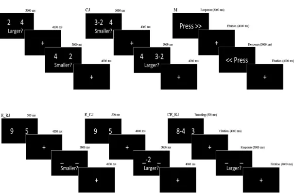
Figure 2.1: Representative trial sequence for each condition. (J) Estimate without WM. (CJ) Compute/Estimate without WM. (M) Control Task. (E RJ) Estimate after Encoding. (E CJ) Compute/Estimate after Encoding. (CE RJ) Estimate after Computing during Encoding.
psychotic cognitive impairment as indicated by worse performance not only in patients but also in healthy subjects.
The working memory paradigm was designated as event-related fMRI design to avoid the predictability of the events. A brief presentation was given which explains the task and the points to take into consideration before the participants performed the task in the MRI scanner. The task was based on computationally and temporally varying information loads including five different events and a control task. Each of the events had a common response phase lasted in 3 s in which the subjects were expected to make a decision by pressing buttons either on the right side or the left side (see Fig. 2.1).
The variants of the events were as the followings: the motor task (M) where the subjects only saw a command on the screen “Press” and they were pressing the response button in which direction the arrows show; the estimate without WM
task (J) in which two digit numbers appeared on the screen and participants were asked to decide which one is larger or smaller; unlike the former one the compute/estimate without WM task (CJ) had a manipulation either on the left or right number and participants were expected to make a decision after subtraction. In the WM tasks, the only difference was they all include encoding phases (E) where two digits were first displayed on the screen and each encoding phase appeared on the screen just 0.5 s and it was followed by a 4 s fixation point. In the estimate after encoding task (E RJ), the participants were expected to hold the information presented during the encoding phase and made their choice in the response phase based on an instruction either the larger or smaller remembered number. In the compute/estimate after encoding task (E CJ), differently from preceding one, the subjects performed a subtraction of 2 or 3 from the retrieved number on the left or right side as stated. In the estimate after computing during the encoding task (CE RJ), the participants were asked to compute a simple subtraction during the encoding phase and like the preceding ones they were decided their choice in the response phase. In each event, only single digits from 0 to 9 were used and the two numbers appeared on the screen left and the right side was equally balanced across 0 to 9. The difference between the numbers was no bigger than 3 and they differed by either 1 or 3 units. The manipulation was performed equally likely on the left or right number. The correct and incorrect responses also equally distributed on the left or the right side and there was an equal number of the larger or smaller instructions. All the events had 10 trials and each event was conducted depending on an order which was optimized by Wager and Nichols in 2003 [116]. But, the displayed numbers of the events on each trial were randomly generated for each subject.
2.5
MR Apparatus
The collection of MR images was performed with a 3-Tesla MRI scanner (Siemens MAGNETOM Trio) to generate high-resolution anatomical images and whole brain blood oxygen level-dependent (BOLD) functional MRI data with echo-planar imaging pulse sequence. Radio-frequency (RF) pulses were applied using
a 32-channel head coil (birdcage) to provide full brain coverage. Flexible ear protectors were used to protecting participants from the disruptive noises arising out of the nature of RF pulses generation. Head stabiliser vacuum cushions placed both sides of the head were used to meet the exclusion criteria for head movement.
Stimuli were presented via a 31.5” telemedicine LCD screen (1920 x 1080 pixel resolution and 59 Hz refresh rate) while the participants were lying on the table inside the MR scanner. A mirror was placed onto the head coil and thus the participants were able to see the screen by looking at the mirror. A stimulus delivery and experiment control program Presentation (Neurobehavioral Sys-R
tems) was used for the stimulus generation and the measurement of reaction time and accuracy for each trial. Each response given by participants during WM task was recorded through a fiberoptic response box.
2.6
MR Protocol
2.6.1
Structural MR Imaging
Before acquiring structural images, a localizer sequence which lasts for 13 seconds was performed for each participant to obtain three orthogonal slices in sagittal, coronal and transverse planes. Thereafter, the T1-weighted images were acquired with a 7 min 18 s long scan to screen anatomical structure of the brain in high resolution [176 slices, echo time, 3.02 ms; repetition time, 2.6 s; slice thickness, 1 mm; flip angle, 8◦; field of view, 256 mm; matrix, 256 x 256; voxel dimensions, 1.0 x 1.0 x 1.0 mm,].
2.6.2
Functional MR Imaging
The images for functional analysis were acquired in a sequence where the par-ticipants perform working memory task in the scanner. The scans were taken 11 min 42 sec to complete the whole task and the participants were expected to make a decision in each trial. To avoid any confounding effect, the participants were in the totally dark room while they were performing the task. T2* weighted echo-planar imaging pulse sequence for acquisition of 24 contiguous slices was ap-plied to collect whole-brain BOLD functional MRI data. The slices were acquired in an ascending interleaved pattern [echo time, 30ms; repetition time, 2 s; slice thickness, 5 mm; flip angle, 90◦; field of view, 235 mm; matrix, 64 x 64, voxel dimensions, 3.7 x 3.7 x 5.0 mm]. To allow for signal saturation, the first four scans where an 8 s long fixation point appears on the screen at the beginning of the task were discarded.
2.7
Analysis of Imaging Data
2.7.1
Image Preprocessing
The anatomical and functional MRI data were stored as raw data by the software system Snygo MR B17 developed by Siemens Healthineers installed in the MR scanner computer. A software program SPM12 [117, 118, 119] (Wellcome Depart-ment of Cognitive Neurology, London, UK; https://www.fil.ion.ucl.ac.uk/spm/) implemented in Matlab (Mathworks, Natick MA) were used to preprocess the neuroimaging data in order to use in analyses. The raw data stored as DICOM (.ima extension) files were converted to NIFTI (.nii extension) files in the batch of DICOM Import implemented in SPM to be able to process in the further steps. Converted images for each subject were realigned to the middle volume in the series and slice time corrected. Exclusion criteria for excessive movement were determined as less than 2 mm translation and less than 1.5◦ rotation based on literature [113, 115]. None of the subjects were excluded due to the excessive
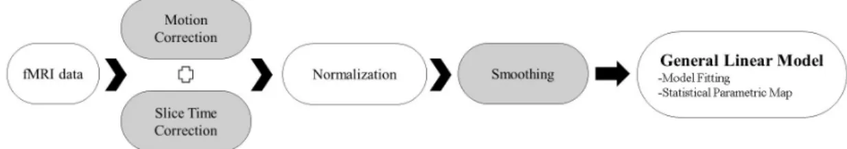
Figure 2.2: The fMRI data processing steps.
movement, they all met the criteria. After individual examination of datasets for motion, each anatomical slice were coregistered with the mean functional image as a reference. Then, a segmentation process was conducted in order to acquire deformation fields which was subsequently used in normalization step.
The low-resolution functional images were warped to high resolution anatom-ical images and then spatially normalized into standard stereotaxic space (Mon-treal Neurological Institute template) by resampling to voxel size 3.7 x 3.7 x 3.7 mm. Additionally, each subject’s functional images were superimposed onto their own anatomy with 1.0 mm isometric voxels. A Gaussian filter set at 8 mm full-width at half-maximum was used for spatial smoothing of the images.
2.7.2
Statistical Analysis
The functional data which was underwent all the preprocessing steps were taken to first-level analysis for each subject. A general linear model (GLM) [120] imple-mented in SPM12 was used for analysing the preprocessed functional data. The responses acquired (the onsets and the accuracy) from event-related designated cognitive paradigm were modelled using the canonical hemodynamic response function. To control the systematic differences in global activity, the ratio was normalized to the whole-brain global mean.To avoid noises in the system, each individual temporally filtered using a high-pass filter of 128 s.
Figure 2.3: Cognitive subtraction for each contrast.
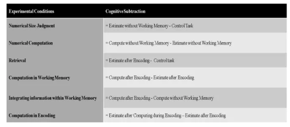
performed using the response phase onsets of each task trials. Without discrimi-nation, the correct and incorrect responses were modelled explicitly as regressors of interest. The movement parameters derived from realignment preprocessing step were also modelled implicitly as regressors of no interests.The following con-trasts were performed to infer putative cognitive processes by using the events such as M, J, CJ, E, E RJ, E CJ, and CE RJ (see Fig. 2.3): numerical size judge-ment (J>M; to remove the activation of sensorimotor regions and also the visual effect of the text appeared on the screen during the task, e.g. ”larger”, ” smaller” or ”press”); numerical computation (CJ>J; performed to obtain simple comput-ing activation); retrieval from WM (E RJ>M; acquired only simple retrieval and also engaged as number processing); computation in WM (E CJ>E RJ; rather than rapid update of the relevant information, it also isolated integrating probe, e.g., ”-2”); integrating information within WM (E CJ>CJ; it reflected a compar-ison including the manipulation of information in WM and simple computation and beyond that temporal effect of encoded probe); computation in encoding (CE RJ>E RJ; represented the manipulation effect on temporally encoded infor-mation).
The second-level group analysis was performed to reveal arithmetic and tem-poral transformations of WM activation differences during the task between the
group identified as prone to psychosis and the subjects who are not prone to psychosis. To demonstrate that a statistical computation method two sample t-test was performed. The statistical matrix design as the first and second col-umn are the no prone to psychosis subjects and the prone to psychosis subjects respectively. Additionally, age and proneness to psychosis levels were included as covariates in statistical calculation to see whether there were any significant effect on the number of activated voxels and they were defined as third and fourth columns of the designated statistical matrix respectively. The above mentioned putative cognitive subtractions was evaluated subsequently to show the main ef-fect of each them. A threshold level of p<0.05 was applied within the whole brain search volume and it was corrected for family-wise error (FWE) [121] in order to prevent false positives (alpha errors). By using Gaussian Random Field theory [122], the correlated voxels were acquired. More leniently revealed voxels p<0.01 uncorrected were also reported.
Among the six contrasts mentioned above, the computation in WM and the integrating information within WM contrasts had a larger effect on prefrontal cortex as we expected. Hence, a further cluster correction analysis was applied over these contrasts at the second-level analysis in order to find significant clus-ters. A toolbox developed by Gaser et al. from University of Jena, threshold-free cluster enhancement (TFCE) [123] was used to generate 5000 permutations of contrasts images to test against the null distribution. The advantage which the method serves is boosting the amplitudes of spatially distributed signals without making any change on the location of their maxima while the strict control over family-wise error maintained. The resulting significant (p<0.05) clusters were reported.
Chapter 3
Results
3.1
Demographic and Behavioural Results.
Regarding the behavioural performance based on accuracy level, subjects that are not prone to psychosis have a slight tendency to show more accurate performance compared to subjects that are prone to psychosis. Considering the accuracy, all the task conditions which are numerical size judgement, numerical computation, retrieval, computation in working memory, integrating information within work-ing memory, and computation in encodwork-ing were averaged per subject to calculate the individual performance. However, this result for performance across psychosis level is not statistically significant (F =2.75, p>0.05).
Age, gender and accuracy averaged over the task conditions interaction were examined to see if any group differences exist. There were no significant psy-chosis level group differences in age (F =0, p=0.96), gender (F =0.15, p=0.35) and overall accuracy (F =2.75, p=0.10).
Therefore, the group differences depending on psychosis classification at the level of BOLD responses only reflected information processing physiologically, which shows no influence on the working memory performance.
Table 3.1: The demographic and behavioural results.
3.2
Task-Related Activation
The affected regions by each cognitive subtraction were identified independently for the prone to psychosis group and the control group. The significantly altered activations in the respective contrast of interest were reported at the second-level group analysis applying the two-sample t-test (all the clusters reported could be found in Appendix B).
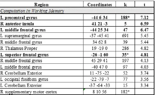
In the working memory tasks, the activations observed were similar for both groups and they were clustered around prefrontal cortices and parietal lobules. In the contrast correlated with computation in working memory (E CJ>E RJ), the significant effects for prone to psychosis subjects were observed at left pre-central gyrus (peak -44 6 34; t =7.12; p<0.05, corrected), right anterior insula (peak 41 21 -3; t =6.59; p<0.05, corrected), left middle frontal gyrus (peak -44 25 34; t =6.47; p<0.05, corrected), and left superior frontal gyrus (peak -26 -1 60; t =4.81; p<0.05, corrected) (see Fig. 3.1 ). When the threshold was decreased to more lenient level, larger but on the other hand weaker effects were detected at left supramarginal gyrus (peak -37 -45 41; t =5.45; p<0.01, uncorrected), right tha-lamus proper (peak 19 -19 0; t =4.82; p<0.01, uncorrected), right middle frontal gyrus (peak 45 29 41; t =4.13; p<0.01, uncorrected). For the control group, the activated regions different than prone to psychosis subjects were observed at left superior parietal lobule (peak -26 -64 45; t =6.95; p<0.05, corrected), right supra-marginal gyrus (peak 41 -38 41; t =6.16; p<0.05, corrected), right superior parietal lobule (peak 26 -68 41; t =6.14; p<0.05, corrected), right thalamus proper (peak 11 -16 8; t =6.04; p<0.05, corrected), right anterior cingulate gyrus (peak 8 25 34; t =5.68; p<0.05, corrected) and left thalamus proper (peak -14 -16 4; t =5.64;
p<0.05, corrected) (see Fig. 3.2 ). By applying Threshold Free Enchancement Method (TFCE) to avoid the strict nature of FWE correction, a small cluster at right middle frontal gyrus (peak 34 10 56; k =16; p<0.05, corrected) was also associated with the computation in working memory task (see Appendix A).
In the contrast examining integrating information within working memory (E CJ>CJ), weaker activations were observed compared to computation in work-ing memory task.In the prone to psychosis subjects, at the more lenient thresh-old, small clusters located at left middle frontal gyrus (peak -40 3 37; t =4.99; p<0.01, uncorrected) and right superior frontal gyrus medial segment (peak 8 40 34; t =4.02; p<0.01, uncorrected) were observed. The only cluster acquired differ-ently in the control group during the task was located at right superior parietal lobule (peak 15 -68 60; t =3.02; p<0.01, uncorrected).
The significantly activated voxels related to numerical computation task (CJ>J) were observed at the right supramarginal gyrus (peak 41 -38 45; t =5.82; p<0.05, corrected) in the control group (see Fig. 3.3 ). After the application of TFCE method, largely activated cluster located at the left superior parietal lobule (peak -26 -60 45; k =121; p<0.05, corrected) and a small activated cluster found at the left precuneus (peak -7 -68 52; k =2; p<0.05, corrected) were also detected at the end of the statistical analysis (see Appendix A). In prone to psy-chosis subjects, no significantly activated voxels were observed rather than small clusters distributed throughout the whole brain at the more lenient threshold (p<0.01).
In the numerical size judgement task (J>M), there were no affected regions at the chosen threshold in both groups. However, when threshold was decreased to more lenient level, a small effect in the right cun cuneus (peak 8 -79 23; t =4.17; p<0.01, uncorrected) was observed in prone to psychosis subjects. On the other hand, the control group had largely affected area located at left cerebellum exterior (peak -29 -68 -22; t =5.64; p<0.01, uncorrected).
The contrast requiring computation during a brief period (0.5 s) in the encoded information (CE RJ>E RJ) was resulted with a weak effect on the brain in both
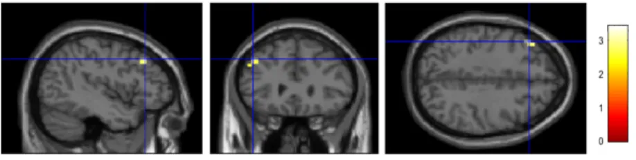
Figure 3.1: The results of computation in working memory contrast for the prone to psychosis group (p<0.05, corrected): sagittal (on the left side), coronal (at the middle) and transverse (on the right side) planes are illustrated above. The activated regions are represented as (A) left precentral gyrus, (B) left middle frontal gyrus, (C) right anterior insula, (D) left superior frontal gyrus and dis-played on standard stereotaxic (MNI152) space. The right hemisphere of the brain corresponds to the right side of the images.
Figure 3.2: The computation in working memory contrast results for the control group (p<0.05, corrected). The activated regions are represented as (A) left superior parietal lobule, (B) right supramarginal gyrus, (C) right superior parietal lobule, (D) right anterior cingulate gyrus.
Figure 3.3: The numerical computation contrast results for the control group (p<0.05, corrected).
Table 3.2: The activated regions during the computation in working memory task. While Table A represents the activated regions for prone to psychosis group, Table B lists the activated regions for the control group. The columns represent the regions activated, the coordinates, the number of voxels (k) and t values respectively from left to right. The rows marked as bold show the activated regions at the threshold p<0.05 corrected, the rest of the rows are thresholded at p<0.01 uncorrected. The asterisk represents the clusters in which the TFCE method applied.
Table 3.3: The activated regions during the numerical computation contrast: (A) The prone to psychosis group, (B) The control group.
groups. While the left middle frontal gyrus (peak -44 47 -3; t =4.61; p<0.01, uncorrected) was activated weakly in prone to psychosis subjects, the activation located at the right superior parietal lobule (peak 26 -64 41; t =3.08; p<0.01, uncorrected) was observed in the control group.
At the retrieval task (E RJ>M), no effect was observed rather than a small fraction at the left middle cingulate gyrus (peak 0 21 30; t =3.46; p<0.01, uncor-rected) in the prone to psychosis subjects and small clusters located at bilateral cerebellum exterior (for the left hemisphere; peak -7 -68 -18; t =3.12; p<0.01, un-corrected; for the right hemisphere; peak 19 -60 -22; t =2.83; p<0.01, uncorrected) in the control group.
3.3
Proneness to Psychosis Effects in Cognitive
Processes of Interest
A second-level group analysis where inter-subject variability treated as a random effect was performed by applying the two-sample t-test to demonstrate the sig-nificant difference between the two groups. Each contrast identified above were examined to detect any altered activation due to the proneness to psychosis. Be-sides that in order to demonstrate whether any positive or negative correlation, the ages and psychosis levels of the participants were used as covariates in the analysis.
At the chosen threshold (p>0.05, corrected), there were no altered activa-tion due to proneness to psychosis. On the other hand, when the threshold was decreased to more lenient level (p>0.01, uncorrected), the slightly altered ac-tivation for every contrast except the computation in encoding (CE RJ>E RJ) was observed (see Table 3.4). In the cognitive process of computation in working memory (E CJ>E RJ), an increased activation at left middle frontal gyrus (peak -44 29 41; t =3.43; p<0.01, uncorrected) was observed (see Fig. 3.4). On the other hand, the integrating information in working memory task (E CJ>CJ) had
Figure 3.4: The increased activation due to proneness to psychosis for the com-putation in working memory contrast result (PP>NPP).
three increased activation regions located at the left middle frontal gyrus (peak -40 3 37; t =3.45; p<0.01, uncorrected), the right superior frontal gyrus (peak 19 40 52; t =2.86; p<0.01, uncorrected) and the left superior temporal gyrus (peak -55 -34 4; t =2.68; p<0.01, uncorrected) (see Fig. 3.5). Besides those, the right supramarginal gyrus (peak 60 -23 37; t =2.74; p<0.01, uncorrected) showed a tendency to decrease in activation for prone to psychosis subjects (see Fig. 3.6).
Figure 3.5: The increased activation due to proneness to psychosis for the inte-grating information in working memory contrast result (PP>NPP). The regions illustrated above were (A) left middle frontal gyrus, (B) right superior frontal gyrus (C) left superior temporal gyrus.
Figure 3.6: The decreased activation due to proneness to psychosis for the inte-grating information in working memory contrast result (NPP>PP).
Table 3.4: The regions which had altered activations during each contrast. (A) The prone to psychosis group>The control group, (B) The control group>The prone to psychosis group.
Chapter 4
General Discussion and
Conclusion
The primary objective of this thesis was to present evidence that the subjects mildly prone to psychosis have altered brain activations in a well-established verbal WM task in which consist of computationally and temporally varying information loads. Even though, there are studies on the subjects who are at the high-risk level of psychosis in the literature [124], the effect of mild proneness to psychosis in WM impairment has not been examined before in the literature to our knowledge. The task-related activation results in our study may interpreted as computation and integrating requiring processes in the brain are weakly altered due to mild proneness to psychosis. It is needed to be underlined that under the strict conditions of FWE analysis, these alterations has not been observed. Nevertheless, the WM impairment in prone to psychosis is a progressive symptom as demonstrated in the subjects varying from healthy controls to first episode psychosis patients [98], thus our results could be claimed as consistent with the literature. According to our findings, computation requiring processes have a stronger influence on specific regions located in prefrontal and parietal cortices compared to numerical computation without WM in both groups. However, the effect of mild proneness to psychosis has caused to a weak alteration on a specific region which is the middle frontal gyrus. The integrating information of WM
processes have not shown a strong effect in both groups, on the other hand, the mild proneness to psychosis have exhibited weak alterations but a larger effect on the prefrontal cortex (e.g. left middle frontal gyrus and right superior frontal gyrus). The alterations have also observed in the left superior temporal gyrus and the right supramarginal gyrus which are out of the PFC. These findings suggest that while the computation requiring information processes in WM is disrupted in a specific pattern which affects the frontal lobe, the integration of information processes in WM is altered the activation in a larger network in the brain due to mild proneness to psychosis.
Age-associated developmental transformations are considered as an important factor which influence the progress of working memory deficits [124, 125]. Pre-vious works conducted on age-related WM activations have concluded with the idea that in regions where the activation increased (e.g. higher order cortices) are performance enhancing regions, whereas decreased network activation is in-terpreted as a circuitry needed to be pruned during the developmental period of adolescent [78, 126]. In our study, the influence of ageing was investigated as a covariate in the statistical analysis of functional images. The age-association has not shown any significant effect on each contrast. This inconsistency with the literature may have emerged from our sample size. A strong statistical power requires a meaningful sample size for each age group and we were not able to assess enough number of subjects (e.g. age of 15) for each group. Additionally, we have used the proneness to psychosis score in SIS-R as a covariate as well and investigated that whether the WM activation would be altered directly or inversely proportional to the proneness scores. However, the effect of the prone-ness scores have not affected the activation level of WM processes in a significant manner.
One of the most commonly investigated factor in the psychopathology of psy-chosis is the gender differences. Nevertheless, many of these studies considering the first episode psychosis or at risk mental state individuals resulted with in-consistency. In a study which specifically focused on the gender differences in psychosis has demonstrated that the gender difference is not an effective fac-tor among neither in at high-risk mental state or first episode psychosis group
[127]. The only difference was while the female subjects had higher scores in the positive symptom assessments, the male subjects had higher scores in nega-tive symptom assessments. In the behavioural results of our study, we have not observed any significant correlation between gender differences and proneness to psychosis scores in parallel with the study of Gonzalez et al.
Additionally, the working memory deficiency is a well-described cognitive im-pairment in first episode psychosis and ultra high risk individuals [128]. The sub-jects who developed later on psychosis are shown poorer performance in working memory tests [61]. The working memory accuracy between the prone to psychosis group and the control group did not differ significantly compared to their psy-chosis scores in SIS-R. Even though we have observed greater difference compared to other factors in the behavioural analysis, there was no meaningful correlation.
In this study, we have used a very well-established proneness to psychosis as-sessment (SIS-R) in order to classify the mildly prone group and the subjects for control. The subjects who diagnosed with psychiatric illnesses or using psychi-atric drugs were excluded. The working memory task used was a well-designated paradigm to stimulate the network associated with working memory activation and to detect alterations related to proneness to psychosis. Each individual was analysed separately to prevent the performance differences among the subjects and the whole brain was examined applying the strict voxelwise correction to acquire the most powerful statistical results. While these features represent the strengths of our study and there were the weak sides which have to be empha-sized at the same time. Firstly, the sample size for both group was small to obtain a stringent statistical analysis. A larger sample pool would lead to more meaningful results. We have also performed a cross-sectional analysis to observe the alterations in the brain due to proneness to psychosis. The development of the same subjects should be followed, and a within-subject analysis performed to observe change over time.
The future aspect of this study is determining the developmental trajectories of the working memory. In order to achieve this, the brain development of the subjects should be followed by performing the same experiment one year later.
Thus, a within-subject would be able to be performed to test the change over time. With the collection of blood or saliva samples, the polygenic risk scores and miRNA profiling of each subject could be generated, and this will allow to investigate the reciprocal causation models for the psychosis. Due to the fact that our subjects are healthy volunteers, an in silico analysis would be appropriate to compare the profiles of our subjects and the psychotic patients.
Moreover, a widely used approach in the analysis of fMRI data is the regions of interest (or ROI) analysis in which the signals are extracted from a specified region [129]. The ROI analysis is often useful for the designs which have multiple levels. By plotting each condition and comparing against other variables of interest, the signal of the interested region becomes discernible. Another advantage which ROI analysis serves is controlling the Type I error by limiting the number of statistical tests to a few ROIs [129]. In a previous study where working memory performance investigated, the left DLPFC was chosen as a region of interest in order to avoid the type I error [130]. Furthermore, in the study of Tan et al. [113], an additional functional ROI analysis was conducted to isolate the signal more distinguishable around the prefrontal cortex, posterior parietal cortex and striatum. As an additional future aspect for this study, an ROI can be determined based on the results of the study (e.g. middle frontal gyrus). A functional ROI could not be appropriate because, when the activation signal is not strong, the signals acquired could be very noisy. Alternatively, estimating the region of interest using the anatomical images would be more convenient.
Another future aspect of this study might be conducting an effective connec-tivity analysis to reveal the correlation between impaired regions due to proneness to psychosis and the strongly activated intact regions. While functional connec-tivity refers to the simple temporal correlation between activation of spatially segregated neural events, the effective connectivity based on a mathematical model which reveals that how one neuronal system is influential over another [131]. In a study which investigates the causal relationships between cortical-cortical and cortical-cortical-subcortical-cortical networks selectively engaged during maintenance and manipulation operations in working memory has been used dynamic causal
modelling (DCM) [132] to demonstrate correlations between the associated net-works [115]. Another study has been conducted a psychophysiological interaction analysis (PPI) [133] to test whether patients show a de-coupling of regions that are functionally connected during WM demands (e.g. DLPFC with parietal re-gions) [125]. On the other hand, both of the methods require a strongly activated seed region to demonstrate the correlation with regions. When the sample size could be enlarged in the future, both methods are capable of to demonstrate the impairment of working memory network caused by the proneness to psychosis.
As a conclusion, we have used proneness to psychosis as a measure of network signalling alteration in healthy human volunteers to investigate transformation of working memory. Manipulating and rapid updating of information processes are involved in a larger network of prefrontal cortex and superior parietal lobule. This study suggests that the mild proneness to psychosis has a weak effect on the working memory processes and that specific alterations in the prefrontal and parietal lobules may be associated with the onset of psychosis.
Bibliography
[1] A. D. Baddeley, “The psychology of memory,” New York, 1976.
[2] A. Baddeley, Essentials of human memory (Classic Edition). Psychology Press, 2013.
[3] V. S. Mattay, F. Fera, A. Tessitore, A. R. Hariri, K. F. Berman, S. Das, A. Meyer-Lindenberg, T. E. Goldberg, J. H. Callicott, and D. R. Wein-berger, “Neurophysiological correlates of age-related changes in working memory capacity,” Neuroscience letters, vol. 392, no. 1-2, pp. 32–37, 2006.
[4] L. Nyberg, E. Dahlin, A. Stigsdotter Neely, and L. B¨ackman, “Neural cor-relates of variable working memory load across adult age and skill: Disso-ciative patterns within the fronto-parietal network,” Scandinavian journal of psychology, vol. 50, no. 1, pp. 41–46, 2009.
[5] A. M. Owen, K. M. McMillan, A. R. Laird, and E. Bullmore, “N-back working memory paradigm: A meta-analysis of normative functional neu-roimaging studies,” Human brain mapping, vol. 25, no. 1, pp. 46–59, 2005.
[6] Z. U. Khan and E. C. Muly, “Molecular mechanisms of working memory,” Behavioural brain research, vol. 219, no. 2, pp. 329–341, 2011.
[7] A. Baddeley, “Working memory,” Current biology, vol. 20, no. 4, pp. R136– R140, 2010.
[8] W. J. Chai, A. I. Abd Hamid, and J. M. Abdullah, “Working memory from the psychological and neurosciences perspectives: A review,” Frontiers in psychology, vol. 9, p. 401, 2018.
[9] P. Goldman-Rakic, “Circuitry of primate prefrontal cortex and regulation of behavior by representational memory. hoboken,” 1987.
[10] E. K. Miller and J. D. Cohen, “An integrative theory of prefrontal cortex function,” Annual review of neuroscience, vol. 24, no. 1, pp. 167–202, 2001.
[11] A. Baddeley, “Working memory: looking back and looking forward,” Nature reviews neuroscience, vol. 4, no. 10, p. 829, 2003.
[12] J. M. Fuster and G. E. Alexander, “Neuron activity related to short-term memory,” Science, vol. 173, no. 3997, pp. 652–654, 1971.
[13] S. Funahashi, C. J. Bruce, and P. S. Goldman-Rakic, “Dorsolateral pre-frontal lesions and oculomotor delayed-response performance: evidence for mnemonic” scotomas”,” Journal of Neuroscience, vol. 13, no. 4, pp. 1479– 1497, 1993.
[14] F. A. Wilson, S. Scalaidhe, and P. S. Goldman-Rakic, “Dissociation of ob-ject and spatial processing domains in primate prefrontal cortex,” Science, vol. 260, no. 5116, pp. 1955–1958, 1993.
[15] R. Levy and P. S. Goldman-Rakic, “Segregation of working memory func-tions within the dorsolateral prefrontal cortex,” in Executive control and the frontal lobe: Current issues, pp. 23–32, Springer, 2000.
[16] G. Rainer, W. F. Asaad, and E. K. Miller, “Selective representation of relevant information by neurons in the primate prefrontal cortex,” Nature, vol. 393, no. 6685, p. 577, 1998.
[17] E. K. Miller, C. A. Erickson, and R. Desimone, “Neural mechanisms of visual working memory in prefrontal cortex of the macaque,” Journal of neuroscience, vol. 16, no. 16, pp. 5154–5167, 1996.
[18] K. Sakai, J. B. Rowe, and R. E. Passingham, “Active maintenance in pre-frontal area 46 creates distractor-resistant memory,” Nature neuroscience, vol. 5, no. 5, p. 479, 2002.
[19] S. M. Courtney, L. Petit, J. M. Maisog, L. G. Ungerleider, and J. V. Haxby, “An area specialized for spatial working memory in human frontal cortex,” Science, vol. 279, no. 5355, pp. 1347–1351, 1998.
[20] E. Zarahn, G. K. Aguirre, and M. D’Esposito, “Temporal isolation of the neural correlates of spatial mnemonic processing with fmri,” Cognitive Brain Research, vol. 7, no. 3, pp. 255–268, 1999.
[21] C. E. Curtis, V. Y. Rao, and M. D’Esposito, “Maintenance of spatial and motor codes during oculomotor delayed response tasks,” Journal of Neuro-science, vol. 24, no. 16, pp. 3944–3952, 2004.
[22] R. H. Bauer and J. M. Fuster, “Delayed-matching and delayed-response deficit from cooling dorsolateral prefrontal cortex in monkeys.,” Journal of comparative and physiological psychology, vol. 90, no. 3, p. 293, 1976.
[23] S. Funahashi, M. V. Chafee, and P. S. Goldman-Rakic, “Prefrontal neuronal activity in rhesus monkeys performing a delayed anti-saccade task,” Nature, vol. 365, no. 6448, p. 753, 1993.
[24] N. G. M¨uller, L. Machado, and R. T. Knight, “Contributions of subregions of the prefrontal cortex to working memory: evidence from brain lesions in humans,” Journal of Cognitive Neuroscience, vol. 14, no. 5, pp. 673–686, 2002.
[25] A. Tsuchida and L. K. Fellows, “Lesion evidence that two distinct regions within prefrontal cortex are critical for n-back performance in humans,” Journal of Cognitive Neuroscience, vol. 21, no. 12, pp. 2263–2275, 2009.
[26] B. Voytek and R. T. Knight, “Prefrontal cortex and basal ganglia contri-butions to visual working memory,” Proceedings of the National Academy of Sciences, p. 201007277, 2010.
[27] S. P. Wegener, K. Johnston, and S. Everling, “Microstimulation of monkey dorsolateral prefrontal cortex impairs antisaccade performance,” Experi-mental brain research, vol. 190, no. 4, pp. 463–473, 2008.
[28] T. J. Druzgal and M. D’Esposito, “A neural network reflecting decisions about human faces,” Neuron, vol. 32, no. 5, pp. 947–955, 2001.
[29] C. E. Curtis and M. D’Esposito, “Persistent activity in the prefrontal cortex during working memory,” Trends in cognitive sciences, vol. 7, no. 9, pp. 415– 423, 2003.
[30] B. R. Postle, T. J. Druzgal, and M. D’Esposito, “Seeking the neural sub-strates of visual working memory storage,” Cortex, vol. 39, no. 4-5, pp. 927– 946, 2003.
[31] C. Ranganath, J. DeGutis, and M. D’Esposito, “Category-specific modu-lation of inferior temporal activity during working memory encoding and maintenance,” Cognitive Brain Research, vol. 20, no. 1, pp. 37–45, 2004.
[32] K. K. Sreenivasan, C. E. Curtis, and M. D’Esposito, “Revisiting the role of persistent neural activity during working memory,” Trends in cognitive sciences, vol. 18, no. 2, pp. 82–89, 2014.
[33] K. K. Sreenivasan, C. Gratton, J. Vytlacil, and M. D’Esposito, “Evidence for working memory storage operations in perceptual cortex,” Cognitive, Affective, & Behavioral Neuroscience, vol. 14, no. 1, pp. 117–128, 2014.
[34] B. R. Postle, “The cognitive neuroscience of visual short-term memory,” Current opinion in behavioral sciences, vol. 1, pp. 40–46, 2015.
[35] F. Collette, M. Van der Linden, S. Laureys, G. Delfiore, C. Degueldre, A. Luxen, and E. Salmon, “Exploring the unity and diversity of the neural substrates of executive functioning,” Human brain mapping, vol. 25, no. 4, pp. 409–423, 2005.
[36] M. Koenigs, A. K. Barbey, B. R. Postle, and J. Grafman, “Superior parietal cortex is critical for the manipulation of information in working memory,” Journal of Neuroscience, vol. 29, no. 47, pp. 14980–14986, 2009.
[37] E. Awh, E. Vogel, and S.-H. Oh, “Interactions between attention and work-ing memory,” Neuroscience, vol. 139, no. 1, pp. 201–208, 2006.
[38] D. E. Nee, J. W. Brown, M. K. Askren, M. G. Berman, E. Demiralp, A. Krawitz, and J. Jonides, “A meta-analysis of executive components of working memory,” Cerebral cortex, vol. 23, no. 2, pp. 264–282, 2012.
[39] C. J. Stoodley and J. D. Schmahmann, “Functional topography in the hu-man cerebellum: a meta-analysis of neuroimaging studies,” Neuroimage, vol. 44, no. 2, pp. 489–501, 2009.
[40] T. D. Wager and E. E. Smith, “Neuroimaging studies of working memory,” Cognitive, Affective, & Behavioral Neuroscience, vol. 3, no. 4, pp. 255–274, 2003.
[41] R. C. O’reilly, “Biologically based computational models of high-level cog-nition,” science, vol. 314, no. 5796, pp. 91–94, 2006.
[42] U. Ekman, J. Eriksson, L. Forsgren, S. J. Mo, K. Riklund, and L. Nyberg, “Functional brain activity and presynaptic dopamine uptake in patients with parkinson’s disease and mild cognitive impairment: a cross-sectional study,” The Lancet Neurology, vol. 11, no. 8, pp. 679–687, 2012.
[43] N. Axmacher, F. Mormann, G. Fern´andez, M. X. Cohen, C. E. Elger, and J. Fell, “Sustained neural activity patterns during working memory in the human medial temporal lobe,” Journal of Neuroscience, vol. 27, no. 29, pp. 7807–7816, 2007.
[44] I. R. Olson, K. Page, K. S. Moore, A. Chatterjee, and M. Verfaellie, “Work-ing memory for conjunctions relies on the medial temporal lobe,” Journal of Neuroscience, vol. 26, no. 17, pp. 4596–4601, 2006.
[45] A. Gazzaley, J. Rissman, and M. D’esposito, “Functional connectivity dur-ing workdur-ing memory maintenance,” Cognitive, Affective, & Behavioral Neu-roscience, vol. 4, no. 4, pp. 580–599, 2004.
[46] K. E. Stephan, T. Baldeweg, and K. J. Friston, “Synaptic plasticity and dysconnection in schizophrenia,” Biological psychiatry, vol. 59, no. 10, pp. 929–939, 2006.
![Figure 1.1: An illustration of the multicomponent working memory model pro- pro-posed by Baddeley, 2010 [7] from the study of Chai et al](https://thumb-eu.123doks.com/thumbv2/9libnet/5571961.108909/13.918.242.721.404.766/figure-illustration-multicomponent-working-memory-model-posed-baddeley.webp)