ICHC 2017
ICHC 2017
Kervansaray Lara Hotel, ANTALYA
May 18 - 21, 2017
www.ichc2017.com
15
th
International Congress of
Histochemistry and Cytochemistry
15
th
International Congress of
Histochemistry and Cytochemistry
“From Molecules to Diseases”
ABSTRACT BOOK
ICHC 2017
ICHC 2017
Kervansaray Lara Hotel, ANTALYA
May 18 - 21, 2017
3
15
th
International Congress of
Histochemistry and Cytochemistry
15
th
International Congress of
Histochemistry and Cytochemistry
“From Molecules to Diseases”
ICHC 2017
ICHC 2017
Kervansaray Lara Hotel, ANTALYA
May 18 - 21, 2017
ICHC 2017
ICHC 2017
Kervansaray Lara Hotel, ANTALYA
May 18 - 21, 2017
www.ichc2017.com
4
Dear Colleagues,
On behalf of the Turkish Society for Electron Microscopy, it is my pleasure to welcome all the participants of 15th
International Congress of Histochemistry and Cytochemistry (ICHC 2017) in Kervansaray Lara Hotel and Exhibition
Center, Antalya, Turkey.
First of all, as the Organization Committee of ICHC 2017, we would like to share our deep sorrow about the loss of our
beloved Vice Chair of the Congress, Past President of Turkish Society for Electron Microscopy, Dearest Friend Prof. Dr.
Suzan Dağlıoğlu, who passed away on April 2 nd 2017. We would like to extend our sincerest condolences to her family
and to her colleagues. ICHC 2017 is dedicated to the memory of Prof. Dr. Suzan Dağlıoğlu.
During the organization period of ICHC 2017, we had an excellent collaboration with members of the Organization Committee, Scientific Programme Committee and
International Scientific Advisory Board to whom we would like to extend our special appreciations.
We gratefully acknowledge the excellent support and teamwork provided by Organization Secretariat, FIGUR INTERNATIONAL, PCO of ICHC 2017.
We wish to express our gratitude to all the sponsoring companies for their financial supports.
A special thank is for Mr. Ibrahim Zaman, a Master of Photography, for his fabulous Photography Show ‘’ My Beloved Turkey’’, during the Opening Ceremony.
During planning of ICHC 2017, we have tried to bring the worldwide histochemists together by creating an environment for close cooperation, collaboration
and exchange of information. We thoroughly believe that the congress program, under the theme’’ From Molecules to Diseases’’ is inspiring with a considerable
amount of keynote, plenary and invited speakers in the field of Histochemistry and Cytochemistry.
The program will include: 1 Keynote, 5 Plenary, 34 Invited Lectures (including Society- sponsored and Journal-sponsored sessions), 21 Scientific sessions with
59 Short Oral Presentations and 2 Poster Sessions 275 Poster Presentations.
As the highlights of The Opening Ceremony: David Glick Lecture, by Prof. Dr. John Couchman, and Keynote Lecture by Prof. Dr. Ron van Noorden will be
delivered. Paul Nakane Prize will be presented to Prof. Dr. Toyoshi Fujimoto during the Opening Ceremony. Photography Show ‘’ My Beloved Turkey’’, by Mr.
Ibrahim Zaman will be performed during the Opening Ceremony.
‘’Let the Seniors share their expertise with Juniors’’ and ‘’Young Histochemists Awardees’’ Sessions, as well as 3 Workshops will be within the Scientific
Programme and provide a unique opportunity for young histochemists to get acquainted with outstanding researchers.
A sponsored company-based exhibition with technical presentations will display the latest advancements in this field.
Welcome Reception, Gala Dinner and Closing Ceremony with Farewell Party are among the Social Programme of ICHC 2017. Post- congress tours will be an excellent
opportunity to discover the beautiful city of Antalya and its surroundings during spring time.
On behalf of the Organization Committee of ICHC 2017, we would like to wish you a compelling and successful ICHC 2017 with lively discussions in a stimulating
scientific atmosphere by scientific contributions of more than 300 distinguished experts in the field of histochemical and cytochemical techniques and their
applications.
As stayed by Mustafa Kemal ATATURK, the Founder of the Republic of Turkey, ‘’Our True Mentor In Life is Science’’ , we believe in the strength of science in
uniting people and nations in the whole world.
We sincerely hope you will have a delightful stay in the beautiful city of Antalya.
Best Regards,
On behalf of the Organization Committee of ICHC 2017,
Serap ARBAK
President
ICHC 2017 & International Federation of Societies for Histochemistry and Cytochemistry
(IFSHC) & Turkish Society for Electron Microscopy
ICHC 2017
ICHC 2017
Kervansaray Lara Hotel, ANTALYA
May 18 - 21, 2017
www.ichc2017.com
15
thInternational Congress of
Histochemistry and Cytochemistry
“From Molecules to Diseases”
www.ichc2017.com
5
Dear Colleagues,
I would like to welcome you all to the 15 th International Congress of Histochemistry and Cytochemistry in Antalya, Turkey. The Turkish Society of Electron
Microscopy first applied for the organization of this meeting under the auspices of the International Federation of Societies for Histochemistry and Cytochemistry
(IFSHC) at the ICHC 2004 in San Diego, USA. Thanks to the persistence of Professor Serap Arbak, the President of the IFSHC, and other members of the
Organizing Committee, and despite many unforeseeable events, our Turkish hosts have succeeded in organizing this well-planned congress distinguished by
highly attractive scientific and social programs.
The mission of the IFSHC is to promote communications and co-operation among scientists throughout the world who are interested in the fields of
microscopy, histochemistry, immunocytochemistry and cell biology. The IFSHC supports the David Glick lecture, Paul Nakane Prize and Young Histochemists
which highlight the achievements of cell imaging sciences. The scientific program of the Congress based on the theme “From Molecules to Diseases” covers
new applications of microscopic techniques both in basic cell reasearch and clinical studies that will be presented by the leading world experts.
I am sure that ICHC 2017 will provide an opportunity not only to attend the sessions and symposia organized by prominent scientists but also to interact and
network with other participants both on formal occasions such as sessions or poster presentations and during informal gatherings at the exhibitions and social
events.
Zbigniew Kmiec
Secretary-General
International Federation
ICHC 2017
ICHC 2017
Kervansaray Lara Hotel, ANTALYA
May 18 - 21, 2017
www.ichc2017.com
ICHC 2017
ICHC 2017
Kervansaray Lara Hotel, ANTALYA
May 18 - 21, 2017
www.ichc2017.com
15
thInternational Congress of
Histochemistry and Cytochemistry
“From Molecules to Diseases”
www.ichc2017.com
7
INDEX
Committees 8
Program at a Glance
10
Scientific Program
11
David Glick Lecture
19
Keynote Lecture
20
Planery Lectures
22
Invited Speakers
28
Young Histochemist Awardees
64
Workshops 72
Oral Presentations
74
Poster Presentations
148
Sponsoring Societies & Journals
496
ICHC 2017
ICHC 2017
Kervansaray Lara Hotel, ANTALYA
May 18 - 21, 2017
www.ichc2017.com
8
COMMITTEES
President
Serap ARBAK
(Acıbadem University)
Vice President
Suzan DAĞLIOĞLU (Istanbul University)
Vice President
Melek ÖZTÜRK
(Istanbul University)
Secretary General
Selma YILMAZER
(Istanbul University)
International Scientific Programme Committee
Serap ARBAK
Turkey
John COUCHMAN
Denmark
Petek KORKUSUZ
Turkey
Ron van NOORDEN
The Netherlands
Melek OZTURK
Turkey
Selma YILMAZER
Turkey
Local Organization Committee
Abit AKTAŞ
Istanbul University
Ranan Gülhan AKTAŞ
Maltepe University
Serap ARBAK
Acıbadem University
Alp CAN
Ankara University
Nur ÇAKAR
TOBB University
Yurdagül CANBERK
Istanbul University
Suzan DAĞLIOĞLU
Istanbul University
Feriha ERCAN
Marmara University
Elif GÜZEL
İstanbul University
Sevinç İNAN
Izmir Economy University
Mehtap KUTLU
Anadolu University
Melek ÖZTÜRK
Istanbul University
Serap ŞİRVANCI
Marmara University
İsmail SEÇKİN
Istanbul University
Matem TUNÇDEMİR
Istanbul University
Selma YILMAZER
Istanbul University
Deniz YÜCEL
Acıbadem University
ICHC 2017
ICHC 2017
Kervansaray Lara Hotel, ANTALYA
May 18 - 21, 2017
www.ichc2017.com
15
thInternational Congress of
Histochemistry and Cytochemistry
“From Molecules to Diseases”
www.ichc2017.com
9
International Scientific Advisory Board
President
Serap ARBAK
Turkey
Secretary-General
Zbigniew KMIEC
Poland
Treasurer
Hinke MULTHAUPT
Denmark
Councilor
Raymond COLEMAN
Israel
Councilor
Tetsuro TAKAMATSU
Japan
Susan BROOKS
United Kingdom
John COUCHMAN
Denmark
Ramazan DEMİR
Turkey
Charles FREVERT
USA
Qutayba HAMID
Canada
Pavel HOZAK
Czech Republic
Bruno HUMBEL
The Netherlands
Erdal KARAÖZ
Turkey
Agnes KITTEL
Hungary
He LI
China
Rossana C. N. MELO
Brazil
C J van NOORDEN
The Netherlands
Paolo ONORI
Italy
Robert Y. OSAMURA
Japan
Margit PAVELKA
Austria
Reinhard RACHEL
Germany
Sanja STIFTER
Croatia
Gulgun ŞENGÜL
Turkey
Çevik TUFAN
Turkey
Zahra ZAKERI
USA
J van ZOOLEN
The Netherlands
Jury Members for ‘’Best Poster Award ICHC 2017’
Moderators: Mehtap KUTLU (Turkey) • Engin YENILMEZ (Turkey) • Deniz YÜCEL (Turkey)
Belgin CAN
Turkey
Jerry Edward CHIPUK
USA
Çiğdem ELMAS
Turkey
Süheyla GONCA
Turkey
Figen KAYMAZ Turkey
Serçin KARAHUSEYİNOĞLU Turkey
Irina KOLOTUEVA
Switzerland
Emel KOPTAGEL
Turkey
Nesrin ÖZFILIZ
Turkey
Engin YENILMEZ
Turkey
Melda YILMAZ
Turkey
Jury Members for ‘’Best Micrograph Award ICHC 2017’
Moderator: Melek ÖZTÜRK (Turkey)
Alp CAN
Turkey
İsmail SEÇKİN
Turkey
Zahra ZAKERI
USA
İbrahim ZAMAN
Turkey
Servet SEZGİN
Turkey
ICHC 2017
ICHC 2017
Kervansaray Lara Hotel, ANTALYA
May 18 - 21, 2017
www.ichc2017.com
10
09:00 18:30 09:45 10:30 10:30 11:00 11:00 12:00 12:00 13:00 13:00 14:00 14:00 14:45 14:45 15:45 15:45 16:45 16:45 17:30 17:30 18:30 20:00 21:00
08:00 18:00 09:00 13:00 12:30 13:00 14:00 15:00 15:45 15:45 16:45 16:45 17:45 17:45 18:00 18:00 19:30
S-1 Epigenetics and Molec
ular Cyt ogenetics Oral pr esenta tion S-4 Glycobiology S-2 Cellular A ging and Cell Dea th-I
S-3 Developmental and Repr
oduc tiv e Biology-I Oral pr esenta tions S-6 Calcified T issues , Bioma terials and Regenera tiv e Medicine Oral pr esenta tions S-9 Cellular A ging and Cell Dea th-II Oral pr esenta tions CL OSING CEREMON Y & F ARE WELL P ART Y EX CURSION SA TURD AY / 20.05.2017 REGISTR ATION FRID AY / 19.05.2017 REGISTR ATION PLENAR Y LEC TURE 3 HALL 1 PLENAR Y LEC TURE 1 HALL 1 PLENAR Y LEC TURE 4 HALL 1 LUNCH PLENAR Y LEC TURE 2 HALL 1 HALL 1 HALL 2 HALL 3 S-10 Corr ela ting Light and E lec tron Micr oscop y S-13 Struc tur e and Func tion of the C ell
S-11 Developmental and Repr
oduc tiv e Biology-II Oral pr esenta tions
S-12 Pathology and Clinic
al Medicine -I Oral pr esenta tions LUNCH PLENAR Y LEC TURE 5 HALL 1 HALL 1 HALL 2 HALL 3 HALL 1 HALL 2 HALL 3 HALL 1 HALL 2 HALL 3 IFSCH - (HALL 4) SEC OND DELEGA TE'S MEETING S-14 Techniques in Immunohist ochemistr y Oral pr esenta tions S-15 Neur oscience Oral pr esenta tions S-16 Pathology and C linic al Medicine -II Oral pr esenta tions
POSTER SESSION - I & C
OFFEE BREAK SOCIAL PR OGR AMME THURSD AY / 18.05.2017 REGISTR ATION WORKSHOP -1 HALL 1 WORKSHOP -2 HALL 2 WORKSHOP -3 HALL 3
IFSHC - FIRST DELEGA
TE'S MEETING
LUNCH
POSTER SESSION - II & C
OFFEE BREAK GAL A DINNER S-17 Stem C ells-II Oral pr esenta tions
S-18 Developmental and Repr
oduc tiv e Biology-III Oral pr esenta tions Let The S eniors Shar e Their Exper tise W ith Juniors Technic al Pr esen ta tion Young Hist ochemist Aw ardees
S-19 Advances in Image Anal
ysis
Oral pr
esenta
tion
S-20 Cancer Biology-II Oral pr
esenta tions HALL 2 HALL 3 HALL 1 COFFEE BREAK COFFEE BREAK COFFEE BREAK S-5 Cancer Biology -I S-8 In V iv o Imaging OPENING CEREMON Y HALL A D
AVID GLICK LEC
TURE HALL A KEY NO TE LEC TURE HALL A Paul Nak ane P riz e A w ar d C er omon y WEL COME RECEPTION SUND AY / 21.05.2017 REGISTR ATION S-7 Stem C ells-I Technic al P resen ta tion
ICHC 2017
ICHC 2017
Kervansaray Lara Hotel, ANTALYA
May 18 - 21, 2017
www.ichc2017.com
15
thInternational Congress of
Histochemistry and Cytochemistry
“From Molecules to Diseases”
www.ichc2017.com
11
08:00 18:00 09:00 13:00 12:30 13:00 14:00 15:00 15:45 15:45 16:45 16:45 17:45 17:45 18:00 18:00 19:30 THURSDAY 18.05.2017 REGISTRATIONOPENING CEREMONY - HALL A
Opening Remarks Serap Arbak
President - ICHC 2017 & IFSHC & TSEM Welcome Speech
Zbigniew Kmiec IFSHC - Secretary-General
Photography Show Ibrahim Zaman – My Beloved Turkey
Photographer
DAVID GLICK LECTURE - HALL A John Couchman ( Denmark)
Syndecans: receptors with signalling functions and roles cell adhesion and disease
Chair: Ron van Noorden (The Netherlands) KEYNOTE LECTURE - HALL A
Ron van Noorden (The Netherlands) What is new in histochemistry and cytochemistry
Chair: Serap Arbak (Turkey)
Paul Nakane Prize Award Ceromony
WELCOME RECEPTION
HALL 1 WORKSHOP-1
Immunogold staining Lecturers: S. Şirvancı, D. Akakın, Ö.T.
Kaya (Marmara University, Turkey)
IFSHC
FIRST DELEGATE'S MEETING
(HALL 4)
HALL 2 WORKSHOP-2
Introduction to three-dimensional modeling and animation in histology and
embryology
Lecturer: T. Peker (Gazi University, Turkey)
LUNCH
HALL 3 WORKSHOP-3
Basic stem cell culture techniques and applications
Lecturers: I. Tuglu, S. Inan, E. Turkoz Uluer, I. Aydemir, P. Kilicarslan Sonmez (Celal
Bayar University, Turkey)
ICHC 2017
ICHC 2017
Kervansaray Lara Hotel, ANTALYA
May 18 - 21, 2017
www.ichc2017.com
12
09:00 18:30 09:45 10:30 10:30 11:00 11:00 12:00 12:00 13:00 13:00 14:00 14:00 14:45 LUNCH PLENARY LECTURE 2 Vasıf Hasırcı (Turkey)The Dialog Between Biomaterials and Cells
Chair: Giuseppe Musumeci (Italy) HALL 1
FRIDAY 19.05.2017 REGISTRATION
PLENARY LECTURE 1 Christopher Cremer (Germany)
Single molecule localization microscopy of nuclear genome nanostructure
Chair: Ron van Noorden (The Netherlands) HALL 1
COFFEE BREAK
HALL 1 S-1
Epigenetics and Molecular Cytogenetics Chairs: Christopher Cremer (Germany) &
Marion Cremer (Germany)
Marion Cremer (Germany) (Invited speaker) Distinct 3D nuclear topography of active and inactive regulatory sequences studied with super-resolution fluorescence microscopy
Milena Georgieva (Bulgaria ) (Invited speaker)
Epigenetic significance of higher-order chromatin organization in health and disease
Oral presentation
Odontoclastic differentiation ability of human dental pulp cells in the presence of triethylene glycol dimethacrylate.
Zeynep Öncel Torun, Deniz Torun, Barış Baykal, Ali Oztuna, Fatih Yesildal, Ferit Avcu (Turkey)
S-4 Glycobiology
Chair: John Couchman (Denmark)
Gunnar Pejler (Sweden) (Invited speaker) Serglycin proteoglycan: regulating apoptosis and protease-dependent epigenetic events
HALL 2 S-2
Cellular Aging and Cell Death-I
(Sponsored by International Cell Death Society)
Chairs: Zahra Zakeri (USA) & Richard Lockshin (USA)
Zahra Zakeri (USA) (Invited speaker) Viral infection and cell death Richard Lockshin (USA) (Invited speaker) Relationship of cell death and aging Jerry Edward Chipuk (USA) (Invited speaker) Mitochondrial division and melanoma: Causes and Consequences
Raymond Birge (USA) (Invited speaker)
Phosphatidylserine sensing by TAM receptors regulates AKT-dependent chemo-resistance and PD-L1 expression
HALL 3 S-3
Developmental and Reproductive Biology-I Chairs: Çiler Çelik Özenci (Turkey) & Oya Evirgen (Turkey)
Çiler Çelik Özenci (Turkey)(Invited speaker)
Blastocyst and the receptive endometrium: it takes two to tango
Oral presentations
The role of trophoblastic items in oocyte culture
Hilal Kabadayı, Kemal Özbilgin, Hafize Seda Vatansever (Turkey)
Influence of estrogen receptor α phosphorylation at serine 309 on male reproductive function of mice
Zhen Li, Jinhua Wei, Binfang Ma, Pang Cheng, Jie Zhao, Xiao Feng, Yuanqiang Zhang (China)
Two-dimensional and three-dimensional endometrial co-cultures:novel approaches for {In-vitro} implantation models
Sercin Karahüseyinoğlu, Deniz Yücel, Gizem Nur Şahin, Kübra Sarı, Ahmet Kocabay, Ali Cihan Taşkın (Turkey)
The relation of Adam2 (Fertilin), catsper, Sox5, septin 12 and histone 3 expressions with the function of spermatozoa in male infertility
Sercin Karahüseyinoğlu, Şule Ayla, Özge Biçeroğlu, Kübra Sarı, Gizem Nur Şahin, Tuba Varli Yelke (Turkey)
The effect of different vitrification solutions and devices on cat ovarian tissue
Ferda Topal Çelikkan, Murside Ayşe Demirel, Duygu Baki Acar, Sinan Özkavukcu, Seçkin Salar, Esra Atabenli Erdemli, Ayhan Bastan (Turkey)
The role of CX3CL1 in fetal-maternal interaction during human gestation
Elif Kervancıoğlu Demirci (Turkey),
ICHC 2017
ICHC 2017
Kervansaray Lara Hotel, ANTALYA
May 18 - 21, 2017
www.ichc2017.com
15
thInternational Congress of
Histochemistry and Cytochemistry
“From Molecules to Diseases”
www.ichc2017.com
13
HALL 1 S-5
Cancer Biology -I
Chairs: Gülperi Öktem (Turkey) & Agnes Kittel (Hungary) Agnes Kittel (Hungary ) (Invited speaker)
Extracellular vesicles and their potential applications in cancer research
Nihal Karakaş (Turkey) (Invited speaker)
In vivo imaging of therapeutic efficacy in stem cell implanted brain tumors.
S-8 In Vivo Imaging
Chairs: Ralph Meuwissen (Turkey) & Alper Özgür Karaçalıoğlu
(Turkey)
Ralph Meuwissen (Turkey) (Invited speaker)
Advances in noninvasive imaging: Implications for pre-clinical research
Alper Özgür Karaçalıoğlu (Turkey) (Invited speaker)
In vivotracking of biologic molecules: from the cell to small laboratory animals 14:45 15:45 15:45 16:25 16:45 16:45 17:30 17:30 18:30 21:00 HALL 2 S-6
Calcified Tissues, Biomaterials and Regenerative Medicine Chairs: Petek Korkusuz (Turkey) & Giuseppe Musumeci (Italy)
(Sponsored by Acta Histochemica)
Petek Korkusuz (Turkey ) (Invited speaker)
Cell gate of osteoporosis
Giuseppe Musumeci (Italy) (Invited speaker)
(Sponsored by Journal of Functional Morphology and Kinesiology) The effects of physical activity, tissue engineering and mechanobiology on articular cartilage
Yannis Missirlis (Greece) (Invited speaker)
All the appropriate signals are necessary for engineering proper tissues: a prerequisite for successful tissue engineering
Oral presentations
Differential proteomics analysis of axolotl’s regenerating tail
Turan Demircan, İlknur Keskin, Gürkan Öztürk, Ahmet Tarık Baykal (Turkey)
The behavior of menstrual blood-derived stem cells on electrospun polymeric fibers
Deniz Yücel, Hazal Gezmiş, Seçil Demir, Gamze Torun Köse, Nesrin Hasirci, Vasif Hasirci (Turkey)
The effect of electro magnetic fields on stem and cancer cell
İbrahim Mehmet Tuğlu, Mehmet Gümüşay, Suna Sayğili, Şamil Öztürk, Mahmut Özkut, Işıl Aydemir, Adnan Kaya (Turkey) S-9
Cellular Aging and Cell Death-II Chairs: Sevda Müftüoğlu (Turkey) &
Marianne Pedersen (Denmark)
Oral presentations
Biological activities of natural compounds extracted from {rhamnus alaternus} plant
Wissem Bhouri, Ines Bouhlel, Leila Chekir Ghedira (Tunisia)
Endocannabinoid induced apoptotic cell death on endometriotic cells
Elif Bilgiç, Emine Elif Güzel Meydanlı, İrem Akar, Sevil Köse, Eda Karaismailoğlu, Alp Usubütün, Petek Korkusuz (Turkey)
Effect of folic acid on testicular toxicity induced by bisphenol A in male Wistar rats
Özay Güleş, Mustafa Yıldız, Zahid Naseer (Turkey)
The role of p97/VCP (Valosin containing protein) in mouse Sertoli cell’s autophagy and protein degradatio
Sevil Çayli, Tuba Ozdemir Sanci, Cansu Şahin, Hilal Nakkas, Seda Ocakli (Turkey)
Stem cell-induced differential gene expression profiles associated with apoptosis, necrosis and anoikis: Novel insights into cell death mechanisms in prostate cancer
Cuneyd Parlayan, Şule Ayla, Guvanch Ovezmyradov, Gunel Mukhtarova, Gülperi Öktem
FRIDAY 19.05.2017
POSTER SESSION - I & COFFEE BREAK
SOCIAL PROGRAMME
HALL 3 S-7 Stem Cells-I
Chairs: Marius Z. Ratajczak (USA) & Erdal Karaöz (Turkey) Marius Z. Ratajczak (USA) (Invited speaker)
Current status and future of stem cell therapies - novel view on stem cells isolated from adult tissues
Erdal Karaöz (Turkey) (Invited speaker)
Stem cell applications on neuro-muscular degenerative disorders: bench to bedside
Alp Can (Turkey) (Invited speaker)
Stem cell approaches contributing to the therapy of ıschemic cardimyopathy
Technical Presentation-I Chair: Gülnur Kızılay Özfidan (Turkey)
Cytation™ 5 Cell Imaging, BioTek, Andreas Riger (USA)
ICHC 2017
ICHC 2017
Kervansaray Lara Hotel, ANTALYA
May 18 - 21, 2017
www.ichc2017.com
14
09:00 18:30 09:00 09:45 09:45 10:30 10:30 11:00 11:00 12:00 12:00 13:00 13:00 14:00 LUNCH IFSCHSECOND DELEGATE'S MEETING (HALL 4)
SATURDAY 20.05.2017 REGISTRATION
PLENARY LECTURE 3 Bruno M. Humbel (Switzerland)
Correlative Light and Electron Microscopy Approaches in Cell Biology
Chair: Alberto Luini (Italy) HALL 1 PLENARY LECTURE 4 Nadine Peyrieras (France)
3D+time imaging data and image analysis for the reconstruction of multilevel dynamics in animal morphogenesis
Chair: Gunnar Pejler (Sweden) HALL 1
COFFEE BREAK
HALL 1 S-10
Correlating Light and Electron Microscopy Chairs: Bruno M. Humbel (Switzerland) &
Irina Kolotueva (Switzerland)
Albero Luini (Italy) (Invited speaker)
Visualizing multi-protein complexes by correlative video-light and electron microscopy
Irina Kolotueva (Switzerland) (Invited speaker)
Array tomography for small asymmetric samples: a direct 3D correlative light and electron microscopy method
S-13
Structure and Function of the Cell Chairs: Hinke Multhaupt (Denmark) &
Arzu Karabay Korkmaz (Turkey)
Hinke Multhaupt (Denmark) (Invited speaker)
Heparan sulfate synthesis enzymes are distributed across the Golgi apparatus
Arzu Karabay Korkmaz (Turkey) (Invited speaker)
Septins as the fourth elements of the cytoskeleton and a new substrate for microtubule severing enzyme p60-katanin
HALL 2 S-11
Developmental and Reproductive Biology-II Chairs: Cristina Pujades (Spain) & Necdet Demir (Turkey) Cristina Pujades (Spain) (Invited speaker)
Building the inner ear: cellular dynamics of neurosensory progenitors during embryonic development
Oral presentations
Pregnancy with an embryo derived from a germinal vesicle stage oocyte and birth of a healthy baby in a stimulated IVF cycle
Barış Baykal, Cem Korkmaz, Cihangir Mutlu Ercan, Seyit Temel Ceyhan, Salih Kozan (Turkey)
The effect of antioxidants to angiogenesis on uterine transplantation
Tuğba Uğurlu, Candan Özoğul, Gülistan Sanem Sarıbaş, Seren Gülşen Gürgen, Seda Nur Akyol, Bahar Kartal (Turkey)
Immunohistochemical localization of certain nervous system markers on the testis and epididymis of rat during postnatal period
Feyzullah Beyaz, Emel Ergün, Mehmet Özbek, Levent Ergün (Turkey)
Investigation of obstructive uropathy for histopathologic changes and apoptosis ın urinary system of adriamycin-exposed rat fetuses
Şule Yalçın, Işık Ünal, Elif Kırlı, Pergin Atilla, Sevda Müftüoğlu, İbrahim Karnak (Turkey)
Histochemical study of the ovaries in {Piezodorus lituratus} (Fabricius, 1794) (Heteroptera: Pentatomidae)
Zekiye Suludere, Selami Candan, Irmak Polat, Damla Amutkan, Nurcan Özyurt Koçakoğlu (Turkey)
Cytochemical aspects of sporoderm development in {Pancratium Maritimum} L
Sevil Tütüncü Konyar (Turkey)
HALL 3 S-12
Pathology and Clinical Medicine-I
Chairs: Elisabetta Falcieri (Italy) & Mara Pilmane (Latvia) Elisabetta Falcieri (Italy) (Invited speaker)
How many types of cell death? The crucial role of morphology
Mara Pilmane (Latvia) (Invited speaker)
The 16 years follow-up results of cleft research in Latvian population
Oral presentations
The influence of different fixatives and preparation methods on morphology, immunohistochemistry and molecular analyses
Lone Bojesen, Dorte Skriver Jensen, Wojciech Skovrider Ruminski, Nadja Beekhuijen, Reema Butt, Anne Mette Wenning, Birgitte Bols, Estrid Høgdall, Hanne Bjørn (Denmark)
Structural effects of microneedling and topical retinol palmitate on burn wounds
Elçin Servet Alpat, Burak Kaya, Hilal Nakkaş, Pınar Bayram, Şule Kızıl, Belgin Can, Serdar Mehmet Gültan (Turkey)
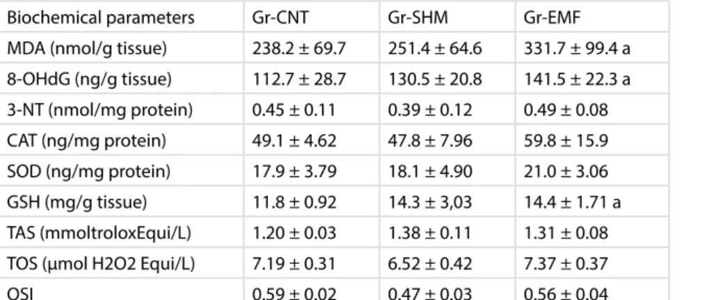
Exposure to a continuous 900-MHz electromagnetic field for 1 hour a day throughout middle and late adolescence alters the morphological structure and some biochemical markers of the female rat kidney at postnatal day 60
Derya Öztürk Okatan, Ersan Odacı (Turkey)
Functional characterization of p.T10M and p.S345Y mutations in HNF1A gene in MODY patients
Özlem Yalçın Çapan, Oğuzhan Fatih Baltacı, Ece Selçuk Şahin, Ergül Berber (Turkey)
Impairments of oxidant/antioxidant levels and some morphological changes in the adult female rat heart following exposure to a continuous 900-MHz electromagnetic field for 1 hour a day throughout middle and late adolescence
Hatice Hancı, Derya Öztürk Okatan, Ersan Odacı (Turkey)
Effects of propolis against cisplatin induced experimental kidney damage in rats
Esin Yuluğ, Sibel Türedi, Engin Yenilmez, Yüksel Aliyazıcıoğlu, Selim Demir, Serap Özer Yaman, Ahmet Menteşe (Turkey)
ICHC 2017
ICHC 2017
Kervansaray Lara Hotel, ANTALYA
May 18 - 21, 2017
www.ichc2017.com
15
thInternational Congress of
Histochemistry and Cytochemistry
“From Molecules to Diseases”
www.ichc2017.com
15
14:00 14:45 14:45 15:45 15:45 16:45 16:45 17:30 17:30 18:30 20:00 23:30 SATURDAY 20.05.2017 PLENARY LECTURE 5 Pavel Hozak (Czech Republic)Uncovering roles of lipids in genome regulation
Chair: Melek Öztürk (Turkey) HALL 1
HALL 3 S-14
Techniques in Immunohistochemistry
Chairs: Marija Plodinec (Switzerland) & Lone Bojesen (Denmark) Marija Plodinec (Switzerland) (Invited speaker) Mechanobiology of epithelia on native basement membranes - relevance for cancer cell invasion & clinics
Bilal E. Kerman (Turkey) (Invited speaker)
An in vitro approach to understanding myelination and myelin disorders
Oral presentations
Differences in ımmunohistochemical localization and distribution of galectin-1 and -3 in rat testes and epididymis during postnatal development
Emel Ergün, Mehmet Özbek, Feyzullah Beyaz, Levent Ergün, Hikmet Altunay, Nevin Kurtdede, Nuh Yıldırım, Özge Özgenç (Turkey)
HALL 2 S-15 Neuroscience
Chairs: Selma Yılmazer (Turkey) & Marc Davenne (France) Marc Davenne (France) (Invited speaker)
The axon initial segment: a novel site of neuronal dysfunction in multiple sclerosis?
Erdinç Dursun (Turkey) (Invited speaker)
Vitamin D perspective in neurodegeneration and Alzheimer’s disease: the genetic background and the cellular mechanisms
Özhan Eyigör (Turkey) (Invited speaker)
Immunonohistochemical Assessment of Neuronal Activation in Neuroendocrine Systems
Oral presentations
Revealing physical and functional ınteraction between p60-katanin and p53
Şirin Korulu (Turkey)
Immunohistochemical evaluation of the autoantibodies in surgically treated MTLE-HS patients
Ayşegül Fırat, Fadime Irsel Tezer Filik, Işık Ünal, Burçak Bilginer, Figen Kaymaz, Figen Söylemezoğlu, Serap Saygı (Turkey)
Investigation of dose-dependent ultrastructural alterations after topical lithium treatment in peripheral nevre injuries by transmission electron microscope
Emre Kocman, İlknur Dağ, Tayfun Sengel, Erdem Söztutar, Mediha Canbek
HALL 3 S-16
Pathology and Clinical Medicine-II
Chairs: Hale Kırımlıoğlu (Turkey) & Emel Koptagel(Turkey) Hale Kırımlıoğlu (Turkey) (Invited speaker) Rejection Pathology In Liver Transplantation Oral presentations
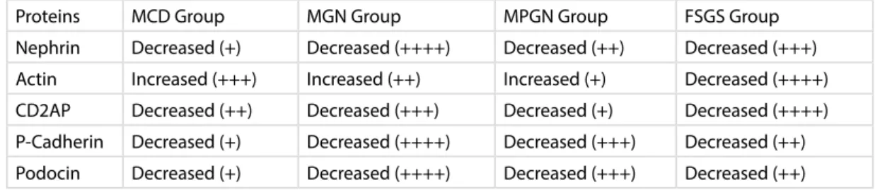
The ultrastructural ınvestigation of slit diaphragm protein expressions and filtration barrier features in some human podocytopathies
Mustafa Yilmaz, Kezban Kibar, Banu Coşkun Yilmaz, Iclal Gürses, Ahmet Kıykım (Turkey)
The role of curcumin in streptozotocin-induced hepatic damage and the trans-differentiation of hepatic stellate cells
Hesham Noaman Abdelraheem Mustafa (Saudi Arabia)
Changes in histological structure and biochemical markers in the adult male rat pancreas following exposure to a continuous 900-MHz electromagnetic field for 1 hour a day throughout adolescence
Gökçen Kerimoğlu, Ersan Odacı (Turkey)
Acute alterations in the morphology and biochemistry of the female rat liver following continuous 900-megahertz electromagnetic field (1 hour per day) administration throughout middle and late adolescence
Derya Öztürk Okatan, Ali Kulaber, Ersan Odacı (Turkey)
A histopathological and biochemical evaluation of oxidative injury in the sciatic nerves of male rats exposed to a 900-megahertz electromagnetic field throughout all periods of adolescence
Gökçen Kerimoğlu, Canan Güney, Şafak Ersöz, Ersan Odacı (Turkey)
The risk assessment of Gd2O3:Yb3+/Er3+nanocomposites as dual-modal nanoprobes for magnetic and fluorescence imaging
Long Huang, Fukang Xie, Li Li (China)
Lipopolysaccharide-induced liver damage is prevented by ginkgo biloba extract 761 and flunixin meglumin in septicemia
Tuba Parlak Ak, Burcu Gül Baykalır, Ismail Seven, Gürdal Dağoğlu, Mine Yaman (Turkey)
Immunohistochemical evaluation of effects of bevacizumab on radiation injury after stereotactic radiosurgery
Ayfer Aslan, Alp Ozgun Borcek, Zeynep Bengisu Kaya, Ozgur Ocal, Erkut Baha Bulduk, Ozge Petek Erpolat Tater, Murat Uçar, Figen Kaymaz (Turkey)
POSTER SESSION - II & COFFEE BREAK
GALA DINNER
S-17 Stem Cells-II
Chairs: İbrahim Tuğlu (Turkey) & Aydan Özgörgülü (Turkey) Oral presentations
The role of stem cell on the reproductive organs
İbrahim Tuğlu, Işıl Aydemir, Mahmud Özkut, Fatma Öztürk, Alican Gümürdü, Rehime Ablumiti, Dila Sal (Turkey)
Optimizing hydroxy apatite based scaffold with harmony of stem cells for tooth tissue engineering
Alev Cumbul, P. Neslihan Taşlı, Gül Merve Yalçın Ülker, Ünal Uslu, Fikrettin Şahin (Turkey)
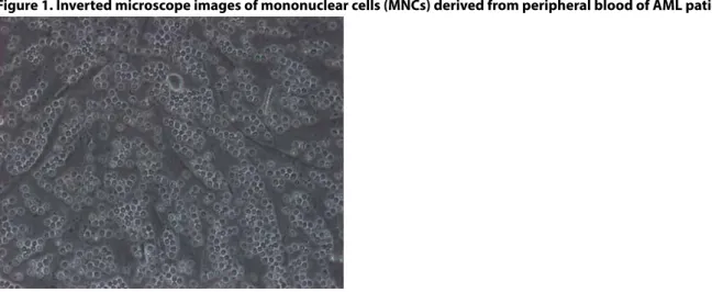
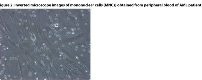
Comparison of immunological properties of mononuclear cells between acute myeloid leukemia patients and healthy donors
İlkay Pişkin, Yasin Köksal, Fatma Karaca Kara, Neşe Yaralı, Meltem Özgüner (Turkey)
Amniotic Fluid derived stem cells and its multilineage differentiation
A S M Golam Kibria, Özlem Özden Akkaya, Korhan Altunbaş, Metin Erdoğan, Artay Yağcı (Turkey)
ICHC 2017
ICHC 2017
Kervansaray Lara Hotel, ANTALYA
May 18 - 21, 2017
www.ichc2017.com
16
09:00 15:30 08:45 09:45 09:45 10:15 10:30 10:30 11:00 11:00 13:00 13:00 14:00 15:00 SUNDAY 21.05.2017 REGISTRATION HALL 1Young Histochemist Awardees
Chairs: Hinke Multhaupt (Denmark) & Zbigniew Kmiec (Poland)
Debora Burini (Italy)
Morphological study of chondrocyte cell death in patients affected by chondrocalcinosis
Let the Seniors Share their Expertises with Juniors
Sara Escudeiro-Lopes (Czech Republic)
Characterization of Lamin A / Phosphatidylinositol-4,5-bisphosphate complex Özlem Tuğçe KAYA (Turkey)
Ultrastructural Examination of Doublecortin, GABA and VGLUT1 Levels in the Dentate Gyrus of Absence Epileptic Rats
HALL 2
Technical Presentation-II Chair: Şahin Sırmalı (Turkey)
New Tools for 3D Electron Microscopy, ZEISS, Joerg Lindenau (Germany)
HALL 3
S-18
Developmental and Reproductive Biology-III Chairs: Ramazan Demir (Turkey) & Ayşegül Uysal (Turkey) Oral presentations
T-2 toxin disrupts sertoli cell barrier via changes in tight and adherent junctional proteins in SerW3 cells
Elif Karacaoğlu, Güldeniz Selmanoğlu (Turkey)
Nesfatin-1 protects against torsion-induced testicular oxidative injury in rats
Sevil Arabacı Tamer, Alper Yıldırım, M. Kutay Köroğlu, Özge Dağdeviren Çevik, Feriha Ercan, Berrak Ç. Yeğen (Turkey)
Histological examination of the effect of {lycium barbarum} (Goji Berry) on testis and epididymis of acrylamide treated young male rats
Havva İmran Özdemir, Aysel Kükner (Turkey)
Protective effects of melatonin on ovarian structures of rats exposed to environmental toxic agent
2,3,7,8-tetrachlorodibenzo-p-dioksin (TCDD)
Semir Gül, Mehmet Gül, Birgül Yiğitcan (Turkey)
Investigation of the effects of vitamin D treatment on the uterine structural changes in the experimental model with polycystic ovary syndrome: an ultrastructural and immunohistochemical study
Yurdun Kuyucu, Latife Seyran Çelik, Ebru Dündar Yenilmez, Abdullah Tuli, Ufuk Özgü Mete (Turkey)
HSP 70 Histological examination of the effect of {lycium barbarum} (Goji Berry) on testis and epididymis of acrylamide treated young male rats
Bülent Celpkulu, Mete Köksal, Halil Çiftçi, Fuat Dilmeç (Turkey)
Morphology, morphometric and biochemical parameters changes in the adult rat ovarium following continuous 900-megahertz electromagnetic field applied in middle and late-adolescence
Derya Özturk Okatan, Ersan Odacı (Turkey)
S-19
Advances in Image Analysis
Chairs: Nurhan Özlü (Turkey) & Çağdaş Son (Turkey) Nurhan Özlü (Turkey) (Invited Speaker)
Proteomic analysis of Epithelial Mesenchymal Changes
Çağdaş Son (Turkey) (Invited Speaker)
Applications of Föster Resonance Energy Transfer and Split EGFP Techniques at Protein-protein Interaction Studies
Oral presentation
Effect of Systemic Magnesium Sulfate on Retina in Neonatal Rats with Hypoxic Ischemic Encephalopathy
Serhat Imamoğlu, Ebru Yalın Imamoğlu, Serkan Erdenöz, Lorina Haziri, Yağız Özdağ, Alev Cumbul, Ünal Uslu, Şamil Aktaş, Gökhan Pekel, Mehmet Şahin Sevim, Mustafa Nuri Elçioğlu, Fahri Ovalı (Turkey)
S-20 Cancer Biology-II
Chairs: Ayhan Bilir (Turkey) & Gamze Tanrıöver (Turkey) Ayhan Bilir (Turkey) (Invited Speaker)
The Autophagıc Activity of LICL on Different Tumor Cell Lines using Electron Microscopy
Oral presentations
Effects of paclitaxel, bevacizumab and metformin on PI3K/AKT pathway and angiogenesis in MDA-MB 231 breast cancer cell line
Fatma Firat, Elgin Turkoz Uluer, Sevinç Inan (Turkey)
The Evaluation of the Distribution of CD133, CXCR1 and the tumor associated macrophages in different molecular subtypes of the breast cancer
Can Ilgın, Erdem Çomut, Çağlar Sarıgül, Selçuk Korkmaz, Enver Vardar, Sevda Fatma Müftüoğlu (Turkey)
Loss of histone H4K20 trimethylation predicts poor prognosis in bladder cancer
Nuray Varol, Cem Karaosmanoglu (Turkey)
Association between CAT C-262T polymorphism and CAT enzyme activity in patients with leukemia
Nazan Eras, Anıl Tombak, Naci Tiftik, Mehmet Berköz, Gözde Türköz, Etem Akbaş (Turkey)
Adjuvant therapeutic effect of cold atmospheric plasma on endometrium cancer cells
Işıl Aydemir, Utku Kürşat Ercan, Tülay Oludağ Mete, Sevinç Inan, Mehmet Ibrahim Tuğlu (Turkey)
Label-free investigation of cancer cell behavior using quantitative phase imaging
Vratislav Kostal, Jan Balvan, Michal Masarik (Czech Republic)
The effects of atmospheric plasma and oleocanthal on cancer cell migration
Işıl Aydemir, Utku Kürşat Ercan, Pınar Kılıçaslan Sönmez, Mesut Mete, Mehmet Ibrahim Tuğlu (Turkey)
COFFEE BREAK
CLOSING CEREMONY & FAREWELL PARTY EXCURSION
Jacek Kiezun (Poland)
The expression of galanin receptors (GALR1, GALR2 and GALR3) in colorectal cancer Mohammed Khurshed (The Netherlands)
IDH1-mutated gliomas rely on anaplerosis of glutamate and lactate whereas IDH1 wild-type gliomas rely on glycolysis and acetate anaplerosis
ICHC 2017
ICHC 2017
Kervansaray Lara Hotel, ANTALYA
May 18 - 21, 2017
www.ichc2017.com
15
thInternational Congress of
Histochemistry and Cytochemistry
“From Molecules to Diseases”
ICHC 2017
ICHC 2017
Kervansaray Lara Hotel, ANTALYA
May 18 - 21, 2017
www.ichc2017.com
18
ICHC 2017
ICHC 2017
Kervansaray Lara Hotel, ANTALYA
May 18 - 21, 2017
www.ichc2017.com
15
thInternational Congress of
Histochemistry and Cytochemistry
“From Molecules to Diseases”
www.ichc2017.com
19
Syndecans: receptors with signalling functions and roles cell adhesion and disease
John R. CouchmanUniversity of Copenhagen, Denmark
Over 30 years ago, evidence accumulated that cell surface proteoglycans, some with hydrophobic properties, were involved in cell adhesion to extracellular matrix. Eventually, four members of the syndecan family of transmembrane proteoglycans were characterized, but their modes of action remained elusive. We have utilized a combination of imaging, immunochemistry, molecular and structural biology to address this question. Ligands for the syndecans can bind either their heparan sulphate polysaccharide chains, or the external portion of the core protein. Signaling and intracellular interactions are complex, but can be resolved into three parts. For example, syndecan-4 interacts with actin-associated proteins, protein kinase C and PDZ proteins. Interactions with α-actinin are essential for cytoskeletal organization, while signaling through protein kinase C impacts the activity of small G proteins of the Rho family, as well as regulating the gating of ion channels belonging to the TRPC (transient receptor potential canonical) group. In turn, therefore, syndecans regulate intracellular calcium levels, with impact on adhesion and migration. PDZ protein interactions regulate the trafficking of syndecans and may be essential in exosome biology. Finally, dysregulation of syndecan expression accompanies many diseases, including some tumours, cardiovascular disease and fibrosis.
ICHC 2017
ICHC 2017
Kervansaray Lara Hotel, ANTALYA
May 18 - 21, 2017
www.ichc2017.com
20
ICHC 2017
ICHC 2017
Kervansaray Lara Hotel, ANTALYA
May 18 - 21, 2017
www.ichc2017.com
15
thInternational Congress of
Histochemistry and Cytochemistry
“From Molecules to Diseases”
www.ichc2017.com
21
What’s new in histochemistry and cytochemistry
Cornelis J.F. Van NoordenDepartment of Medical Biology Academic Medical Center at the University of Amsterdam
Since Gomori stained activity of phosphatase in tissue sections in the 1930s, histochemistry and cytochemistry (H&C) developed into true disciplines that cannot be done without in science. Especially, in the second half of last century, tremendous progress was achieved in specificity, sensitivity and precision of localization of H&C methods. H&C can be defined as the disciplines that localize specific compounds, such as a specific sequence of DNA or RNA using in situ hybridization, a specific protein using immunoH&C or a product of activity of a specific enzyme using enzyme H&C. The latter method was developed by Gomori to visualize phosphatase activity in tissue sections or cell preparations, respectively, and this was in fact the initiation of H&C .
Especially immunoH&C has become indispensable for microscopic imaging, flow cytometry and cell sorting on the basis of fluorescent or coloured staining of one or more specific proteins in tissue or cell samples. Besides, in situ hybridization has become a valuable technology for the staining of specific sequences of DNA (e.g. DNA of viruses in a human tissue) or RNA (idem of RNA viruses or mRNAs to visualize where in a tissue or in which types of cells a specific gene is expressed). The third major approach, enzyme H&C or metabolic mapping1,2, that visualizes the product of a specific enzyme reaction as a fluorescent or coloured end product, became almost obsolete in the last 2 decades of last century. Metabolism was considered old fashioned in that time. How did this opinion change in the beginning of this century by the awareness that metabolism is very much involved in major complex diseases such as cancer and diabetes. This awareness initiated studies that targeted therapeutically metabolic pathways that are crucial for diseased tissue or cells. For example, specific mutations in genes encoding for enzymes such isocitrate dehydrogenase 1 and 2 are important steps in the development of a number of types of cancer such as glioma (primary brain tumors), acute myeloid leukemia, cholangiocarcinoma (tumors of bile ducts) or chondrosarcoma (tumors of cartilage)3. Metabolic mapping plays an important role in the search for therapies of types of cancer that have this mutation4. It is only one example of metabolic mapping where it has experienced a revival in the present century showing its new role in H&C.
The other major development is 3D histochemistry. Histochemistry has always been performed on thin tissue sections, but since the group of Deisseroth published their Clarity method to look through brain, 3D histochemistry has become the talk of the scientific world5. The principles of 3D histochemistry are clearing of the tissue, fluorescence labelling of specific proteins or structures, and visualization of the entire organs or tissue sample using confocal microscopy or light-sheet microscopy. Basically, two approaches exist for clearing of organs or tissue samples. The first one is removal of lipids of cell membranes from the organ or tissue. The lipids cause the opaqueness of tissues and removal of the lipids renders tissues transparent or clear which is the principle of Clarity. The other clearing method is based on matching the refractive index of the tissue sample and the solution in which the tissue sample is embedded . This method works for tissues that contain significant amounts of extracellular matrix (ECM)6. The central nervous system hardly contains ECM but large amounts of cell membranes and this makes Clarity ideal for brain but not for tissues that contain considerable amounts of ECM. These tissues can be cleared in solutions that have a similar refractive index such as in iDISCO and BABB methods6. The group of Erturk recently showed that an entire rat can be cleared using iDISCO methodology and fluorescence imaging can be performed in 3D in the intact animal7. Moreover, iDISCO and BABB can also be used to clear human tissues6, because perfusion of the tissue is not needed as is the case with Clarity. Therefore, Clarity can only be applied to experimental animals. The advantages of 3D histochemistry are enormous as molecular and cellular interactions can be studied directly in 3D instead of indirectly using reconstructions on the basis of images of large amounts of serial sections.
These developments in H&C in combination with the rapid developments in microscopy and nanoscopy make imaging of tissues and cells increasingly important tools in the life sciences.
Van Noorden CJF (2010) Imaging enzymes at work: Metabolic mapping by enzyme histochemistry. J Histochem Cytochem 58:481-497 Van Noorden CJF (2014) Metabolic mapping by (quantitative) enzyme histochemistry. Pathobiology of Human Disease: A Dynamic Encyclopedia of Disease Mechanisms, pp 3760-3774
Molenaar RJ et al (2014) The driver and passenger effects of isocitrate dehydrogenase 1 and 2 mutations in oncogenesis and survival prolongation. Biochim Biophys Acta 1846:326-341
Molenaar RJ et al (2015) Radioprotection of IDH1-mutated cancer cells by the IDH1-mutant inhibitor AGI-5198. Cancer Res 75:4790-4802 Chung K et al. (2013) Structural and molecular interrogation of intact biological systems. Nature 497:332-337
Azaripour A et al. (2016) A survey of clearing techniques for 3D imaging of tissues with special reference to connective tissue. Progr Histochem Cytochem 51:9-23
ICHC 2017
ICHC 2017
Kervansaray Lara Hotel, ANTALYA
May 18 - 21, 2017
www.ichc2017.com
22
ICHC 2017
ICHC 2017
Kervansaray Lara Hotel, ANTALYA
May 18 - 21, 2017
www.ichc2017.com
15
thInternational Congress of
Histochemistry and Cytochemistry
“From Molecules to Diseases”
www.ichc2017.com
23
Single Molecule Localization Microscopy of Nuclear Genome Nanostructure
Christoph Cremer1,2,31Institute of Molecular Biology (IMB), D-55128 Mainz/Germany;
2Institute for Pharmacy and Molecular Biotechnology (IPMB), University Heidelberg & Kirchhoff-Institute for Physics (KIP), D-69120
Heidelberg/Germany;
3Department of Physics, University Mainz (JGU), D-55128 Mainz/Germany
The human genome has been decoded, but we are still far from understanding the regulation of all gene activities. A largely unexplained role in these regulatory mechanisms is played by the spatial organisation of the genome in the cell nucleus which has far-reaching functional consequences for gene regulation. Until recently, it appeared to be impossible to study this problem on the nanoscale by light microscopy. However, novel developments in optical imaging technology have radically surpassed the limited resolution of conventional far-field fluorescence microscopy (ca. 200 nm). These limits have been overcome by various super-resolution fluorescence microscopy (SRM) methods, such as Stimulated Emission Depletion (STED), Photoactivated Localization Microscopy (PALM), Structured Illumination Microscopy (SIM), or Stochastic Optical Reconstruction Microscopy (STORM)1. Here we report on a complementary SRM approach to study nuclear genome structure at the single cell/single molecule level, Spectral Precision Distance/Position Determination Microscopy (SPDM). SPDM, a variant of localization microscopy, makes use of conventional fluorescent proteins or single standard organic fluorophores in combination with standard or appropriately modified specimen preparation conditions, allowing to use the same laser frequency for both photoswitching and fluorescence read out2. Presently, this approach allows us to optically resolve nuclear structures in individual cells down to few tens of nanometer, and to perform quantitative analyses of individual small chromatin domains; of the nanoscale distribution of histones, chromatin remodeling proteins, and transcription, splicing and repair related factors. In addition, it has become possible to combine localization microscopy (SPDM) of nuclear DNA distribution, positioning up to around one million of individual DNA-bound fluorophore signals in an optical section within a 3D intact cell nucleus, with simultaneous measurements of the spatial positions of individual epigenetic histone marker molecules. The experimental results support recent models of functional nuclear nanostructure3. As a translational application, using dual-color SPDM, it became possible to monitor in mouse cardiomyocyte cells quantitatively the effects of ischemia conditions on the chromatin nanostructure (DNA)4,5. These novel “molecular optics” approaches open an avenue to study the nuclear landscape directly on the individual cell level at unprecedented optical and structural resolution.
1C. Cremer, B.R. Masters (2013) Resolution enhancement techniques in microscopy.Eur. Phys. J. H, Eur. Phys. J. H 38: 281–344. 2C. Cremer et al. (2011) Superresolution Imaging of Biological Nanostructures by Spectral Precision Distance Microscopy (SPDM), Biotechnology Journal 6: 1037 – 1051. 3T. Cremer et al. (2015) The 4D nucleome: Evidence for a dynamic nuclear landscape based on coaligned active and inactive nuclear compartments. FEBS LettersFEBS Letters 589: 2931–2943. 4I.Kirmes et al. (2015) A transient ischemic environment induces reversible compaction of chromatin. Genome Biology 16:246. 5A.Szczurek et al. (2017) Imaging chromatin nanostructure with binding-activated localisation microscopy based on DNA structure fluctuations. Nucleic Acids Research 2017, 1–11.doi: 10.1093/nar/gkw1301.
ICHC 2017
ICHC 2017
Kervansaray Lara Hotel, ANTALYA
May 18 - 21, 2017
www.ichc2017.com
24
The Dialog Between Biomaterials and Cells
Vasıf HasırcıBIOMATEN, METU Center of Excellence in Biomaterials and Tissue Engineering, and Dept. of Biological Sciences, Ankara, Turkey The interactions between the cells and the artificial substrates they are deposited on involve constant mobility and feeling of the cell microenvironment. The structural and functional macromolecules constituting the extracellular matrix are a source of complex chemical and physical signals that guide cell morphology and fate. The deformability of the cells is defined by their phenotype, level of differentiation and also the of state of health and the final form they take is very much dependent on the topography and chemistry of their environment. Over the last decade we created a large number of micropatterned platforms with different chemistry, spacing and systematically varied feature organization to induce morphological changes in the cells and then later on the nuclei. As a result, certain clues about the cell-substrate relations have been obtained.
ICHC 2017
ICHC 2017
Kervansaray Lara Hotel, ANTALYA
May 18 - 21, 2017
www.ichc2017.com
15
thInternational Congress of
Histochemistry and Cytochemistry
“From Molecules to Diseases”
www.ichc2017.com
25
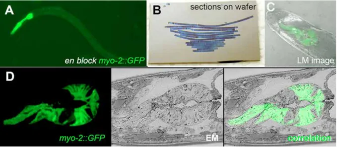
Correlative Light and Electron Microscopy in Cell Biology
Bruno M Humbel1, Céline Loussert Fonta3, Caroline Kizilyaprak1, Heinz Schwarz2, Jean Daraspe1, Willy Blanchard1
1Electron Microscopy Facility, University of Lausanne, Lausanne, Switzerland 2Max-Planck-Institute for Developmental Biology, Tuebingen, Germany 3Nestle Research Center, Lausanne, Switzerland
In recent years correlative microscopy, combining the power and advantages of light and electron microscopy, has become an important tool for biomedical research.
Light microscopy has the advantage of easily searching large areas, even volumes, for the cells of interest, e.g., a special cell type in tissue, astrocytes in brain2 or for cells that have been modified either by transfections or by RNAi in a large population of non-modified cells. Also on thin sections, the low magnification of light microscopy and therefore ease of searching large areas are very beneficial to speed-up the analysis of rare events3. The predominant disadvantage of this technique, however, is that only the fluorescently labelled structures can be imaged in relation to each other.
Electron microscopy reveals the cellular ultrastructure a high resolution and individual organelles, even large protein polymers like cytoskeletal filaments or ribosomes can unequivocally identified. Proteins of interest can be labelled with colloidal gold. Searching for a few gold particles within a few cells of a large tissue, however, is very cumbersome and can be extremely time consuming if not impossible. Seen the advantages of light and electron microscopy suggests that the optimal approach is to combine both techniques for cell biology research. Localisation of rare cellular events are followed and identified by (fluorescence) light microscopy, the high resolution data and fine localisation to cellular substructures are done by electron microscopy.
In this presentation we will describe the approach we have chosen to follow the cell(s) of interest from sampling the tissue until the analysis by electron microscopy4.
1Tsien, R.Y., Annu Rev Biochem 67 (1998). 509-544. 2Loussert Fonta, C., et al., J Struct Biol 189 (2015). 53-61.
3Schwarz, H. and B.M. Humbel, Methods Mol Biol 1117 (2014). 559-592. 4Loussert Fonta, C. and B.M. Humbel, Arch Biochem Biophys 581 (2015). 98-110. Keywords: correlative microscopy, electron microscopy, light microscopy
ICHC 2017
ICHC 2017
Kervansaray Lara Hotel, ANTALYA
May 18 - 21, 2017
www.ichc2017.com
26
3D+time imaging data and image analysis for the reconstruction of multilevel
dynamics in animal morphogenesis
Nadine Peyriéras
Head of the BioEmergences laboratory CNRS-USR3695, Gif-sur-Yvette, France
We approach the understanding of Deuterostome early embryogenesis through the quantitative analysis and biomechanical modeling of cell dynamics from multiscale in vivo imaging data. The automated reconstruction of the cell lineage tree, annotated with nucleus and membrane segmentation, provides measurements for cell behavior: displacement, division, shape and contact changes. This quantitative data is used to derive statistical models for key parameters and calculate descriptors for tissue deformation. Confronting numerical simulations derived from multi-agent based biomechanical models with empirical measurements extracted from the reconstructed digital specimens is the basis for testing biological hypotheses. Further correlating cell behavior, tissue biomechanics and biochemical activities by comparing the patterns revealed by cell fate, kinematic descriptors or gene expression, is a step toward the integration of multi-level dynamics underlying morphogenetic processes.
ICHC 2017
ICHC 2017
Kervansaray Lara Hotel, ANTALYA
May 18 - 21, 2017
www.ichc2017.com
15
thInternational Congress of
Histochemistry and Cytochemistry
“From Molecules to Diseases”
www.ichc2017.com
27
Nuclear lipids contribute to intranuclear order and efficient DNA transcription
Pavel HozakBiology of the Cell Nucleus, Institute of Molecular Genetics ASCR, Prague,
Phosphatidylinositol 4,5-bisphosphate (PIP2) functions in the cell nucleus as a regulator involved in chromatin remodelling, transcription, and splicing. Since its involvement in RNA polymerase II (Pol II) transcription is still little understood, we studied the role of nuclear PIP2 in the organization of Pol II transcription complexes. We show that nuclear PIP2 associates with Pol II, transcriptional factors, nascent transcripts, and NM1 at the periphery of PIP2 islets. Integrity of PIP2 islets as well as the interaction of PIP2 with NM1 are necessary for Pol II transcription. We demonstrate that the transcriptionally active foci are preferentially positioned on the surface of PIP2 islets. We show that PIP2 is a major component of PIP2 islets, while RNA, ceramide and cholesterol constitute the minor part of their periphery. We suggest that PIP2 islets provide a platform for the proper arrangement of the complexes involved in Pol II transcription. We hypothesize that PIP2 islets, due to their heterogeneous multi-component nature, have a role in the spatial formation and maintenance of transcription factories and they thus participate in nuclear organization.
This work was supported by MEYS CR (LM2015062), GACR (GA15-08835Y), TACR (TE01020118)[b3] . Keywords: cell nucleus, DNA transcription, phosphoinositides, lipids
ICHC 2017
ICHC 2017
Kervansaray Lara Hotel, ANTALYA
May 18 - 21, 2017
www.ichc2017.com
28
ICHC 2017
ICHC 2017
Kervansaray Lara Hotel, ANTALYA
May 18 - 21, 2017
www.ichc2017.com
15
thInternational Congress of
Histochemistry and Cytochemistry
“From Molecules to Diseases”
www.ichc2017.com
29
[Epigenetics & molecular cytogenetics]
Distinct 3D nuclear topography of active and inactive regulatory sequences studied
with super-resolution fluorescence microscopy
Marion Cremer1, Volker J. Schmid2, Felix Kraus1, Yolanda Markaki1, Andreas Maiser1, Heinrich Leonhardt1, Sam John3,
John Stamatoyannopoulos3, Thomas Cremer1
1Department Biology II, Biocenter, Ludwig Maximilians University (LMU), Martinsried, Germany 2Institute of Statistics, LMU Munich, Munich, Germany
3Departments of Genome Sciences and Medicine, University of Washington, Seattle, WA 98195, USA
We describe the nuclear topography of transcription regulatory elements (TREs) in two human cell lines employing 3D-fluorescence in situ hybridization (3D-FISH), 3D structured illumination microscopy (3D-SIM) and a novel tool that enables quantitative 3D mapping of DNA targets on chromatin compaction defined nuclear landscapes. DNA probe sets of different complexity are targeted to sites harboring either active TREs identified by their DNAse I hypersensitivity (DHS+) or inactive TREs, which lack hypersensitivity (DHS-). The results of our initial study fit well within a recently proposed model for a functional nuclear organization, based on co-aligned active and inactive nuclear compartments, called the ANC-INC network model (Cremer et al., 2015. FEBS Letters 589). According to this model, chromatin domain clusters (CDCs) constitute the basic subunits of chromosome territories. CDCs pervade the nuclear space in a network-like manner and are organized as shell-like structures with layers of different chromatin compaction levels. The compacted inner core of CDCs represents the inactive nuclear compartment (INC) while active domains with decondensed chromatin form the peripheral layer of CDCs, named the perichromatin region (PR) which represents the nuclear compartment where transcription occurs. The PR lines the interchromatin compartment (IC), a contiguous channel system, which is connected to nuclear pores, permeates between CDCs and harbors nuclear bodies required for functions occurring within the PR. IC and PR together are therefore considered as the active nuclear compartment (ANC). We demonstrate that DNA segments harboring active TREs (DHS+) are highly significantly enriched within the ANC while larger segments lacking active TREs (DHS-) are enriched in the INC, suggesting positional changes of TREs between ANC and INC depending on their functional state.
From the methodological side issues of chromatin preservation after 3D-FISH experiments recorded at the level of super-resolution microscopy will be addressed in the talk.
Keywords: transcription regulatory sequences (TRE), DNAse I hypersensitive sites, super-resolution microscopy, chromatin domain, nuclear architecture, active / inactive nuclear compartment
ICHC 2017
ICHC 2017
Kervansaray Lara Hotel, ANTALYA
May 18 - 21, 2017
www.ichc2017.com
30
[Epigenetics & molecular cytogenetics]
Epigenetic significance of higher-order chromatin organization in health and
disease
Milena Georgieva1, Dessislava Staneva1, Bela Vasileva1, Milena Draganova Filipova3, Plamen Zagorchev2, George Miloshev1
1Laboratory of Molecular Genetics, Institute of Molecular Biology, Bulgarian Academy of Sciences, Sofia, Bulgaria 2Faculty of Pharmacy, Department of Medical Physics, Biophysics and Mathematics, Medical University, Plovdiv, Bulgaria 3Department of Biology, Medical University, Plovdiv, Bulgaria
Introduction & Objectives: Chromatin is a DNA multi-protein complex in which the genome of eukaryotes is organized. Its main role is to store and protect the molecule of DNA, but also to genuinely modulate gene expression. Chromatin structure and dynamics are very important epigenetic mechanisms through which different genetic programs are performed in response to intrinsic and extrinsic environmental signals. This allows the cells and the organisms to response to these signals by changing its expression program. Multiple levels of chromatin organization exist and they have always been a challenge for the molecular biologists. The most elusive and yet sparsely known are the higher-order chromatin structures - the 30 nm fiber and structures above it like blobs and loops. In addition, chromatin is not static but is a very dynamic structure which through changes in its compaction controls gene expression.
Our main objective is to study in detail chromatin remodeling in response to different stress factors by which adaptation of cells and organisms to the surrounding environment is performed. Particularly we intend to reveal the main players in these processes and to spot crucial protein contacts within and with chromatin which are prerequisite for the process of remodeling.
Materials & Methods: Numerous different model organisms have been used in our studies including lower and higher eukaryotic cells, tissue biopsies and several laboratory animals. The methods used include standard molecular biology, biochemistry and biophysics methods like DNA and chromatin analyzing techniques, PCRs, techniques for genetic manipulations, Yeast two-hybrid system, differential scanning calorimetry, high resolution light and fluorescent microscopy and atomic force microscopy.
Results: We have shown that the linker histones are among the main players in chromatin remodelling. Especially, we have detected an important contact between the linker histones and chromatin remodelling complexes. Moreover, this contact proved to be crucial for chromatin remodelling in response to UV stress. By utilizing the developed by us Chromatin Comet Assay (ChCA) we have analyzed chromatin loop dynamics during ageing and stress adaptability of the cells. Using ChCA we have shown that chromatin loop structures have been abolished in mutants without the gene for the linker histone. Notably, these cells exhibited premature ageing phenotypes and inability to adequately react to stress.
Conclusions: Our results prove the significance of higher-order chromatin structure organization as a major epigenetic factor which controls the way cells react to stress. We have proved that cellular viability, adaptability and stress resistance are dependent on the proper chromatin organization, especially at its higher levels of compaction.