nv
estiga
tion
ali Erdem yılDırım1, ali DalGıC1, Denizhan DıvaNlıoGlu1, rifat aKDaG2, Nuri Eralp CETıNalP3, Fatih alaGoz1, Fatma hElvaCıoGlu4, Gulnur TaKE5, yahya GuvENC6, ısmet KoKSal7, ahmed Deniz bElEN1
1Ankara Numune Research and Education Hospital, Department of Neurosurgery, Ankara, Turkey 2Bursa Sevket Yilmaz Education and Research Hospital, Department of Neurosurgery, Bursa, Turkey 3Cukurova University Hospital, Department of Neurosurgery, Adana, Turkey
4Baskent University Hospital, Department of Histology and Embryology, Ankara, Turkey 5Gazi University Hospital, Department of Histology and Embryology, Ankara, Turkey 6Sincan State Hospital, Department of Neuorusurgery, Ankara, Turkey
7Yenimahalle State Hospital, Department of Ortophedia, Ankara, Turkey Presented in: 15th WFNS World Congress of Neurosurgery, Seoul – Korea, 2013.
Clinical Trial Registration Number: Ankara Training and Research Hospital Ethics Board decision no 2282
Corresponding Author: Ali Erdem YIlDIrIM / E-mail: [email protected]
ABSTRACT
AIM: Catechin is a type of polyphenol, along with epicatechin, epigallocatechin, and epigallocatechin-gallate (EGCG). This study aims to investigate the effect of EGCG, a major metabolite of catechin, which is the principle bioactive compound in green tea, on rats with peripheral nerve injury.
MATErIAL and METHods: A total of 74 rats were divided into six groups, namely the control, the trauma, the normal saline, a 25mg/kg EGCG, a 50mg/kg EGCG and a daily consumption group (10mg/kg EGCG was given intraperitoneally for 14 days before the trauma). Except the first group, the other groups underwent a 1-minute sciatic nerve compression by clip with 50gr/cm2 pressure. Nerve samples were obtained at 28
day after trauma for the biochemical and histopathological analysis.
rEsuLTs: Our study showed that the Daily consumption, 25mg/kg EGCG and 50mg/kg EGCG groups demonstrated statistically significant decreased lipid peroxidation levels and particularly daily consumption, and the 25mg/kg EGCG group showed a favourable reduction of degeneration and edema histologically.
CoNCLusIoN: This study shows that Catechin and its derivatives have a protective effect on peripheral nerve injury.
KEywords: Axonal degeneration, Epigallocatechin-gallate, Electron microscope, Light microscope, Lipid peroxidation, Peripheral nerve injury
ÖZ
AMAÇ: Kateşinler polifenol grubundan olup başlıca kateşin, epikateşin, epigallokateşin, epigallokateşingallat (EGKG) gibi alt grup maddeler içermektedir. Çalışmada kateşinin temel metaboliti olan EGKG’ın, periferik sinir hasarı yapılmış sıçanlardaki etkisinin gösterilmesi amaçlanmıştır. yÖNTEM ve GErEÇLEr: Toplam 74 Albino-Wistar cinsi rat kontrol, travma, serum fizylojik (SF), 25 mg/kg, 50 mg/kg ve günlük tüketim ( travma öncesi 14 gün boyunca intraperitoneal 10 mg/kg EGKG verildi) EGKG grupları olmak üzere 6 gruba ayrıldı. İlk grup hariç diğer gruplarda siyatik sinire 50 gr/cm2 basıncı ile kapanan klip ile 1 dakika kompresyon hasarı yapıldı. Bütün travma gruplarında, 28 gün sonra sinir örnekleri alınarak
biyokimyasal ve histopatolojik olarak incelendi.
BuLGuLAr: Çalışmamızda günlük tüketim EGKG, 25mg/kg EGKG, 50mg/kg EGKG gruplarında istatistiksel olarak anlamlı düzeyde lipid peroksidasyonunun azaldığı ve özellikle travma+günlük tüketim EGKG, travma+25 mg/kg EGKG grularında histolojik olarak da dejenerasyon ve ödemin olumlu yönde azaldığı saptanmıştır.
soNuÇ: Bu çalışma kateşin ve türevlerinin periferik sinir hasarı gibi nöronal hasarlanmalarda koruyucu etkisinin olduğu gösterilmiştir. ANAHTAr sÖZCÜKLEr: Aksonal dejenerasyon, Epigallokateşingallat, Elektron mikroskobu, Işık mikroskobu, Lipid peroksidasyon, Periferik sinir hasarı
biochemical and histopathological Effects of
Catechin on Experimental Peripheral Nerve Injuries
Deneysel Periferik Sinir Hasarında Kateşin Etkisinin Biyokimyasal ve
Histopatolojik Olarak İncelenmesi
InTRoduCTIon
Peripheral nerve injury causes loss in the labor force, economic loss and psychological problems. It is a topic of much interest, due to lack of available treatment options. The severity of a nerve compression lesion depends on several factors, including compression pressure, length of pressure, and the area which the pressure affects. Neuronal tissue damage continues after the compression is resolved (25). Compression injury to the nerve induces Wallerian degeneration and axon regeneration, whereas after central nervous system (CNS) injury axons fail to regenerate. These findings were first described by Ramon Y. Cajal under light microscope examinations in 1928 (33). Similarly, the process has also been shown by electron microscopic studies. The post-traumatic nerve tissue changes, observed by electron microscope, include intracytoplasmic edema, and changes in the nucleus, mitochondria, axon and myelin sheath (24). Similar to the blood–brain barrier in the CNS, there is a blood– nerve barrier in the peripheral nervous system. The blood-nerve barrier regulates the endoneurial microenvironment. Many studies have examined the effect of local experimental compression and intraneural microcirculation, and concluded that 20 – 30 mm Hg external pressure induces a blockage of venous blood flow in the epineurium. Pressure of 80 mm Hg leads to complete cessation of intraneural blood flow (19). Injury to a peripheral nerve triggers an initiation of a response that incorporates a sequence of biochemical alterations. Severe injury can lead to neuronal edema, more intense neutrophil infiltration, and apoptosis. Increased neutrophil infiltration, myeloperoxidase activity, and the level of tissue malondialdehyde (MDA) lead to an increased level of lipid peroxidation (15). Lipid peroxidation is a toxic process and a self-propagating chain-reaction (6, 15). Lipid peroxidation can impair membrane function directly and damage cell components indirectly. A marked increase in lipid peroxidation is observed after injury. Hall and Braugler reported that lipid peroxidation increases to a peak level at 1, 24 and 48 hours after spinal cord trauma (17).
The tea plant, Camellia sinensis, is a member of the Theaceae family (4, 14, 37). Tea leaves contain polyphenol and poly-phenol oxidase enzymes. The main component of green tea extract polyphenol is called catechins. Black tea contains 250 mg/L of catechin, compared to green tea which contains 420 mg/L (40). Approximately 25-35% of the dry weight of tea leaves is catechins. A single cup of green tea contains 100-200 milligrams of catechin (39, 41). The anticancer, antitumor, an-timutagenic, chemopreventive, antiproliferative, antiinflam-matory, antioxidant, antidiabetic, antiallergic, antihyperten-sive, antiplatelet, antiobesity, hypocholesterolemic, and neu-roprotective effects of catechins have been shown in various in vivo and in vitro studies (36).
The flavonoids contain a double bond and eight isomers. The main catechin group consists of eight polyphenolic flavonoid- type compounds, namely, catechin (C), epicatechin (EC),
gallo-catechin (GC), epigallo-gallo-catechin (EGC), gallo-catechin-gallate (CG), epicatechin-gallate (ECG), gallocatechin-gallate (GCG) and epigallocatechin gallate (EGCG). EGCG is the most prominent flavonoid compound in tea leaves, and has the highest antioxidant activity of all the green tea catechins (ECG > EGCG > EGC > EC) (1, 7, 8, 27, 28, 32). Tea catechins and polyphenols are effective scavengers of physiologically relevant reactive oxygen and nitrogen species, including superoxide, peroxyl radicals, singlet oxygen, and peroxynitrite (10, 13, 16, 29). The best treatment approach for peripheral nerve injuries remains unclear. In the treatment of peripheral nerve injury, non-steroidal anti-inflammatory and steroids can be used to reduce inflammation, and nerve growth factors, thyroid hormones, growth hormone, ACTH, and insulin like peptides to improve regeneration (11, 20, 31, 35, 38). The aim of this study was to examine favourable antioxidant and anti-inflammatory effects of catechin on peripheral nerve injury.
mATeRIAl and meThodS
Study guidelines and experimental protocol was approved by Ethical Committee of Ankara Training and Research Hospital (Decision # 2282) and all experimental procedures were performed at Animal Laboratory of the same hospital. In this study, 74 healthy adult male Albino Wistar rats, with body weight of 180–210 g and 3–5 months old, were used. The rats were put in a standard laboratory cage with standardized conditions and sufficient food and water, at 18-21 oC. The rats were exposed to 12 hours of light and 12 hours of dark cycle (26, 30).
The rats were randomized to six groups, including the control group, the trauma group, the normal saline group, 25 mg/kg EGCG group, 50mg/kg EGCG group, and daily consumption group (10mg/kg EGCG). The control and trauma groups were include 11 rats, the others grops were include 13 rats. The control group received no injury. The trauma group received compression-induced injury caused by clips, which closed with 50gr/cm2 pressure, for 1 minute. The third group, the normal saline group, received trauma and daily intraperitoneal injection of 0.25cc normal saline for 7 days. The treatment groups (group 4 and group 5) were given therapeutic dosages that have been used previously for the treatment of cerebrovascular stroke (36). The fourth group received trauma and intraperitoneal injection of 25 mg/ kg EGCG (Sigma-Aldrich, Catalog No E4268®) for 7 days. The fifth group, 50 mg/kg EGCG group, received intraperitoneal injection of 50 mg/kg EGCG for 7 days after trauma. The sixth group, the daily consumption group, received trauma and 10 mg/kg EGCG for 14 days.
The samples were harvested from rats in all groups at 28 days after trauma, and dry tissue samples were transported in a -4oC cold-chain for biochemical examination, and the samples,
fixed with glutaraldehyde solution, were transported to the laboratory center for histopathologic examination.
The statistical analysis was performed by using the t-test and X2-test for SPSS Windows 13.
Standard Preparation
Anesthesia and Surgical procedure: The rats, which were left hungry for one night, were weighed, and anaesthetized with Xylocaine (Rompun®, 2% solution, Bayer, Istanbul, Turkey) 10mg/kg and Ketamine Hydrochloride (Ketalar®, 5% solution, Parke Davis-EWL, Eczacibasi, Levent, Istanbul, Turkey) 50 mg/kg intraperitoneally prior to surgery (42). Rats were placed in the prone position, and the sciatic nerve, going under the gluteus maximus muscle, was explored through a longitudinal skin incision, at the level of the greater trochanter, proximal to the right lower extremity, and dissected by avoiding tractional damage. Neural injury was produced by compression of the sciatic nerve of each rat for 1 min using an aneurysm clip (Yasargil FE 693 temporary aneurysm clip, Aesculap) with a closing pressure of 50 g/cm2 for axonotmesis nerve injury. Then the incision was closed anatomically. Four weeks later, all rats were anesthesized, and specimens of the damaged sciatic nerves, including 0.5 cm proximally and distally nerve segments, were collected from the prior incision sites. At the end of the study period the rats were sacrificed by administering phenobarbital.
Biochemical examination: The lipid peroxidation value was calculated for each rats in terms of nanomoles per gram of tissue.
examination by light and electron microscope: Tissue samples were minced into 1 mm-3 pieces, and fixed in 0.1 M phosphate buffer (pH 7.4) containing 2.5% glutaraldehyde for 2 hours. The samples were washed 3 times with buffer, and 1% osmium tetroxide was used for post fixation. The fixed tissues were dehydrated in alcohol. Finally, the tissues were treated with propylene oxide and then mounted as tissue blocks using the Araldite CY212 kit. The tissues were then polymerized at 56 0C in an incubator for 48 hours, and the cured block was trimmed and made into semi-thin sections and stained in Toluidine Blue solution to be examined with a light microscope. Thin-sections, acquired from marked areas, were stained by uranyl acetate and lead citrate, and observed with Carl Zeiss EVO LS 10+ ED transmission electron microscope (TEM), and were illustrated by appropriate magnifications.
Histopathological specimens were examined by light microscopy, and the changes in myelinated fibers were assessed by histological scoring (Table I). 12 or more microscopic fields were selected at random from sciatic nerve of each rat and degenerated axons were counted according a protocol which starts from the first right corner of the rectangular field to the last left corner. All samples were evaluated by 2 independent histopathologists blind to the present study.
ReSulTS Biochemical Results
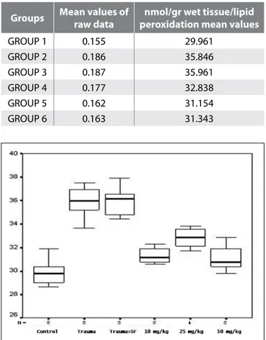
There was no significant difference between group 1 and group 6, and the treatment groups of 4 and 5 in the statistical examination of biochemical values (p>0.05) (Table II).
However, there was a significant difference between group 2 and 3 as well as groups 4, 5 and 6 (p<0.05). In other words, EGCG administered in daily consumption and in therapeutic doses decreased the level of lipid peroxidation in a statistically significant manner (Figure 1).
Histological Results light microscopy results:
The samples were harvested from rats in all groups at traumatized area. In Group 1, Schwann cells, and myelinated and non-myelinated nerve fibers were evaluated. Duplication and undulation of the myelin sheath, axonal
Table I: Histological Grading Score of Nerve Injury
histology
Score grade
0 Normal
1 Duplication of the myelin sheath 2 Undulation of the myelin sheath
3 Axonal degenerations (axonal withdrawal) 4 Nerve fibers with severe degeneration
Table II: Distribution of Biochemical Values by Groups
groups mean values of raw data peroxidation mean valuesnmol/gr wet tissue/lipid
GROUP 1 0.155 29.961 GROUP 2 0.186 35.846 GROUP 3 0.187 35.961 GROUP 4 0.177 32.838 GROUP 5 0.162 31.154 GROUP 6 0.163 31.343
Figure 1: Multiple Comparison of Lipid Peroxidation Values
between the groups Trauma, trauma + saline > control, 10mg/kg, 25 mg/kg, 50 mg/kg respectively.
withdrawal of myelinated nerve fibers, Schwann cell hypertrophy, and widespread tissue edema were detected in Group 2 (Figure 2A, B). Group 3 had similar results to group 2. In trauma groups was seen that qualitatively, siatic nerve injury consisted of Wallerian degeneration characterized by variable degree of degeneration in great myelinated axons and endoneural edema. Group 4 and Group 6 showed withdrawal of myelin sheath, duplication of myelin sheath, and undulation especially in great myelinated nerve fibers, as in the trauma group. Although these groups also had Schwann cell hypertrophy, edema in myelinated nerve fibers was not observed in these groups. Group 5 showed decreased
Figure 2: A) Sciatic nerve semi-thin light microscopy sections of normal structured myelinated nerve fibers (4) as well as hypertrophic
Schwann cells (à), myelinated sheath duplication (:) and undulation (è), withdrawal of axonal structures on some myelinated fibers
(8), and generalized edema throughout the tissue (+) is seen (Toluidine Blue x400). B) Figure of the same group showing normal
structured myelinated nerve fibers (4), duplication in myelinated sheath (:) and undulation (è), withdrawal of axonal structures on some myelinated fibers (8), and edema of nerve fibers (+) is seen (Toluidine Blue x400).
Figure 3: A) The sciatic nerve semi-thin light microscopy sections in the myelinated nerve fibers showing a decrease of the duplication (:) and undilated appearance (è) but the myelin sheath is sheltering the axon in some areas (u) and forming blebs (´) through axoplasm
(Toluidine Blue x400). B) Semi-thin sections of the same group showing the normal observed structure of myelinated nerve fibers (4), myelinated sheath duplication (:), undulated appearance (è) and intense degeneration (u) of axonal structures (Toluidine Blue x400).
duplication in myelinated nerve fibers, undulated appearance and Schwann cell hypertrophy, which were observed in the trauma group (Group 2) (Figure 3A, B).
Light microscopy observations showed that based on the distribution of groups related to histopathological changes of myelinated fibers, multiple comparison procedure indicated a statistically significant decrease in myelin sheath separation, myelin sheath undulation and axonal degeneration in Group 4 and Group 6 compared to Group 2 and Group 3 (p<0.05). In Group 5, intensive degeneration in myelin and axons were found to be significantly higher compared to other groups (p<0.05).
A B
fibers with small diameter, whereas great myelinated nerve fibers showed myelin sheath separation, axon withdrawal, and swelling and crystolysis in the mitochondria. The cells were hypertrophic. Myelinated nerve fibers were in normal structure through the tissue. Edema, which was observed in trauma groups, was not detected in endoneurium (Figure 6A, B).
Finally, application of the appropriate doses of catechin was observed to suppress endoneurial edema and increase of collagen fibers and wallerian degenerations (Table III) caused by trauma. Although catechin was observed to reverse undulation and myelin duplication in myelinated nerve fibers, it was considered insufficient to prevent prominent degeneration in myelinated nerve fibers with great diameter. B- electron microscopy results:
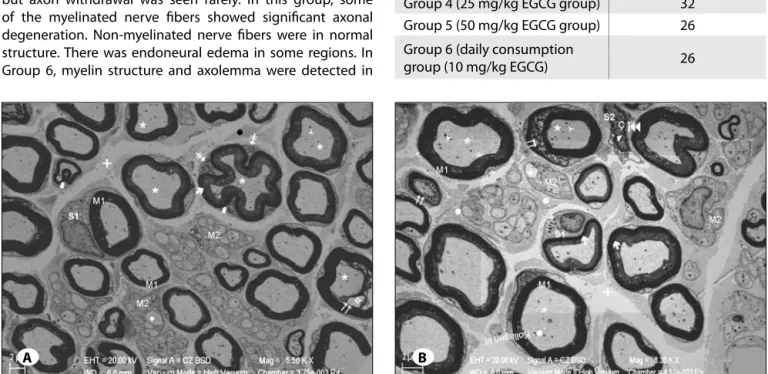
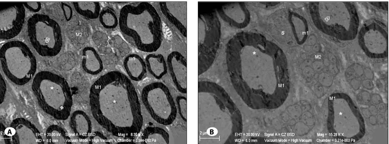
In Group 1, the general structure was considered normal. The endoneural edema and fibrosis, and an increase in type 3 col-lagen fibers were detected in Group 2. Schwann cells were determined to be hypertrophic. This study showed that there was an increase in lysosomes, expansion in the tubulus sys-tem of granular endoplasmic reticulum (GER), mitochondria with dense matrix, and chromatin condensation and dual-membrane separation in the nucleus in Schwann cell cyto-plasm. Great myelinated nerve fiber undulation, separation of myelin sheath, and axon withdrawal were detected. Also, the mitochondria with dense matrix were increased in Schwann cell cytoplasm, which comprises the outer boundary of the myelin sheath in great myelinated cells (Figure 4A, B). The findings of Group 3 were similar to that of trauma group. In Group 4, small myelinated nerve fibers were in normal morphology and structure. Myelin separation and rarely axon withdrawal were observed in great fibers. Non-myelinated nerve fibers were in normal structure. There was no endoneural edema in this group contrary to other groups. Schwann cells were recognized with their normal structure and organelle content (Figure 5A,B). In Group 5 small myelinated nerve fibers had normal structure. Some of the fibres with great diameter showed myelin separation up to duplication, and undulation, but axon withdrawal was seen rarely. In this group, some of the myelinated nerve fibers showed significant axonal degeneration. Non-myelinated nerve fibers were in normal structure. There was endoneural edema in some regions. In Group 6, myelin structure and axolemma were detected in
Table III: Distribution of Wallerian Degenerations Measurement
in All Groups
groups Axons with wallerian The number of
degenerations
Group 1 (control group) 7
Group 2 (trauma group) 21
Group 3 (saline group) 25
Group 4 (25 mg/kg EGCG group) 32
Group 5 (50 mg/kg EGCG group) 26
Group 6 (daily consumption
group (10 mg/kg EGCG) 26
Figure 4: A) Electron microscopy of the Trauma Group sciatic nevre. m1: Myelinated nerve fiber, ´: Myelinated nerve fiber with undulation, è: Decrease of the myelinated nerve fiber electron density before separation, m2: Unmyelinated nerve fibers, u: Unmyelinated nerve
fiber with degeneration, S1: Hypertrophic Schwann cell, /: Mitochondria with dense matrix inside Schwann Cell cytoplasm located on the border of myelin sheath, I: Separation of myelin sheath, *: Axoneme, ñ: Mitochondria with dense matrix inside the axoneme, v: Axon withdrawal, +: Endoneurium edema, î: Collagen fiber. B) Large magnification electron microscopy images of the trauma group sciatic nerve. m1: Myelinated nerve fiber, è: Decrease of the myelinated nevre fiber electron density before separation, m2: Unmyelinated nerve fibers, u: Unmyelinated nerve fiber with degeneration, S1: Hypertrophic Schwann Cell, l: Lysosom, †: Mitochondria with dense matrix inside Schwann Cell cytoplasm, S2: Schwann Cell with Nucleus (Ç) like deformation, 4: Chromatin condensation, :: Separations in the nuclear membrane, I: Separation of myelin sheath up to duplication, +: Endoneurium edema, î: Collagen fiber, *: Axoneme, ñ: Mitochondria with dense matrix inside the axoneme, Ö: Mitochondrion in the axoneme with cristolysis (Uranyl Acetate & Lead Citrate).
degeneration was detected particularly in axon and myelin sheath with the treatment dose of 50mg/kg.
Injury to peripheral nerves leads to microvascular damage, which can cause to endoneural edema and promote the increase of endoneural fluid pressure. The removal of compression lead to the resumption of blood flow into nerve cells which not only provide nutrients, including oxygen, but also increases the formation of free oxygen radicals and lipid peroxidation, and this process is called reperfusion injury (24). The central and peripheral nervous systems are rich in dISCuSSIon
If the compression pressure on peripheral nerves is higher than capillary perfusion pressure, oxidative stress-induced cell impairment develops after ischemia (9, 24). Antioxidants have potentials to counter the tissue-damaging effect of the inflammatory response. In our study, EGCG, a polyphenol with antioxidant properties, possessed substantial antioxidant activity measured by several biochemical assays in a rat sciatic nerve damage model at daily consumption dose (10mg/kg) and treatment doses (25mg/kg and 50mg/kg), but dense
Figure 5: A) m1: Large diameter myelinated nerve fiber, m1: Small diameter myelinated nerve fiber, m2: unmyelinated nerve fibers, *:
Axoneme, Ö: Mitochondrion with cristolysis in the axoneme, v: Axon withdrawal. B) m1: Large diameter myelinated nerve fiber m1: Small diameter myelinated nerve fiber, m2: Unmyelinated nerve fibers, *: Axoneme, Ö: Mitochondrion in the axoneme with cristolysis,
S: Schwan Cell (Uranyl Acetate & Lead Citrate).
Figure 6: A) m1: Large diameter myelinated nerve fiber, m1: Small diameter myelinated nerve fiber, B) Myelin blebs extending through
axoneme, m2: Unmyelinated nerve fibers, u: Unmyelinated nerve fiber with degeneration, S1: Hypertrophic Schwann Cell, †: Swollen mitochondria with cristolysis inside the Schwann Cell cytoplasm, *: Axoneme, ñ: Mitochondria with cristolysis inside the axoneme, v: Axon withdrawal, +: Endoneurium edema. B) m1: Myelinated nerve fiber, S1: Hypertrophic Schwan Cell, Ç: Nucleus of the Schwann Cell, †: Swollen mitochondria with cristolysis inside the Schwann Cell cytoplasm, *: Axoneme, Ö: Mitochondrion with cristolysis in the Schwann Cell cytoplasm, li: Lipid droplet, geR: Network of rough endoplasmic reticulum, v: Axon withdrawal, +: Endoneurium edema (Uranyl Acetate & Lead Citrate).
A B
EGCG attenuates secondary ischemic damage that follows peripheral nerve injury as it was shown by Sutherland et al. that EGCG also reduces neurodegeneration associated with cerebral ischemic process.
Detailed histopathological examination including myelin sheath separation, ondulation of the myelin sheath, axonal degeneration, and intensive degeneration in myelins and axons were performed. Particularly these values in Group 4 and 6 were found to be lower as compared to that of Group 2 and 3. Intensive degeneration in myelins and axons detected in the treatment group, Group 5, indicated to increased degeneration. As a matter of fact, Sutherland et al. reported that the effective dose of EGCG for ischemia protection and early treatment of cerebral ischemia was 25-50 mg/kg, however, since EGCG given at this dose needs to cross the blood-brain barrier to be effective, lower doses of EGCG may also have both therapeutic and toxic effects in a dose-dependent manner, in well vascularized tissues such as peripheral nerves.
ConCluSIon
Our study showed that EGCG, might be effective in the prevention and treatment of secondary damage related to peripheral nerve injury in daily consumption and low-dose treatment groups. Nevertheless it might induce degenerative process particularly when given at a high dose which necessitates dose dependent further investigations. Despite the absence of concrete correlation of histological recovery and neurological findings, the present study demonstrated that EGCG might have inhibitory effects on neurodegeneration and potentially neuroprotective which might be a critical guide to future studies in this field.
ReFeRenCeS
1. Baek WK, Jang BC, Lim JH, Kwon TK, Lee HY, Cho CH: Inhibitory modulation of ATP-sensitive potassium channels by gallate-ester moiety of (-)-epigallocatechin-3-gallate. Biochem Pharmacol 70: 1560-1567, 2005
2. Bastianetto S, Yao ZX, Papadopoulos V, Quirion R: Neuroprotective effects of green and black teas and their catechin gallate esters against beta-amyloid induced toxicity. Eur J Neurosci 23: 55-64, 2006
3. Brown JE, Khodr H, Hider RC, Rice-Evans CA: Structural dependence of flavonoid interactions with Cu2_ ions: Implications for their antioxidant properties. Biochem J 330: 1173–1178, 1998
4. Cadenas E, Packer L: Handbook of antioxidants. New York: Marcel Dekker İnc, 1996
5. Chaturvedi RK, Shukla S, Seth K, Chauhan S, Sinha C, Shukla Y, Agrawal AK: Neuroprotective and neurorescue effect of black tea extract in 6- hydroxydopamine-lesioned rat model of Parkinson’s disease. Neurobiol Dis 22: 421-434, 2006 6. Cheeseman Kh, Slater TF: An introduction to free radical
biochemistry. British Med Bulletin 149: 481-493, 1993 7. Dalluge JJ, Nelson BC: Determination of tea catechins. J
Chromatogr A 881: 411-424, 2000
myelin, a substance rich in lipids. Release of oxygen-free radicals activated by ischemic-reperfusion injury triggers lipid peroxidation and accelerates tissue damage (34). The products of lipid peroxidation can further damage membrane proteins, including membrane-bound receptors and enzymes by increasing membrane permeability for ions (37). Damage to cell membranes causes ion imbalances, allows extracellular calcium to enter the cell, and leads to edema and necrosis. Free radicals induce oxidative stress, which is balanced by the body’s endogenous enzymatic and nonenzymatic antioxidant systems. The most efficient enzymatic antioxidants involve glutathione peroxidase, catalase and superoxide dismutase, whereas nonenzymatic antioxidants include vitamins and minerals. The reaction involves the catechin losing a hydrogen atom to a reactive free radical. As chain-breaking antioxidants, tea catechins are thought to interrupt deleterious oxidation reactions (12). Similarly, catechins chelate metal ions such as copper and iron to form inactive complexes and prevent the generation of potentially damaging free radicals (3). Also, catechins have been found to inhibit lipoxygenase and cyclooxygenase activity, enzymes which are capable of increasing oxidative stress or damage in some tissues (18, 21). Catechin appeared to limit neuronal damage. It was shown that 6-hydroxydopamine-induced nuclear factor-kappaB activation and cell death was attenuated by tea extracts in neuronal cultures (23). EGCG, helps to prevent ethanol-associated apoptosis in fetal rhombencephalic neurons, and presents anti-apoptotic effect on liver (22, 32).
Sutherland et al. found in their study that catechins’ actions of attenuating oxidative stress and the inflammatory response may account for their confirmed neuroprotective capabilities following cerebral ischemia. It was found that EGCG had a dose-dependent response with 25 and 50 mg/ kg eliciting significant neuroprotection (36). Rahman et al. reported that EGCG might be an appropriate intervention for the treatment of acute cerebral ischemia (32). Chaturvedi and colleagues said that black tea extract might slow progression of Parkinson disease, a chronic, progressive degenerative disorder (5). Bastianetto et al. showed the protective effect of catechin derivatives in black tea on amyloid-dependent neurotoxicity (2).
Experimental research with mice has shown that EGCG causes severe hepatic necrosis and 67% mortality when given daily at 50mg/kg ip. In fact, clinical preparations of tea extracts have also exhibited hepatotoxic effect (39). In contrast, there is evidence that purified green tea extracts in vivo are hepatoprotective. These results suggest that the route and method of administration may determine whether catechins induce hepatotoxicity or have hepatoprotective effects (39). In the present study, comparison of biochemical values in tissue samples demonstrated that lipid peroxidation levels in treatment and protection groups, namely Groups 4, 5 and 6, were lower at a statistically significant level than that of Group 2 and 3, but were close to the values of Group 1.
25. Lundborg G, Dahlin LB: Anatomy, function, and pathophysi-ology of peripheral nerves and nerve compression. Hand Clin 12: 185-193, 1996
26. Macchi MM, Bruce JN: Human pineal physiology and functional significance of melatonin. Front Neuroendocrinol 25: 177-195, 2004
27. Mendoza-Wilson AM, Glossman-Mitnik D: Theoretical study of the molecular properties and chemical reactivity of (+)-catechin and (-)-epicatechin related to their antioxidant ability. J Mol Struct Theocem 761: 97-106, 2006
28. Nwuha V, Nakajima M, Tong J, Ichikawa S: Solubility study of green tea extracts in pure solvents and edible oils. J Food Eng 40: 161-165, 1999
29. Paquay JB, Haenen GR, Stender G, Wiseman SA, Tijburg LB, Bast A: Protection against nitric oxide toxicity by tea. J Agric Food Chem 48: 5768–5772, 2000
30. Persengiev S, Kanchev L, Vezenkova G: Circadian patterns of melatonin, corticosterone, and progesterone in male rats subjected to chronic stress: Effect of constant illumination. J Pineal Res 11: 57-62, 1991
31. Rabinovsky ED: The multifunctional role of IGF-1 in peripheral nerve regeneration. Neurol Res 26: 204-210, 2004
32. Rahman RM, Nair SM, Helps SC, Shaw OM, Sims NR, Rosengren RJ: Epigallocatechin gallate as an intervention for the acute treatment of cerebral ischemia. Neurosci Lett 382: 227–230, 2005
33. Ramon Y, Cajal S: Degeneration and regeneration of the nervous system. London: Oxford University Press, 1928 34. Schmelzer JD, Zochodne DW, Low PA: Ischemic and
reperfusion injury of rat peripheral nerve. Proc Natl Acad Sci 86: 1639-1642, 1989
35. Seddon HJ: Three types of nerve injury. Brain 66: 237-288, 1943
36. Sutherland BA, Rahman RMA, Appleton I: Mechanisms of action of gren tea catechins, with a focus on ischemia-induced neurodegeneration. J Nutr Biochem 17: 291-306, 2006
37. Tang SZ, Kerry JP, Sheehan D, Buckley DJ, Morrissey PA: Antioxidative effect of dietary tea catechins on lipid oxidation of long term frozen chicken meat Science 57: 331-336, 2001 38. Van der Zee CE, Brakkee JH, Gispen WH: Putative neurotrophic
factors and functional recovery from peripheral nerve damage in the rat. Br J Pharmacol 103: 1041-1046, 1991 39. Xuczaj W, Skrzydlewska E: Antioxidative properties of black
tea. Prev Med 40: 910-918, 2005
40. Yilmaz Y: Novel uses of catechins in foods. Trends in Food Sci Technol 17: 64-71, 2006
41. Zaveri NT: Green tea and its polyphenolic catechins: Medicinal uses in cancer and noncancer applications. Life Sci 78: 2073-2080, 2006
42. Zhang YL, Zhang PB, Qiu SD, Liu Y, Tian YF, Wang Y: Effects of ketamine-midazolam anesthesia on the expression of NMDA and AMPA receptor subunit in the peri-infarction of rat brain. Chin Med J 119: 1555-1562, 2006
8. Doss MX, Potta SP, Hescheler J, Sachinidis A: Trapping of growth factors by catechins: A possible therapeutical target for prevention of proliferative diseases. J Nutr Biochem 16: 259-266, 2005
9. Fern R, Harrison PJ: The contribution of ischaemia and deformation to the conduction block generated by compression of the cat sciatic nerve. Exp Physiol 79: 583-592, 1994
10. Frei B, Higdon JV: Antioxidant activity of tea polyphenols in vivo: Evidence from animal studies. J Nutr 133: 3275– 3284, 2003
11. Fu SY, Gordon T: The cellular and molecular basis of peripheral nerve regeneration. Mol Neurobiol 14: 67-116, 1997
12. Gramza A, Korczak J: Tea constituents (Camellia sinensis L.) as antioxidants inlipid systems. Trends in Food Sci Technol 16: 351-358, 2005
13. Guo Q, Zhao B, Shen S, Hou J, Hu J, Xin W: ESR study on the structure-antioxidant activity relationship of tea catechins and their epimers. Biochim Biophys Acta 1427: 13–23, 1999 14. Gupta S, Saha B,Giri AK: Comparative antimutagenic and
anticlatogenic effect of green tea and black tea: A review. Mutation research 512: 37-65, 2002
15. Gutteridge JMC: Lipid peroxidation and antioxidants as biomarkers of tissue damage. Clin Chem 41: 1819-1828, 1995 16. Haenen GR, Paquay JB, Korthouwer RE, Bast A: Peroxynitrite
scavenging by flavonoids. Biochem Biophys Res Commun 236: 591–593, 1997
17. Hall ED, Braughler M: Effects of intravenous methylpredniso-lone on spinal cord lipid peroxidation and Na-K ATPase activ-ity. J Neurosurg 57: 247- 253, 1982
18. Hong J, Smith TJ, Ho CT, August DA, Yang CS: Effects of purified gren and black tea polyphenols on cyclooxygenase- and lipoxygenase-dependent metabolism of arachidonic acid in human colon mucosa and colon tumor tissues. Biochem Pharmacol 62: 1175–1183, 2001
19. Igarashı T, Yabuki S, Kıkuchı S, Myers RR: Effect of acute nerve root compression on endoneurial fluid pressure and blood flow in rat dorsal root ganglia. J Orthop Res 23: 420-424, 2005 20. Kanje M, Skottner A, Lundborg G: Effects of growth hormone
treatment on the regeneration of rat sciatic nerve. Brain Res 475: 254-258, 1998
21. Katiyar SK, Mukhtar H: Inhibition of phorbol ester tumor promoter 12- tetradecanoylphorbol- 13-acetate-caused inflammatory responses in SENCAR mouse skin by black tea polyphenols. Carcinogenesis 18: 1911–1916, 1997
22. Katunuma N, Ohashi A, Sano E, Ishimaru N, Hayashi Y, Murata E: Catechin derivatives: Specific inhibitor for caspases-3, 7 and 2, and the prevention of apoptosis at the cell and animal levels. FEBS Lett 580: 741-746, 2006
23. Levites Y, Youdim MB, Maor G, Mandel S: Attenuation of 6- hydroxydopamine (6-OHDA)-induced nuclear factor-kappaB (NF-kappaB) activation and cell death by tea extracts in neuronal cultures. Biochem Pharmacol 63: 21-29, 2002 24. Li Y, Bickel KD, Im MJ, Hu L, Dellon AL, Vander Kolk CA, Manson
PN: Effects of deferoxamine on ischemia/reperfusion injury after peripheral nerve compression. Ann Plast Surg 36: 365-369, 1996