139
Clinical Article
Introduction
Chromosome abnormalities are one of the main reasons for congenital defects. The prevalence of chromosomal abnormal-ities in clinically recognized early pregnancy loss is greater than 50%.1The overall aneuploidy rate is 6-11% for all still-births and neonatal deaths.1The incidence of chromosomal ab-normalities is about 0.65% in all live newborns.1Prenatal cy-togenetic diagnosis is crucial for pregnant women having high-risk indicators. By the help of the improvements in prenatal di-agnosis, the evaluation of each woman's risk having a child with chromosomal abnormalities takes place an essential role in obstetric care. Screening for chromosomal abnormalities in-cludes maternal age, ultrasonographic evaluation during the first and second trimester, and serum screening tests. The risk
of aneuploidies increases with advanced maternal age, but nowadays screening by maternal age alone is insufficient, so widespread screening with biochemical and ultrasonographic evaluation has been used. Both second trimester prenatal screening by using biochemical parameters (alpha-fetoprotein and free fraction of human chorionic gonadotropin measure-ments in maternal serum) and maternal age reach a detection rate of 60-80% for Down syndrome.2In recent years, the im-provements in ultrasonographic evaluation help the develop-ment of first trimester screening. By the way the detection rates for Down syndrome reach 90%.2Fetuses with chromosomal abnormalities may have somatic abnormalities that are de-tected by ultrasonographic evaluation. If an aneuploidy is sus-pected in ultrasonographic evaluation, prenatal cytogenetic analysis can provide a definitive diagnosis. Chorionic villus sampling, amniocentesis and umbilical blood sampling during pregnancy are all reliable methods for prenatal diagnosis.
In our country, the demand for prenatal tests and genetic counseling has increased markedly, because of the increase in the pregnancies in aged 35 year and older. We analyzed retro-spectively the cytogenetic results of over thirteen thousand (13.466) AS (amniocentesis), CVS (chorionic villus sam-pling), PUSB (percutaneous umbilical blood samsam-pling), aborted material samples to investigate the changes in the
dis-Prenatal Cytogenetic Findings in 13.466 Cases of High-Risk Pregnant
Women in One Laboratory
Leyla ÖZER1, Evrim ÜNSAL2, Özge AYVAZ2, Aşkın ŞEN3, Volkan BALTACI2
Ankara, Turkey
OBJECTIVE: This study was aimed to evaluate cytogenetic findings of high risk pregnancies according to indications.
STUDY DESIGN: Between years 2001 and 2009, a large series of 13466 pregnant women with various high-risk factors were referred to our genetic laboratory for prenatal genetic diagnosis. 12.124 amnio-centesis, 212 chorionic villus sampling (CVS), 173 percutaneous umbilical blood sampling (PUBS) sam-ples and 809 fetal and placental tissue samsam-ples (from aborted or from stillbirth fetuses) were collected. All of the cytogenetic findings were assessed retrospectively. We compared the cytogenetic results in the distribution of indication groups.
RESULTS: Among all indications advanced maternal age was the most common indication. Chromosomal abnormalities were observed in 1029/13406 cases (7.6%). Trisomy 21 was the most com-mon chromosomal abnormality found in 228/1029 cases (22.2%). Of sex chromosomal abnormalities, monosomies were the most common abnormality (3.3%). Of structural rearrangements translocations were the most common abnormality (2.3%). Balanced chromosome rearrangement carriers had the highest percentage of pregnancies with abnormal chromosomes.
CONCLUSION: This study presents the largest series of cytogenetic findings on prenatal samples per-formed in Turkey. Analysis of samples of high risk pregnancies could provide an important database for prenatal genetic counseling and obstetric management for each indication groups.
Key Words: Prenatal diagnosis, High-risk pregnancy, Chromosomal abnormalities, Cytogenetic, Fetal anomalies, Maternal age
1Mikrogen Genetic Diagnosis Center, Ankara
2Bilim University School of Medicine Division of Medical Biology and Genetics, İstanbul
3Ufuk University School of Medicine Division of Medical Genetics, Ankara
Address of Correspondence: Leyla Özer
Mikrogen Genetic Diagnosis Center Tunalı Hilmi Caddesi 98/1 Kavaklıdere Ankara
[email protected] Submitted for Publication: 15. 07. 2013
Accepted for Publication: 26. 08. 2013
Obstetrics; Maternal-Fetal Medicine and Perinatology
tribution of indications, maternal age and cytogenetic findings and the rate of abnormalities according to indications.
Material and Method
Subjects
Between years 2001 and 2009, 13.466 pregnant women with various high-risk factors were referred to our genetic lab-oratory for prenatal genetic diagnosis. Among those patients 12.124 underwent amniocentesis, 212 underwent chorionic villus sampling (CVS), 173 underwent percutaneous umbilical blood sampling (PUBS) and in 809 cases fetal and placental tissue samples were collected from aborted fetuses or from stillbirth fetuses. All of the findings of cytogenetic analysis were assessed retrospectively. The classification of the pa-tients according to age groups was given in table 1.
Table 1: Classification of the patients to age groups.
Age groups N (%) 20 ages 189 (1.4%) 21-25 ages 1536 (11.4%) 26-30 ages 2559 (19%) 31-35 ages 3502 (26%) 36-40 ages 4700 (34.9%) 40 ages 970 (7.2%) Total 13.466
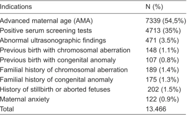
Prenatal sampling criteria were as follows: advanced ma-ternal age (AMA), positive serum screening tests, abnormal ultrasonographic findings, previous birth with chromosomal aberration, previous birth with congenital anomaly, familial history with chromosomal aberration, familial history with congenital anomaly, history of stillbirth or aborted fetuses and maternal anxiety of an anomalous fetus. Table 2 lists the re-ferral indications.
Table 2: Indications for prenatal diagnosis
Indications N (%)
Advanced maternal age (AMA) 7339 (54,5%) Positive serum screening tests 4713 (35%) Abnormal ultrasonographic findings 471 (3.5%) Previous birth with chromosomal aberration 148 (1.1%) Previous birth with congenital anomaly 107 (0.8%) Familial history of chromosomal aberration 189 (1.4%) Familial history of congenital anomaly 175 (1.3%) History of stillbirth or aborted fetuses 202 (1.5%)
Maternal anxiety 122 (0.9%)
Total 13.466
Signed informed consents were obtained from each pa-tient. The study was approved by the local Ethic committee.
Ultrasonographic Findings
All pregnant women underwent ultrasound examination by
referring clinics due to routine obstetric follow-up. Abnormal ultrasonographic findings of patients were cardiac defects, central nervous system abnormalities, cystic higroma, non-im-mune hydrops, urogenital anomalies, abdominal wall defects, hand-foot (extremity) anomalies, intrauterine growth retarda-tion (IUGR), polyhydramnios/oligohydramnios, gastrointesti-nal anomalies, single umbilical artery, increased nuchal translucency, nasal bone hypoplasia, choroid plexus cyst and echogenic intra-cardiac focus.
Cytogenetic analysis
Chorionic villus sampling was performed between 10 and 12 weeks of gestation. The cells were cultured in culture media (Biological Industries) at 37 C0and 5% CO2up to 6-8 colonies were obtained. Cell growth was monitored every day. The cells were collected when multiple clones with numerous metaphase cells were observed using an inverted microscope. G banding was performed and karyotypes were analyzed according to ISCN standards. Amniocentesis was performed between 16 and 20 weeks of gestation. The cells were cultured in culture media (Biological Industries) and growth in incubator for 10-15 days at 37 C0and 5% CO2. Then cells were treated using the same procedure as described for CVS chromosomal samples were prepared following the same procedures used for CVS and AS. Fetal blood was collected between 18 and 24 weeks of gestation. The blood sample was cultured for 72 hours at 37 C0. The cells were harvested and chromosomal samples were pre-pared following the same procedures used for CVS and AS. Tissue biopsy samples from stillbirths or aborted fetuses were cultured as described for CVS.
Statistical analysis
Statistical analysis was performed by using SPSS for Windows 11.5. Nominal variables were analyzed by Pearson’s Chi-square or Fisher’s Chi-square test. p<0.05 was considered to indicate statistical significance.
Results
1. Age distribution
Findings from 13.466 pregnant women with various high-risk factors were analyzed over an 8-year period between 2001 and 2009. The age distribution of the 13.466 patients was de-termined as follows; 34.9% were between 36 and 40 years, 26% were between 31 and 35 years, 19% were between 26 and 30 years, 11.4% were between 21 and 25 years, 7.2% were over 40 years and 1.4%were less than 20 years (Table 1).
2. Clinical Indications
The most common indication for prenatal diagnosis was advanced maternal age (54.5%), followed by positive serum screening tests(35%), abnormal ultrasound findings (3.5%) (Table 2). The frequency of chromosomal abnormalities was higher in patients with advanced maternal age (49.1%) and positive serum screening tests (20%) (Table 3, 4)
3. Cytogenetic Findings
The success rate of cytogenetic analyses was 99.3% (13.466/13.406). Chromosomal abnormalities were observed in 7.6% of the analyzed cases (1029/13.406). Among these, the most common chromosomal abnormality was autosomal tri-somies with a frequency of 42.4% (436/1029). Numerical sex chromosomal abnormalities were found in 6.2%, (64/1029), unbalanced structural rearrangements were found in 4.8% (49/1029) and balanced structural rearrangements were found in 16.6% of the cases (171/1029) (Table 5). The majority of chromosomal abnormalities were autosomal ones. In cases with sex chromosomal abnormalities; monosomies were the most common abnormalities with a frequency of 3.3% (34/1029). In cases with unbalanced structural rearrangements translocations were the most common abnormalities that were found in 2.3% (23/1029) of cases. In cases with balanced re-arrangements inversions were the most common abnormalities with a frequency of 6.9%(71/1029). The frequency of chro-mosomal abnormalities and the classification of chrochro-mosomal abnormalities are shown in Table 6. Among all abnormalities Trisomy 21 was the most common (22.2%, 228/1029) (Table 7). In cases with sex chromosomal abnormalities; monosomies were the most common abnormalities with a frequency of 3.3% (34/1029). In cases with unbalanced structural rearrange-ments translocations were the most common abnormalities that
were found in 2.3% (23/1029) of cases. In cases with balanced rearrangements inversions were the most common abnormali-ties with a frequency of 6.9%(71/1029). The frequency of chromosomal abnormalities and the classification of chromo-somal abnormalities are shown in Table 6.
Table 5: Chromosome abnormalities frequencies
Chromosomal abnormality N (%) Numerical Autosomal 436 (42.4) Trisomies 375 (36.4) Ploidies 42 (4.1) Mosaicism 15 (1.5) Trisomies + Ploidies 2 (0.2) Monosomies 2 (0.2)
Numerical Sex Chromosome 64 (6.2)
Monosomies 40 (3.9)
Trisomies 13 (1.3)
Mosaicism 11 (1.1)
Unbalanced structural rearrangements 49 (4.8)
Translocations 24 (2.3) Other* 11 (1.1) Marker chromosome 5 (0.5) Mosaicism 5 (0.5) Deletion 2 (0.2) Isochromosome 2 (0.2)
Table 3: The relation between chromosomal abnormality and clinical indications
Variables Sensitivity Specificity PPV NPV Accuracy
Advanced maternal age 49.1% 45.7% 5.9% 92.9% 45.9%
Positive serum screening tests 20.0% 64.6% 4.5% 90.6% 61.1%
Abnormal ultrasonographic findings 3.8% 97.1% 9.8% 92.4% 89.9%
Previous birth with chromosomal aberration 0.3% 99.2% 3.1% 92.3% 91.6%
Previous birth with congenital anomaly 0.3% 99.6% 6.3% 92.3% 92.0%
Familial history of chromosomal aberration 8.1% 99.8% 74.8% 92.9% 92.7%
Familial history of congenital anomaly 0.5% 99.3% 5.4% 92.3% 91.7%
History of stillbirth or aborted fetuses 2.2% 99.3% 20.5% 92.4% 91.8%
Maternal anxiety 0.2% 99.5% 3.2% 92.3% 91.9%
PPV: Positive predictive value, NPV: Negative predictive palue
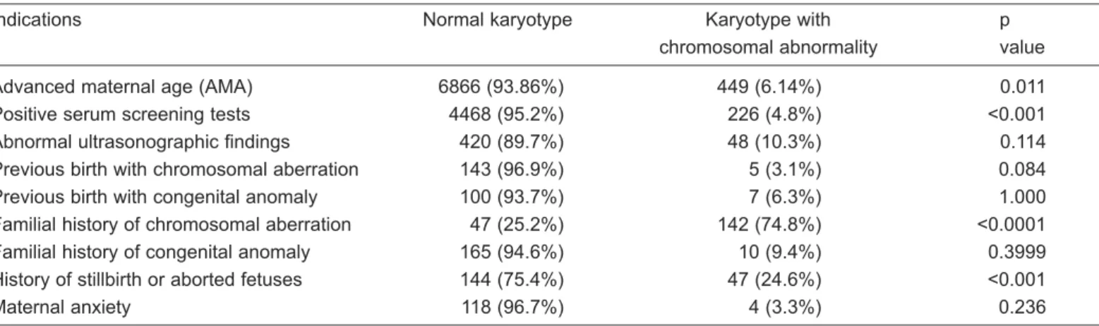
Table 4: Chromosomal abnormality and clinical indications
Indications Normal karyotype Karyotype with p
chromosomal abnormality value
Advanced maternal age (AMA) 6866 (93.86%) 449 (6.14%) 0.011
Positive serum screening tests 4468 (95.2%) 226 (4.8%) <0.001
Abnormal ultrasonographic findings 420 (89.7%) 48 (10.3%) 0.114
Previous birth with chromosomal aberration 143 (96.9%) 5 (3.1%) 0.084
Previous birth with congenital anomaly 100 (93.7%) 7 (6.3%) 1.000
Familial history of chromosomal aberration 47 (25.2%) 142 (74.8%) <0.0001 Familial history of congenital anomaly 165 (94.6%) 10 (9.4%) 0.3999 History of stillbirth or aborted fetuses 144 (75.4%) 47 (24.6%) <0.001
Balanced structural rearrangements 171 (16.6) Inversion 71 (6.9) Robertsonian translocation 45 (4.4) Other** 30 (2.9) Reciprocal translocation 22 (2) Mosaicism 0
*:add(1), add(6), add(11), add(15), add(21), add(22), der(1)(t)(1;?), d er(18)t(18;?), der(22)(?;22)
**: 1qh(+), 9qh(+), 13p(-), 15ps+, 15ps-, 16qh+, 21ps+,Yqh(-)
Table 6: Chromosomal abnormalities of each sample; AS,CVS, PUSB, and Abort material
Chromosomal abnormalities
Numerical abnormalities AS CVS PUSB Abort material Trisomies 241 23 5 107 Ploidies 10 1 31 Mosaicism 12 1 1 7 Trisomies+Ploidies 2 Monosomies 1 3
Numerical Sex Chromosome abnormalities
Monosomies 9 1 31
Trisomies 12 6
Mosaicism 18 1 1 4
Unbalanced structural rearrangements
Translocation 11 22 Marker chromosome 5 1 Mosaicism 2 Deletion 1 1 Isochromosome 1 Other 16 1 2
Balanced structural rearrangements
Robertsonian translocations 20 2 Reciprocal translocations 42 1 2
Mosaicism 2 1 6
Inversion 60
Other 30 1
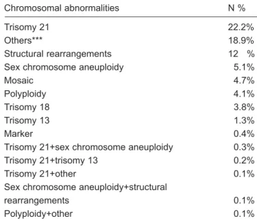
Table 7: Classification of the most common chromosomal ab-normalities according to frequency
Chromosomal abnormalities N %
Trisomy 21 22.2%
Others*** 18.9%
Structural rearrangements 12 %
Sex chromosome aneuploidy 5.1%
Mosaic 4.7%
Polyploidy 4.1%
Trisomy 18 3.8%
Trisomy 13 1.3%
Marker 0.4%
Trisomy 21+sex chromosome aneuploidy 0.3%
Trisomy 21+trisomy 13 0.2%
Trisomy 21+other 0.1%
Sex chromosome aneuploidy+structural
rearrangements 0.1%
Polyploidy+other 0.1%
***: Trisomies of other chromosomes except chromosome 13,18,21, monosomies of autosomal chromosomes, inv(9)(p12q13), additions, 1qh(+), 9qh(+), 13p(-), 15ps+, 15ps-, 16qh+, 21ps+,Yqh(-).
4. Frequency of chromosomal abnormalities by referral indications
Among all the patients cases with advanced maternal age (49.1%) resulted in a chromosomal abnormality, which con-stitutes the group with the highest frequency of the anomaly detected. Among all patients the cases with positive serum screening, 20%, with a familial history of chromosomal aber-ration 8.1%, with an abnormal ultrasound finding 3.8%, with a history of stillbirth or aborted fetuses 2.2%, with a familial history of congenital anomaly 0.5%, with a previous birth with chromosomal aberration 0.3% , previous birth with congenital anomaly 0.3% and maternal anxiety 0,2% were found to have a chromosomal abnormality (Table 4). Familial history of chromosomal aberration has the highest PPV(positive predic-stive value) (Table 4). Familial history of chromosomal aber-ration was highly significant with chromosomal abnormalities (Table 8).
Table 8: Frequency of chromosomal abnormalities by referral indications
Indications Numerical Numerical sex Balanced Unbalanced
autosomal chromosome structural structural
Advanced maternal age (AMA) 173 15 62 5
Positive serum screening tests 94 10 38 7
Abnormal ultrasonographic findings 26 7 3 1
Previous birth with chromosomal aberration 3
Previous birth with congenital anomaly 1 6
Familial history of chromosomal aberration 22 86 7
History of stillbirth or aborted fetuses 11 3 3
A significant correlation between advanced maternal age, positive serum screening tests, history of stillbirth or aborted fetuses and chromosomal abnormality was found (Table 4,5).
5. Frequency of chromosomal abnormalities with abnormal USG findings
Of the cases 168 with increased nuchal translucency 39 (3.8%) resulted in a chromosomal abnormality, which consti-tutes the group with the highest frequency of the anomaly de-tected. Percentage of abnormal USG findings detected in fe-tuses with chromosomal abnormalities was 10.3%. Cardiac defects, CNS abnormalities, non-immune hydrops & immune hydrops, increased NT, hand-foot anomalies, polyhydram-nios-oligohydramnios were significantly related to fetal chro-mosomal abnormalities (Table 9).
Discussion
In this study, we aimed to examine the distribution of chro-mosomal abnormalities according to indications of prenatal diagnosis. During the period of 2001 and 2009 13.466 sam-ples were analyzed in our laboratory and cytogenetic results were obtained in 13.406 of samples.
The frequencies of indications for cytogenetic study that were observed in our report were similar to that found in pre-vious studies.2-7 Advanced maternal age and positive serum screening test are the most common indications for prenatal cytogenetic study and they represent the 89.5% of total indi-cations in our series. Advanced maternal age was main refer-ral indication for prenatal diagnosis in our study. Nowadays
with the improving of prenatal diagnosis procedures pregnan-cies with advanced maternal age were increased. Advanced maternal age is included in the prenatal screening for fetal aneuploidies.
The maternal age between 36-40 years was the most com-mon age group (34.9%) and it was followed by the age group of 31-35 years (26%). The distribution of cytogenetic findings is evaluated in advanced maternal age, high serum screening, positive USG findings groups separately.
Recent studies have shown that advanced maternal age is the most common indication for prenatal diagnosis.6,8,9,10 It has been reported that advanced maternal age (≥35 years) is associated with an increased risk for trisomy 21 and other ane-uploidies3Our findings were compatible with previous studies that advanced maternal age is the most common indication for prenatal diagnosis (54.5%). In this study, among 7.315 ad-vanced maternal age indications, chromosomal abnormality was observed in 449/7.315 (6.14%) and among all indications, the chromosomal abnormality rate of advanced maternal age indication was 47.8%. There was a statistically significant cor-relation between chromosome abnormality rate and advanced maternal age (p=0.011). It is well known that the risk of chro-mosomal abnormality increases with age. Aneuploidies are often caused by nondisjunction during female meiosis and a probability of nondisjunction increases with advanced mater-nal age. Several studies showed that the numerical autosomal chromosome abnormalities were the most frequent abnormal-ity in prenatal samples with advanced maternal age indication. This study's results are compatible with previous reports that Table 9: Abnormal USG findings and chromosomal abnormality frequency
USG findings Normal Karyotype with p value
karyotype chromosomal
abnormality
Cardiac defects 79 (0.6%) 17 (1.7%) <0.001
Central nervous system abnormalities 40 (0.3%) 12 (1.2%) <0.001
Cystic Higroma 27 (0.2%) 14 (1.4%) <0.001
Urogenital anomalies 12 (0.1%) 3 (0.3%) 0.103
Abdominal wall defects 34 (0.3%) 2 (0.2%) 1.000
Non immune-immune hydrops 7 (0.1%) 4 (0.4%) 0.007
Hand-foot anomalies 78 (0.6%) 13 (1.3%) 0.018
Intrauterine growth retardation(IUGR) 21 (0.2%) 1 (0.1%) 1.000
Polyhydramnios/oligohydramnios 23 (0.2%) 5 (0.5%) 0.060
Gastrointestinal anomalies 79 (0.6%) 6 (0.6%) 0.824
Single umbilical artery 98 (0.8%) 8 (0.8%) 0.953
Increased nuchal translucency 168 (%1.4) 39 (%3.8) <0.001
Nasal bone hypoplasia 12 (0.1%) 2 (0.2%) 0.294
Choroid plexus cyst 276 (2.2%) 17 (1.7%) 0.218
Echogenic intracardiac focus 76 (0.6%) 5 (0.5%) 0.605
the numerical autosomal chromosome abnormalities, espe-cially trisomy 21 were the most frequent in prenatal samples (AS, CVS, UCB sampling) with advanced maternal age indi-cation.3
In several studies, abnormal serum screening tests have been accepted as the second main indication for prenatal diag-nosis.2,7Our study was compatible with these studies, the fre-quency of positive serum screening indication was 35%. Chromosomal abnormality was observed in 20% of patients with positive serum screening indication and among all indi-cations, the chromosomal abnormality rate of positive serum screening indication was 4.8%. Several studies reported the frequencies of chromosomal abnormalities in patients with serum screening test as 2.8% and 1.39%, and it was the sec-ond frequent indication.2,7 Among all indications the fre-quency of chromosomal abnormality was 24% in our study. Zhang et al reported that among the all chomosomal abnor-malities the frequency of chromosomal abnormality was 17.12%.7The correlation between the chromosome abnormal-ity rate and abnormal serum screening tests was highly sig-nificant (p<0.001). 63% of cases had numerical autosomal chromosome abnormalities, 6.7% of cases had numerical sex chromosome abnormalities, 25.5% of cases had balanced structural chromosome abnormalities and 4.6% of cases had unbalanced structural chromosome abnormalities. Numerical autosomal chromosome abnormalities were the most frequent abnormality in cases with abnormal serum screening tests.
It was previously reported that in case of a familial history of chromosomal aberration, the probability of fetal chromo-somal abnormality was much higher.2 In our present study, chromosomal abnormality was observed in 74.8% of the pa-tients who referred to our medical center, because of their fa-milial history of chromosomal aberration. Zhang et al reported the chromosomal abnormality rate 67.8% in cases with famil-ial history of chromosomal aberration7 The correlation be-tween the chromosome abnormality rate and familial history of chromosomal aberration was highly significant (p<0.001). Mademont-Soler et al. reported that in relation to the fre-quency of chromosome abnormalities according to the differ-ent indications, pardiffer-ental chromosome rearrangemdiffer-ents had the highest positive predictive value, which is similar to our re-sults.2As expected, these results suggest that prenatal diagno-sis is very important and effective in a group of patients with a familial history of chromosomal aberration. Balanced struc-tural chromosome abnormalities had seen in 74% of cases.
This study also evaluated the role of USG in the detection of chromosomal abnormalities. Prenatal ultrasound screening for chromosomal abnormalities in pregnancy is highly sensi-tive. Abnormal USG finding was the third among the most common indications for prenatal diagnosis (3.5%). In our
study there were 471 cases with abnormal ultrasonographic indications of which 48. (10.3%) had a chromosomal abnor-mality. Zhang et al.7and Bottalico et al.11were reported the frequency of chromosomal abnormality in patients with ab-normal USG finding as 11.81%, which was similar to our re-sults.7,8,10,11Increased choroid plexus cyst had the highest fre-quency in abnormal USG findings followed by nuchal translu-cency but the chromosomal abnormalities were mostly seen in the patients with NT indication. This finding was compatible with the study of Smith-Bindman et al.12They reported that the choroid plexus cyst had the highest frequency among the sonographic findings. According to their study, choroid plexus cysts were not significantly associated with Down syndrome.21 We also found insignificant correlation between choroid plexus cysts and chromosomal abnormalities and this is con-sistent with other previous reports.8,12In our study, there was a significant correlation between NT and chromosomal abnor-mality (p<0.001). Although NT measurement is determined as an USG soft marker, it has become the most common method for fetal chromosomal screening, because of its high detection rate. Smith-Bindman et al reported that isolated soft USG markers except NT were not associated with Down syndrome, therefore NT evaluation is very important for aneuploidy screening.8,13,14 In our study, the other significant correlation was found with cardiac defects (p<0.001). The cardiovascular defects and chromosomal abnormalities had significant corre-lation in previous studies.14-16
Although the ICEF (intracardiac echogenic focus) had the high frequency in our study, there was an insignificant corre-lation between chromosomal abnormalities and ICEF. Some studies reported an increased frequency of chromosomal ab-normalities in fetuses with ICEF, however some studies don't support such findings.13,14Wax et al reported that the ICEF had the highest frequency in abnormal USG findings, but it was an independent risk factor for a fetal chromosomal abnormality.16 Cystic higroma is one of the sonographic findings which was highly significant within chromosomal abnormalities (p<0.001). According to Shimada et al's study, cystic hi-groma, abnormal extremity, cardiovascular abnormality, hy-drops fetalis and advanced maternal age were all significantly related to fetal abnormalities.17In this study, we also found significant correlations between chromosomal abnormalities and cystic higroma, cardiovascular abnormality, and ad-vanced maternal age.
The majority of the medical literature on the prenatal ul-trasonographic evaluation has focused on the detection of Down syndrome, so the frequencies for USG findings were generally associated with Down syndrome.
The cytogenetic success rate of abortion samples was 92.6% (749/809). Our success rate was higher than the rates of
previous studies. The cytogenetic success rate in previous studies varies; Zhang et al.18reported 80.38%(41/51) success rate,3 Zhang et al. reported 79.2% (258/355) success rate. Milunsky reported in series of 13.669 spontaneous abortuses that 48.8% were found to have chromosomal abnormalities.19 The frequency of chromosomal abnormality in abortion sam-ples was 29%. In literature, the rate of chromosome abnor-mality in aborted material was 48.8%.19Of these, 55% were autosomal trisomies, 16% were 45, X, 20% were polyploidy and 8% were other anomalies (structural aberration, mo-saicism, double trisomies, other complex karyotypes). Among the all aborted material samples with chromosomal abnormal-ities autosomal trisomies were the most frequent abnormali-ties (47.3%). Among the autosomal trisomies, trisomy 16 ac-counts for 26% of the aborted material samples. 45, X was the most frequent chromosomal abnormality among all aborted material samples with chromosomal abnormalities (13%).
Overall, the cytogenetic success rate was 99.2% in all sam-ples. The cytogenetic success rate was 99.8% in AS sampling. Several studies reported different success rates (96.9%, 98.81%) but they are similar to our results.20-23
The most frequently detected chromosome abnormalities were classical autosomal aneuploidies, which represented 58.7% of the total number of chromosome abnormalities (AS, CVS, UCB, Aborted samples). Among them, trisomy 21 was the most common abnormality diagnosed. Of the sex chromo-some aneuploidies, monosomy X was the most frequently de-tected abnormality. Mademont-Soler et al revealed that posi-tive serum screening test and advanced maternal age were the most common referral indications for the detection of numer-ical autosomal chromosome abnormalities because prenatal screening including AMA and positive serum screening tests have been basically used for the trisomy 21.2Our study also showed that cases with positive serum screening tests and AMA had the highest frequency of numerical autosomal chro-mosome abnormalities.
Our study was the largest and comprehensive study on cy-togenetic findings of high risk pregnancies performed in Turkey. We present an extensive study including cytogenetic analyses of all prenatal diagnosis procedures (AS, CVS, UCB, and aborted samples culture). Such studies will help to deter-mine the risk of chromosomal abnormalities, depending on the indication for prenatal diagnosis. This study could be an im-portant database for genetic counseling of pregnancies and guidance for further studies.
Bir Genetik Laboratuvarına Başvuran 13.466
Yüksek Riskli Gebe Kadında Değerlendirilen
Prenatal Sitogenetik Bulgular
AMAÇ: Bu çalışmada endikasyonlarına göre yüksek riskli ge-beliklerde gözlenen sitogenetik değişikliklerin değerlendirilme-si amaçlanmıştır.
GEREÇ VE YÖNTEM: 2001 - 2009 yılları arasında genetik la-boratuarımıza yüksek risk faktörleri nedeniyle prenatal tanı amaçlı başvuran 13.466 hamile bireyden oluşan geniş bir seri değerlendirilmiştir. 12.124 amniyosentez, 212 koryon villus, 173 fetal kord kanı örnekleri ve 809 abortus veya stillbirth fe-tuslardan elde edilen fetal veya plasental örnekler değerlendi-rilmeye alınmıştır. Tüm sitogenetik sonuçlar retrospektif olarak değerlendirilmiştir. Sitogenetik sonuçlar endikasyon grupları-nın dağılımına göre karşılaştırılmıştır.
BULGULAR: Tüm endikasyonlar arasında en sık gözlenen artmış anne yaşıdır. Kromozomal anomaliler vakaların %7,6' sında gözlenmiştir (1.029/13.406). Kromomozal anomaliler arasında trizomi 21 %22,2'lik yüzdeyle (228/1.029) en sık rast-lanan kromozomal anomalidir. Cinsiyet kromozomlarına ait anomaliler içinde monozomiler en sık rastlanandır (%3,3). Yapısal kromozomal anomaliler içinde translokasyonlar en sık rastlanan anomalidir (%2,3). Dengeli kromozom değişikliklerini taşıyan bireyler anormal kromozom kuruluşu olan gebelikler içinde en yüksek yüzdeye sahiptir.
SONUÇ: Bu çalışmada Türkiye'de prenatal örneklere ait sito-genetik bulguların yer aldığı en kapsamlı çalışmadır. Yüksek riskli gebeliklere ait örneklerin incelenmesi prenatal genetik da-nışmanlık ve farklı endikasyon gruplarına göre obstetrik yakla-şım açısından önemli bir veritabanı oluşturacaktır.
Anahtar Kelimeler: Prenatal tanı, Yüksek riskli gebelik, Kromozomal anomaliler, Fetal anomaliler, Anne yaşı
References
1. Acog Practice Bulletin. Invasive Prenatal Testing for Ane -up loidy. Obstetrics& Gynecology 2007;110(6):1459-67. 2. Anderson CL, Brown CE. Fetal chromosomal
abnormalities: antenatal screening and diagnosis. Am Fam Physi ci -an 2009;79:117-23.
3. Bottalico JN et al. Second trimester genetic sonogram for detection of fetal chromosomal abnormalities in a com-munity- based antenatal testing unit. Ultrasound Obstet Gynecol 2009;33:161-8.
4. Caron L, Tihy F, Dallaire L. Frequencies of Chromosomal Abnormalities at Amniocentesis: Over 20 Years of Cytogenetic Analyses in One Laboratory. Am J Med Ge -net 1999;82:149-54.
5. Ferguson-Smith MA, Yates JR. Maternal age spesific rates for chromosome aberrations and factors influencing them: report of a collaborative European study on 52 965 am-niocenteses. Prenat Diagn 1984;4:5-44.
6. Flood K, Malone FD. Screening for fetal abnormalities with ultrasound. Curr Opin Obstet Gynecol 2008;20:139-145.
7. Forabosco A, Percesepe A, Santucci S. Incidence of non-age-dependent chromosomal abnormalities: a population-based study on 88965 amniocentesis. Eur J Hum Genet 2009;17:897-903.
8. Geerts L. Prenatal diagnosis of chromosomal abnormali-ties in a resource-poor setting. Int J Gynecol and Obstet 2008;103:16-21
9. Han SH et al. Clinical and Cytogenetic Findings on 31,615 Mid-trimester Amniocentesis. Korean J Lab Med 2008;28:378-85.
10. Hobbins et al. An 8-center study to evaluate the utility of midterm genetic sonograms among high-risk pregnancies. J Ultrasound Med 2003;22:33-38.
11. Jiang J, Fu M, Wang D. Cytogenetic analysis in 61 couples with spontaneous abortions. Chin Med J 2001;114:200-1. 12. Korteweg F et al. Cytogenetic Analysis After Evaluation
of 750 Fetal Deaths. Obstet Gynecol 2008;111:865-74. 13. Liehr T, Weise A. Frequency of small supernumerary
marker chromosomes in prenatal, newborn, developmen-tally retarded and infertility diagnostics. Int J Mol Med 2007;19:719-31.
14. Mademont-soler I et al. Prenatal cytogenetic diagnosis in Spain: analysis and evaluation of the results obtained from amniotic fluid samples during last decade. Eur J Obstet Gynecol Reprod Biol 2011;157:156-60.
15. Milunsky A, Milunsky JM. Prenatal Diagnosis and Fetus Diagnosis, Prevention and Treatment. Boston, Wiley-Blackwell 2011;194-260.
16. Miyake H,Nakai A, Shimada T, Takeshita T. Effect of First-Trimester Ultrasound Examination for Chromoso
-mal Aberrations in Women Undergoing Amniocentesis. J Nippon Med Sch 2006;73:271-6.
17. Park IY, Kwon JY, Kım YH, Kım M, Shin JC. Maternal Age Spesific Rates of Fetal Chromosomal Abnormalities at 16-20 Weeks' Gestation in Korean Pregnant Women ≥ 35 years of Age. Fetal Diagn Ther 2009;27:214-21. 18. Picklesimer AH, Moise KJ, Wolfe HM. The impact of
ges-tational age on the sonographic detection of aneuploidy. Am J Obstet Gynecol 2005;193:1243-7.
19. Shimada S et al. Spesific ultrasound findings associated with fetal chromosome abnormalities. Congenital Anoma -lies 2009;49:61-5.
20. Silver RM. Fetal death. Obstet Gynecol 2007;109:153-67. 21. Smith-Bindman R, Hosmer W, Feldstein VA, Deeks JJ, Goldberg JD. Second-trimester ultrasound to detect fe-tuses with Down syndrome: a meta-analysis. JAMA 2001;285:1044-1055.
22. Warburton D. De novo balanced chromosome rearrange-ments and extra marker chromosomes identified at prena-tal diagnosis: clinical significance and distribution of breakpoints. Am J Hum Genet 1991;49:995-1013. 23. Wax Jr, Guilbert J, Mather J, Chen C, Royer D, Steinfeld
JD, Ingardia CJ. Efficacy of communitybased second tri -mester genetic ultrasonography in detecting the chromoso-mally abnormal fetus. J Ultrasound Med 2000;19:689-94. 24. Zhang L, Zhang XH, Liang MY, Ren MH. Prenatal
cyto-genetic diagnosis study of 2782 cases of high-risk preg-nant women. Chinese Med J 2010;123(4):423-30
25. Zhang L et al. Cytogenetic analysis of 355 cases of fetal loss in different trimesters. Prenat Diagn 2011;31:152-8.