African Journal of Pharmacy and Pharmacology Vol. 6(19), pp. 1417-1427, 22 May, 2012 Available online at http://www.academicjournals.org/AJPP
DOI: 10.5897/AJPP12.267
ISSN 1996-0816 ©2012 Academic Journals
Full Length Research Paper
Comparative anatomical studies on the two Stachys
species (sect. Eriostomum, subsect. Germanicae)
growing in Turkey
Eyüp Erdoğan
1, Ekrem Akçiçek
2, Selami Selvi
3* and Gülendam Tümen
11Department of Biology, Faculty of Arts and Sciences, Balıkesir University, Çağıs Campus, 10145, Balıkesir, Turkey. 2Department of Biology Education, Necatibey Education Faculty, Balıkesir University, 10100, Balıkesir, Turkey. 3Medicinal and Aromatical Plants Programme, Altınoluk Vocational College, Balıkesir University, 10870, Altınoluk,
Edremit-Balıkesir, Turkey.
Accepted 13 April, 2012
In this study, comparative anatomical studies on the two Stachys species (sect. Eriostomum, subsect.
Germanicae) in Turkey were carried out on the plants collected from their type localities. These species
are Stachys balansae Boiss. & Kotschy and Stachys carduchorum (R. Bhattacharjee) Rech.f. which are morphologically very similar to each other. Anatomical properties of Germanicae subsection was reported for the first time. In anatomical studies, the root, stem, leaf and petioles have been examined under light microscope in details; microphotographs was taken and micro-anatomical measurements of cells and tissues was done and presented in table. In anatomical results, there is almost no marked difference in anatomical structures of the roots and stems cross sections belonging to the taxa, however, anatomical differences are clear in vascular bundles present in median veins of the cross sections taken from the leaves and petioles. Anatomic properties of the two species was determined to be similar to the anatomic properties of other species of the genus Stachys.
Key words: Anatomy, Eriostomum, Lamiaceae, Stachys, Turkey.
INTRODUCTION
Stachys, with around 300 species worldwide, is one of the largest genera of Lamiaceae. The genus is concentrated in the warm temperate regions of the Mediterranean and South West Asia, with secondary centres in North and South America and Southern Africa (Bhattacharjee, 1980).
Seventy two species were reported in a revision that R. Bhattacharjee conducted for the Flora of Turkey. Later studies have increased the species number to 90 (115 taxa), 54 of which 47% are endemic to Turkey (Bhattacharjee, 1982; Davis et al., 1982; Sümbül, 1990; Gemici and Leblebici, 1998; Duman, 2000; Dinç and Doğan, 2006; İlçim et al., 2008; Daşkın et al., 2009; Akçiçek, 2010; Yıldırımlı, 2010; Yılmaz et al., 2010;
*Corresponding author. E-mail: [email protected].
Dirmenci et al., 2011; Erdoğan et al., 2011; Akcicek et al., 2012).
The section Eriostomum (Hoffmanns. & Link) Dumort. has 23 species (34 taxa) in Turkey. It is divided into three sub-sections, one of which is subsect. Spectabiles Bhattacharjee that is mainly distributed in oriental and Irano-Turanian regions. On the other hand, subsect. Creticae Bhattacharjee and subsect. Germanicae Bhattacharjee grow widely throughout Europe and Asia (Bhattacharjee, 1974, 1980; Falciani, 1997).
Stachys species are known in Anatolia as Ada çayı and Dağ çayı and used as sage and in popular medicines to treat genital tumours, sclerosis of the spleen, inflame-matory tumours, coughs and ulcers (Potoğlu-Erkaya and Koyuncu, 2007). Teas prepared from the whole plant or leaves are used in phytotherapy, possessing sedative, antispasmodic, diuretic and emmenagogue activities (Jovanovic et al., 2008). In addition, its aerial parts are
Table 1.Collection data of S.balansae and S. carduchorum from Turkey.
Species Collection data and collector number
S. balansae
Turkey, A8 Erzurum: Aşkale-Kop Mountain, gravel field, 40° 00’ 600’’N, 040° 28’ 800’’
E, 2450 m, 03.08.2008, B. Yıldız (BY 16851)
Turkey, A9 Artvin: Ardanuç, Güleş village-Bilan plateau, 2150 m, 06.08.2008, B.Yıldız
(BY 16870).
S. carduchorum
Turkey, B9 Bitlis: KarzDağ, above Kotum, 6500 ft., mashy ground, 28.06.1954,
P.H.Davis 22230 &Q.Polunin, (K)
Turkey, B9 Van: Çatak, Kavuşşahap mountain, Karapet pass, between Çatak and Bahçesaray, limestone ravines, 38° 09’ 154’’N, 042° 52’ 646’’ E, 2750 m, 24.07.2009, Akçiçek 5335, Dirmenci (ISTE)
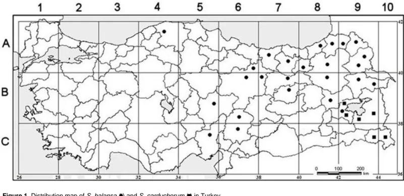
Fig 1. Distribution map of S. balansae ( ) and S. carduchorum ( ) in Turkey. (Fig 1 in proof
will be deleted and this figure will be added.)
Incorrect
Table 1 is mixed with table 2 and Table 1 isn’t in proof. I added Table 1 following.
Correct
Figure 1. Distribution map of S. balansa ( ) and S. carduchorum ( ) in Turkey.
(orally used as herbal tea in the treatment of various infections, asthmatic, antibacterial, antioxidant rheumatic and other inflammatory disorders (Couladis et al., 2003; Grujic-Jovanovic et al,. 2004; Matkowski and Piotrowska 2006; Jovanovic et al., 2008; Ebrahimabadi et al., 2010). Anatomical studies on Stachys taxa are limited (Uysal, 2002, 2003; Potoğlu-Erkaya, 2007; Dinç and Öztürk, 2008). However, there has been no anatomical investigation species from section Eriostomum species as yet.
In a previous study, we investigated the morphological and ecological features of S. balansae and S. carduchorum (Erdoğan et al., 2011). Here, we report on the anatomy of these two species. In this study, the anatomical structure of root, stem, leaf and petiole in the
two species of Eriostomum section grown in Turkey is studied for the first time.
The aim of this paper was carried out in order to provide insight into anatomy two Stachys species. Moreover, a better understanding of systematics helps separate morphologically similar species from each other.
MATERIALS AND METHODS
Plant specimens of the 2 species were collected from their type localities (Table 1, Figure 1). Species collected was examined in the herbaria and were determined using the relevant literature (Ball, 1972; Bhattacharjee, 1982; Davis, 1982; Duman, 2000). The specimens are dried according to standard herbarium techniques and stored in the Herbarium of Necatibey Education Faculty,
Erdogan et al. 1419
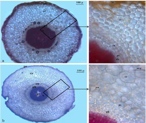
Figure 2. Cross-section of the root of S. balansae (a) and S. carduchorum (b). pe, Periderm; cr,
cortex; en,endodermis; ps. periskl; ph: phloem; x, xylem; t, trache; pt, pith.
Balıkesir University, Turkey.
Anatomical studies were carried on specimens kept in 70% alcohol. Cross-sections of roots, stems, petioles, leaves and surface sections of leaves were taken by hand. Then, sections were stained with Floroglusin and then mounted with Gliserin-Jelatine (Yakar-Tan, 1982). Measurements and photographs of anatomic sections were taken using Olympus CX21 binocular light microscope with an Olympus Camedia camera.
For images of stomata at Scanning Electron Microscope (SEM), leaves of the dried plant samples were mounted on standard SEM pin mount stubs using a double-sided conductive carbon tape. The samples were then coated with a thin layer of gold-palladium using a Cressington 108 Auto sputter coater to reduce charging. The coated samples were imaged using a Hitachi S-4800 Scanning Electron Microscope (SEM) at an accelerating voltage of 5 to 15 kV and working distances ranging from 20 to 22 mm at University of Toledo, Ohio, USA. The SEM micrographs were then analyzed.
RESULTS Root
Cross-sections taken from the root of S. balansae and S. carduchorum have revealed that the periderm layer on the outermost surface of the root is consisting of phellem by 3 to 5 layers and phelloderm and phellogen are not
distinguishable. There is a multi-layered parenchymatic cortex (8 to 13 layers) under the periderm. Endodermis is one rowed and under of the endodermis pericycle is indistinguishable. Phloem is located under endodermis and 4 to 12 layered. Cambium cells are indistinguishable. The xylem is composed of vessels and tracheids. Pith rays comprise 1 to 3-rowed rectangular cells. Pith region of S. balansae consist of xylem elements whereas S. carduchorum consist of polygonal or orbicular parenchymatous cells (Figure 2).
Stem
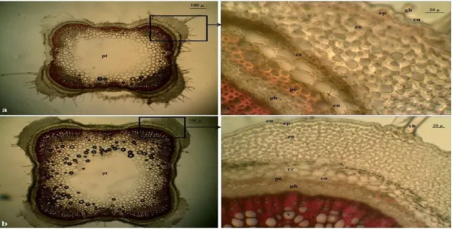
Cross-sections taken from the stem have exhibited a monolayer epidermis covered by a thin cuticle. The epidermis is composed of oval, cubic or rectangular cells. There are glandular and eglandular hairs (simple and unbranched, 1 to 4 cells) on epidermis. On young stems are seen diacytic type stomatas. Underneath the epidermis, multilayered collenchyma cells are located at the corners (7 to 13 layers) and there are 1 to 2 rows of chlorenchyma cells between them. Beneath this is situated the cortex which consists of 2 to 6 layers of oval, ovate or orbicular parenchymatous cells. Endodermis
Figure 3. Cross-section of the stem of S. balansae (a) and S. carduchorum (b). cu, cuticle; gh, glandular hair; eh,
eglandular hair; ep, epidermis; co, collenchyma; cr, cortex parenchyma; en, endodermis; pe, pericycle; ph, phloem; x, xylem; pt, parenchymatic pith.
under the cortex is one rowed. Pericycle is indistinguishable. Sclerenchymatic cell clusters (1 to 4 layers) are situated in beneath endodermis. Phloem is 3 to 6-layered and consists of oval and rectangular cells. Cambium is indistinguishable. The xylem is composed of trachea and tracheids. Trachea is orbicular or ovoid while tracheids are polyhedral. Rays are 1 to 2 rowed. The pith is large and comprised of hexagonal or orbicular parenchymatic cells with intercelular spaces in the centre of stem (Figure 3).
Leaf
The epidermis is composed of a single layer cells and rectangular or oval cells. Adaxial epidermis is larger than lower and covered with a thick undulate cuticle. There are glandular and eglandular hairs (simple and unbranched, 1 to 2 cells) and on the surfaces of both epidermis. Stomata type is diacytic and occurs on the surfaces of both epidermis being more abundant on the lower surface. Stomata is located on the same level with epidermal cells. Leaf is isobilateral. Mesophyll occurs palisade 2 seriate beneath adaxial epidermis and 1 to 2 seriate above abaxial epidermis, at the median region of mesophyll is located spongy parenchyma that is 2 to 5- midrib that is located to beneath the epidermis is seen collenchyma. Parenchyma that is located beneath collenchymas is shaped polygonal and oval and 3 to 6 layered at adaxial surface and 3 to 9 layered at adaxial surface. Vascular bundle is surrounding by parenchyma cells. The xylem faces towards the adaxial surface while phloem faces the abaxial surface. The phloem tissue is 3
to 8 layered and surrounded by parenchymatic cells (Figure 5a and c). In the phloem cells of S. balansae are not seen sclerenchymatic cells while phloem cells of S. carduchorum are seen (1 to 2 rowed) (Figure 4).
Petiole
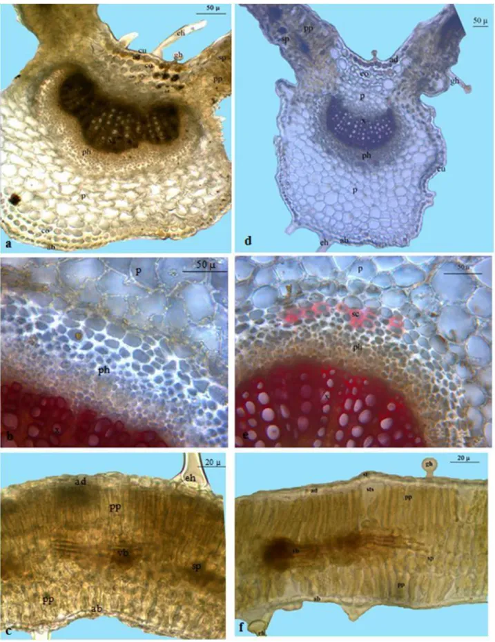
A cross section of the petioles shows that the adaxial surface is concave and slightly curved and the abaxial surface is convex and acute. There are eglandular (simple, 1 to 5 cells) and glandular hairs on both adaxial and abaxial surface. The epidermis is composed of oval and rectangular cells 1 layered. Collenchyma is situated under epidermis. It is 3 to 8 layered on abaxial surface, 1 to 2 layered underadaxial surface and 4 to 12 layered the ends of the petiolar wings. Parenchyma cells are hexagonal oroval shape. They are 2 to 5 layered on nearly flat and abaxial surface is broadly convex while adaxial surface of S. carduchorum deeply concave (V- In the midrib, adaxial surface of S. balansae is shaped) and abaxial surface rounded-convex. 1 to 3 rowed in adaxial surface and 2 to 4 rowed in abaxial surface of collateral vascular bundle is located in the midrib region. seriate and composed of polygonal or rounded cells. The adaxial surface and 4 to 10 layered on abaxial surface. In the midrib, adaxial surface of concave-curved or deeply concave (V-shaped); abaxial surface is convex or rounded-convex. In the middle region of petiole, there are one (S. carduchorum) or two (S. balansae) collateral large vascular bundles crescent shape and one small subsidiary collateral bundle at the ends of petiolar wings. The phloem tissue is 3 to 8 layered and surrounded by
Erdogan et al. 1421
Figure 4. Cross-section of leaf lamina of S. balansae (a-c) and S. carduchorum (d-f). cu, cuticle; gh, glandular hair; eh,
eglandular hair; ad, adaxial epidermis; ab, abaxial epidermis; pp, palisade parenchyma; sp, spongy parenchyma; vb, vascular bundle; st, stomata; sts, stomata space; co, collenchyma; p, parenchyma; sc, sclerenchyma; ph, phloem; x, xylem.
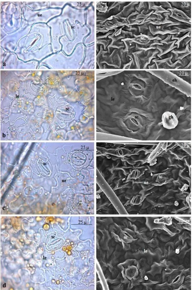
Figure 5. Surface sections of the leaves S. balansae (a, b) and S. carduchorum (c, d). Upper surface (a, c),
lower surface (b, d). Ue, upper epidermis; le, lower epidermis; gh, glandular hair; eh, eglandular hair; sc, stoma cell.
Erdogan et al. 1423
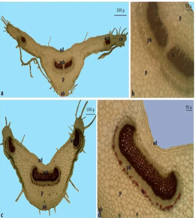
Figure 6. Cross-section of the petiole of S. balansae (a, b) and S. carduchorum (c, d). ab, abaxial epidermis, ad,
adaxial epidermis, co, collenchyma, p, parenchyma, mb, median bundle, acb, accessory bundle, sc, sclerenchyma, ph, phloem, x, xylem.
parenchymati cells. In the phloem cells of S. balansae are not seen sclerenchymatic layer while phloem cells of S. carduchorum are seen (1 to 2 layered). The xylem is composed trachea and tracheids. Xylem faces towards the adaxial surface while phloem faces the abaxial surface (Figure 6).
DISCUSSION
In this study, two taxa belonging to the same subsection (Germanicae) and morphologically difficulty in distinguishing from each other is investigated. Moreover, anatomical properties of Germanicae subsection have
Table 2. Stem anatomical characters of S. balansae and S. Carduchorum.
Species
Stem anatomical characters Collenchyma layers Parenchyma
layer
Sclerenchyma
cluster Phloem layer Pith ray row Corner Between of corner
S. balansae 8-12 1-2 2-6 1-4 4-6 1-2
S. carduchorum 7-13 1-2 2-5 1-3 3-5 1-2
*ad: adaxial surface (upper epidermis) *ab: abaxial surface (lower epidermis
Table 3. Leaf anatomical characters of S. balansae and S. carduchorum.
Species
Leaf anatomical characters
Mesophyll region Median vascular bundle Mesophyll
type
Palisade
Spongy Collenchyma Parenchyma Sclerenchyma Phloem ad* ab* ad* ab* ad ab
S. balansae Isobilateral 2 1-2 2-5 1-3 1-4 3-8 3-6 - 3-6
S. carduchorum Isobilateral 2 1-2 3-4 1-3 2-4 3-7 3-5 1-2 3-8
*ad, Adaxial surface (upper epidermis); *ab, abaxial surface (lower epidermis).
Table 4. Petiol anatomical characters of S. balansae and S. carduchorum.
Species
Petiol anatomical characters
Number of vascular bundle Median Region Median region Wings of petiole Collenchyma Parenchyma Sclerenchyma Phloem ad* ab* ad ab S. balansae 1 1 1-2 4-9 2-5 1-2 2-5 3-5 S. carduchorum 2 1 1-2 3-5 7-12 4-7 2-5 3-5
*ad, Adaxial surface (upper epidermis); *ab, abaxial surface (lower epidermis).
been reported for the first time. Our anatomical results are similar to Metcalfe and Chalk (1950) and Salmaki et al. (2011). The results obtained from anatomical investigation carried out on two species were generally similar. No important differences observed in root and stem anatomical features of S. balansae and S. carduchorum. However, Pithregion (root) of S. balansae consist of xylem elements while S. carduchorum consist of polygonal or orbicular parenchymatous cells.
Of the anatomy of leaf is appeared two significant differences. The first; in the midrib, adaxial surface of S. balansae is nearly flat and abaxial surface is broadly convex while adaxial surface of S. carduchorum deeply concave (V-shaped) and abaxial surface rounded-convex. The second; in the phloem cells of S. balansae are not seen sclerenchymatic cells while phloem cells of S. carduchorum are seen.
Of the anatomy of petiole is appeared significant difference. In the midrib, adaxial surface of S. balansae is slightly concave-curved and abaxial surface is convex
while adaxial surface of S. carduchorum deeply concave (V-shaped) and abaxial surface rounded-convex. Besides, large vascular bundles in the middle region of petiole are one bundle of S. carduchorum and two bundles of S. balansae. Moreover; in the phloem, cells of S. balansae are not seen sclerenchymatic layer while phloem cells of S. carduchorum are seen.
Comparative anatomical characters of stem, leaf and petiol are given (Table 2 to 4). In addition to comparative micro-anatomical measurements, various tissues of S. balansae and S. carduchorum are presented in Table 5.
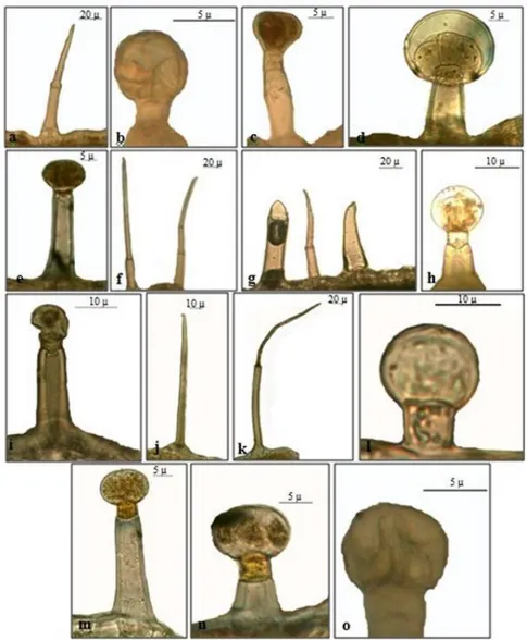
Selected micrographs of common indumentum types of stem, leaf and petiole of S. balansae and S. carduchorum, are presented in Table 6 and Figure 7. Two basic types of trichomes can be distinguished: Glandular and non-glandular trichomes. Nonglandular trichomes are unbranched and unicellular (Figure 7g and j) or multicellular (Figure 7f). Glandular trichomes are two types: Capitate and peltate. Capitate trichomes consist of short stalked (single cell base, short single cell neck, and
Erdogan et al. 1425
Table 5. Comparative micro-anatomical measurements various tissues of S. balansae and S.carduchorum.
Tissues
S. balansae S. carduchorum
Width (µm) Length (µm) Widths (µm) Lenght (µm)
Min Max Min Max Min Max Min Max
Root Peridermis 19 48 - - 28 69 - - Parenchyma layers 184 261 - - 160 250 - - Parenchyma cells 12 45 16 42 16 38 17.5 32.6 Endodermis layers 10 14 - - 8 12 - - Phloem layers 13 45 - - 21 42 - - Xylem layers 140 200 - - 170 220 Trache cells 7 15 8 17 5 21 4 23 Stem Epidermis cell 5 12 5 14 7 16 6 14 Parenchyma layers 50 90 - - 47 85 - - Endodermis layers 13 43 - - 13 30 - - Phloem cells 13 18 10 17 11 19 10 18 Trache cells 4 47 6 24 5.7 22.4 7.3 16.8 Pith cells 21 97 18 102 16 89 15 95 Leaf Cuticle 0.8 2.5 - - 0.5 2 - -
Adaxial epidermis cells 7 13 10 22 9 20 8 23
Abaxial epidermis cells 4 11 9 18 8 18 9 21
Mesophyll layers 80 110 - - 75 120 - -
Palisade cells parenchyma 3 8 13 38 3.3 9.5 5 39
Spongy cells cellparenchyma 6.4 15.06 9.5 14.6 6 15 9 14
Petiole
Adaxial cells 13.8 28.1 13.8 22.8 11 22 10 21
Abaxial cells 11.1 29.4 12.2 26.5 10 31 10 33
Parenchyma cells 14.2 30.6 10.2 24.9 14.2 30.6 10.2 24.9
Trache cells 3 12 5 13 8 19 6 17
Table 6. Characterisation of the trichome types of S. balansae and S. carduchorum.
Trichomes Type Description S. balansae S. carduchorum Tissue Non glandular
trichomes A
Acicular or curved, unbranced with one or more cells, mostly having one to five cells, arranged in a single row and having no cuticle with micropapillae
+ + Stem + + Leaf + + Petiole Glandular trichomes B1
Peltate, 1 short stalk cell, 1 short neck cell and enlarged with 40 to 60 µm ahead of 4 to 8 secretory cells
- + Stem
+ + Leaf
- - Petiole
B2
Peltate; Longer 1 stalk cell, 1 short neck cell and enlarged with 40 to 60 µm ahead of 4 to 8 secretory cells
- + Stem
+ + Leaf
- - Petiole
C Capitate; consisted of a short unicellular stalk and a globose head cell (20 to 30 µm)
+ + Stem
Table 6. Contd.
+ + Petiole
D Capitate; 1 stalk of 2 to 5 cell, a short neck cell and a glandular head.
+ - Stem
- - Leaf
- + Petiole
Figure 7. Trichome morphologies stem, leaf and petiole of S. balansae (a-e) and S.
carduchorum (f-o). Stem (a-b; f-i), leaf (c-d; j-m), petiole (e; n-o).
oval one or two cell head) (Figure 7b, l and o) and long stalked trichom (extended two cell base, single cell neck, and oval two cell head (Figure 7e and i). Peltate trichomes consist of single cell base (short or long), single cell neck and six-ten cell head (Figure 7c, d, m and n)
ACKNOWLEDGEMENTS
We would like to thank Prof. Dr. Hulusi MALYER and Associate Prof. Dr. Fatih SATIL for their valuable comments. We would also like to thank TÜBITAK (The Scientific and Technological Research Council of Turkey)
for supporting this research with Grant number 106T489 and SYNTHESYS Program for the financial support (GB- TAF 4797) that provided us with the opportunity to study at valuable herbaria in Europe.
REFERENCES
Akçiçek E (2010). A new subspecies of Stachys cretica (section Eriostomum, Lamiaceae) from Turkey. Turk J. Bot., 34: 131-136. Akcicek E, Dirmenci T, Dundar E (2012). Taxonomical Notes on
Stachys sect. Eriostomum (Lamiaceae) in Turkey. Turk. J. Bot .,36:
217-234.
Ball PW (1972). Stachys. In Tutin TG, Heywood VH, Burgess NA, Moore DM, Valentine DH, Walter SM, Webb DA Cambridge University Press, Cambridge. (Eds.). Flora Europaea, 3: 151–157. Bhattacharjee R (1974). Taxonomic studies in Stachys L: New species
and infra-specific taxa from Turkey. Notes Royal Bot. Garden Edinburgh, 33(2): 275–292.
Bhattacharjee R (1980). Taxonomic studies in StachysII: A new infrageneric classification of Stachys L.. Notes Royal Bot. Garden Edinburgh, 38(1): 65–96.
Bhattacharjee R (1982). Stachys L. in: Davis, PH (ed) Flora of Turkey and the East Agean Islands. Edinburgh University Press, Edinburgh, 7: 199–262.
Couladis M, Tzakou O, Verykokidou E, Harvala C (2003). Screening of Some Greek Aromatic Plants for Antioxidant Activity. Phytotheraphy. Res., 17: 194–195.
Daşkın R., Yılmaz Ö., Kaynak G. (2009). Stachys ketenoglui sp. nov. (sect. Infrarosularis) (Labiatae/Lamiaceae) from south Anatolia, Turkey. Nordic J. Bot., 27(3): 238-242.
Davis PH (1982). Flora of Turkey and The East Aegean Islands. Edinburg University Press, Edinburgh. p. 7.
Dinç M, Öztürk M (2008). Comparative Morphological, Anatomical, and Palynological Studies on the Genus Stachys L. sect. Ambleia Bentham (Lamiaceae) Species in Turkey. Turk. J. Bot., 32:113-121. Dinç M, Doğan HH (2006). Stachys yildirimlii (Lamiaceae), a new
species from south Anatolia, Turkey. Ann. Bot.. Fenn., 43: 143–147. Dirmenci T, Yıldız B, Akçiçek E, Martin E, Dündar E (2011). Stachys
vuralii (Lamiaceae), a new species from north Anatolia, Turkey. Ann.
Bot. Fenn,, 48: 401-408.
Duman H (2000). Flora of Turkey and East Aegean Islands, Stachys L. — In: Güner, A, Özhatay, N, Ekim, T, Başer, KHC Edinburgh University Press, Edinburgh, (eds.), 2(11): 204–206.
Ebrahimabadi AH, Ebrahimabadi EH, Djafari-Bidgoli Z, Kashi FJ, Mazoochi A, Batooli H (2010). Composition and antioxidant and antimicrobial activity of the essential oil and extracts of Stachys
inflate Benth from Iran. Food Chem., 119: 452-458.
Erdogan et al. 1427
Erdoğan E, Akçiçek E, Selvi S, Tümen G (2011).Comparative morphological and ecological studies of two Stachys species (sect.
Eriostomum, subsect. Germanicae) grown in Turkey. .Afr. J.
Biotechnol., 10(78): 17990-17996.
Falciani L (1997). Systematic Revision of Stachys Sect. Eriostomum (Hoffmanns.&Link) Dumort. in Italy. Lagascalia, 19(1-2): 187-238. Gemici Y, Leblebici E (1998). A new species from Southern Anatolia:
Stachys cydni Kotschy ex Gemici and Leblebici. Turkish. J. Bot., 22:
359–362.
Grujic-Jovanovic S, Skaltsa, HD, Marin P, Sokovic P (2004). Composition and antibacterial activity oil of six Stachys species from Serbia, Flavour. Fragrance. J., 19: 139-144.
İlçim A, Çenet M, Dadandı MY (2008). Stachys marashica (Lamiaceae), a new species from Turk. Ann. Bot. Fenn., 45: 151-155.
Jovanovic SG, Marin PD, Dzamic A, Ristic M (2008). Composition of the Essential oil of Stachys germanica from Serbia. Chem. Nat. Comp., 44(5): 670-672.
Matkowski A, Piotrowska M (2006). Antioxidant and free radical scavenging activities of some medicinal plants from the Lamiaceae. Fitoterapia, 77: 346-353.
Metcalfe CR, Chalk L (1950). Anatomy of the Dicotyledons Oxford University Press, London. p. 2.
Salmaki Y, Zarre S, Lindqvist C, Heubl G, Brauchler C (2011). Comparative leaf anatomy of Stachys (Lamiaceae:Lamioideae) in Iran with a discussion on its subgeneric classification. Plant Syst. Evol., 294 (1-2): 109-125.
Potoğlu-Erkaya İ, Koyuncu O (2007). A Study of the Anatomy and Pollen Morphology of Two Economically Important Species of
Stachys L. (Lamiaceae) in Turkey. J. Appl. Biol. Sci., 1(3): 49-56.
Sümbül H (1990). Two new species from South Anatolia, Turkish. J. Bot., 22: 359-362.
Uysal I (2002). Stachys cretica L. subsp. smyrnaea Rech fil. Endemik Taksonunun Morfolojisi, Anatomisi ve Ekolojisi Üzerinde Araştırmalar. Ecology, 11(42):16-20.
Uysal İ (2003). Stachys thirkei C.Koch (Kekikgiller) türünün morfolojisi, anatomisi ve ekolojisi üzerine araştırmalar. Ot Sistematik Botanik Dergisi, 10: 129-141.
Yakar-Tan N (1982). Bitki Mikroskopisi Klavuz Kitabı. İstanbul Üniv. Fen Fak. Yay. İstanbul, p. 166,
Yıldırımlı Ş (2010). Some new taxa, records and taxonomic treatments from Turkey. Ot Sistematik Botanik Dergisi.,17(2): 1-114.
Yılmaz Ö, Daşkın R, Kaynak G (2010). Stachys pseudobombycina sp. nov. (Lamiaceae) from South Anatolia. Nordic J. Bot., 28: 341-343.