Mitochondrial carrier homolog 1 (Mtch1) antibodies in neuro-Behçet's disease
Burçak Vural
a, Elçin
Şehitoğlu
a, Filiz Çavu
ş
a, Nazl
ı Yalçınkaya
b, Hazal Haytural
b, Melike Küçükerden
b,
Canan Ulusoy
b, Elif U
ğurel
a, Selin Turan
a, Leyla Bulut
c, Recai Türko
ğlu
d, Erkingül Shugaiv
e, Murat Kürtüncü
e,
Şükrü Atakan
f, Ali O. Güre
f, Ahmet Gül
g, Mefkure Eraksoy
e, Gül
şen Akman-Demir
h, Erdem Tüzün
b,⁎
a
Department of Genetics, Institute for Experimental Medicine, Istanbul University, Istanbul, Turkey
bDepartment of Neuroscience, Institute for Experimental Medicine, Istanbul University, Istanbul, Turkey c
Department of Biochemistry, Istanbul Faculty of Medicine, Istanbul University, Istanbul, Turkey
d
Department of Neurology, Haydarpasa Numune Training and Research Hospital, Istanbul, Turkey
e
Department of Neurology, Istanbul Faculty of Medicine, Istanbul University, Istanbul, Turkey
f
Department of Molecular Biology and Genetics, Bilkent University, Ankara, Turkey
g
Division of Rheumatology, Department of Internal Medicine, Istanbul Faculty of Medicine, Istanbul University, Istanbul, Turkey
h
Department of Neurology, Bilim University, Istanbul, Turkey
a b s t r a c t
a r t i c l e i n f o
Article history: Received 20 March 2013
Received in revised form 3 August 2013 Accepted 14 August 2013
Keywords: Behçet's disease Neuro-Behçet's disease Anti-neuronal antibody Mitochondrial carrier homolog 1 Antibody
Autoimmunity
Efforts for the identification of diagnostic autoantibodies for neuro-Behcet's disease (NBD) have failed. Screening of NBD patients' sera with protein macroarray identified mitochondrial carrier homolog 1 (Mtch1), an apoptosis-related protein, as a potential autoantigen. ELISA studies showed serum Mtch1 antibodies in 68 of 144 BD patients with or without neurological involvement and in 4 of 168 controls corresponding to a sensitivity of 47.2% and specificity of 97.6%. Mtch1 antibody positive NBD patients had more attacks, increased disability and lower serum nucleosome levels. Mtch1 antibody might be involved in pathogenic mechanisms of NBD rather than being a coincidental byproduct of autoinflammation.
© 2013 Elsevier B.V. All rights reserved.
1. Introduction
Behçet's disease (BD) is a chronic, recurrent and inflammatory dis-order characterized with oral and genital aphthous ulcerations, uveitis, skin lesions and skin pathergy reaction (Gül, 2005; Yurdakul and Yazici, 2008). The presence of inflammatory lesions in involved tissues, increased levels of cytokines and acute phase reactants and identi fica-tion of autoantibodies [directed against heat shock proteins (HSP)-60, -65 and -70, αB-crystallin, stress-induced-phosphoprotein 1, PTEN-induced putative kinase 1 (PINK1),α-enolase, cyclic citrullinated pep-tide, annexins and Saccharomyces cerevisiae antigens] in circulation of BD patients have suggested an autoimmune as well as an auto-inflammatory pathogenesis (Taşçi et al., 1998; Tanaka et al., 1999; Celet et al., 2000; Dinc et al., 2003; Duygulu et al., 2005; Fresko et al.,
2005; Gül, 2005; Koca et al., 2007; Birtas-Atesoglu et al., 2008; Lee et al., 2009; Vural et al., 2009; Iaccarino et al., 2011; Vural et al., 2011). Central nervous system (CNS) involvement, or neuro-Behçet's dis-ease (NBD), develops in 5–10% of BD patients and generally afflicts the brain parenchyma and less frequently the brain vessels and meninges (Akman-Demir et al., 1999). The cerebral parenchymal lesions are mainly composed of mononuclear and neutrophilic in fil-trates (Hirohata, 2008). Nevertheless, NBD associated serum and cerebrospinal fluid (CSF) antibodies to αB-crystallin and HSP-60, -65 and -70 have also been identified suggesting involvement of antibody-mediated pathogenic mechanisms in NBD (Taşçi et al., 1998; Tanaka et al., 1999; Celet et al., 2000; Birtas-Atesoglu et al., 2008). How-ever, these antibodies have been detected in less than 30% of NBD patients, and are thus, of limited use as biomarkers of the disease. In an attempt to identify NBD-specific anti-neuronal antibodies, sera of NBD patients and controls were screened using a protein macroarray, and confirmatory immunohistochemistry and immunoblotting studies were performed. These studies identified mitochondrial carrier homolog 1 (Mtch1) autoantibody, which appears to be highly sensitive and spe-cific for NBD and BD.
⁎ Corresponding author at: Department of Neurology, Istanbul University, Istanbul Faculty of Medicine, 34390 Çapa, Istanbul, Turkey. Tel.: + 90 212 4142000x32580; fax: + 90 212 5334393.
E-mail address:[email protected](E. Tüzün).
0165-5728/$– see front matter © 2013 Elsevier B.V. All rights reserved.
http://dx.doi.org/10.1016/j.jneuroim.2013.08.007
Contents lists available atScienceDirect
Journal of Neuroimmunology
j o u r n a l h o m e p a g e : w w w . e l s e v i e r . c o m / l o c a t e / j n e u r o i m2. Materials and methods 2.1. Patients and samples
Thirty-two consecutive NBD patients (13 women, 19 men; mean age ± standard error, 36.4 ± 1.7) were included. The average NBD duration (±standard error) of these patients was 9.9 ± 1.3 years. Age- and gender-matched controls included 112 BD patients without neurological involvement (41 women, 71 men; mean age, 37.3 ± 1.6; disease duration 9.6 ± 2.3), 47 patients with relapsing remitting multi-ple sclerosis (25 women, 22 men; mean age, 34.3 ± 1.1; disease dura-tion 9.6 ± 2.1), 21 neuromyelitis optica (NMO) patients (12 women, 9 men; mean age, 32.6 ± 1.5; disease duration 8.7 ± 1.3) and 100 healthy controls (47 women, 53 men; mean age, 35.7 ± 1.9). There were no statistically significant differences between NMO patients and control groups by means of age, gender and disease duration (pN 0.05 by Fisher's exact test or Student's t-test). None of the patients had a history of a concomitant neurological disease. NBD and BD patients fulfilled the diagnostic criteria for BD (International Study Group for Behçet's Disease, 1990), MS patients fulfilled McDonald's criteria for definite MS (Polman et al., 2005) and NMO patients fulfilled the revised Wingerchuk criteria (Wingerchuk et al., 2006). EDSS scores of NBD patients were calculated during serum sampling. An informed consent was obtained from all participants before blood samples were obtained. Sera were kept frozen at−80 °C until assayed. Blood samples were col-lected from all NBD and BD patients prior to the initiation of steroid treatment, especially when the sample was obtained during an attack. However, the interference of immunosuppressive therapy with anti-body levels could not be completely avoided. While all NBD patients were under long term azathioprine treatment when sera were collect-ed, 58 BD patients were under immunosuppressive treatment and 54 BD patients were not receiving any immunosuppressants. The study was approved by the Ethics Committee of Istanbul Faculty of Medicine of Istanbul University.
2.2. Protein macroarray, sequencing of cDNA inserts and protein expression To identify NBD related anti-neuronal antibodies, pooled sera of 10 randomly selected NBD patients were screened using a high-density protein macroarray derived from human fetal brain cDNA expression library, which contains approximately 24,000 clones (ImaGenes, Berlin, Germany) (Preuss et al., 2009). Images were captured and analyzed for signal intensity (VisualGrid, GPC Biotech, Martinsried, Germany). The arrays were scored as 0 (absent), 1 (weak), 2 (moderate) and 3 (strong) confirmed by matched duplicates. Selected expression clones were obtained from ImaGenes. Plasmid DNA from clones was isolated for DNA sequencing (Qiagen, Hilden, Germany) according to the manufacturer's instructions. Cloned cDNAs in the purified plasmid DNA were sequenced by Iontek Laboratory (Istanbul, Turkey). Nucle-otide and translated amino acid sequences were compared with known sequences using BLAST algorithms (National Center for Bio-technology Information, Bethesda, MD). Following the confirmation of the selected clone, His-tagged protein was recombinantly expressed in Escherichia coli, purified by affinity chromatography and the purity of the protein was documented by SDS-PAGE analysis (Fig. 1), as report-ed previously (Preuss et al., 2009).
2.3. Immunoblotting analyses
The purified protein was denatured (100 °C, 5 min), 1 μg purified protein was loaded in each lane, electrophoresed (10% acrylamide gel) and transferred to 0.45-μm polyvinylidene fluoride membranes (100 V, 80 min). Membranes were blocked (5% milk in TBST; 90 min) and incubated with individual human sera (diluted 1:200) or commer-cially available rabbit anti-human antibody (1:200 dilution) (Santa Cruz Biotechnology, Santa Cruz, CA) followed by HRP-conjugated goat
anti-human IgG or goat anti-rabbit IgG (Jackson ImmunoResearch Laboratory) at 1:1000 dilutions. Immunoreactivity was visualized on chemilumines-centfilm using ECL Western blotting substrate (Pierce, Thermo Scientific, USA) according to the manufacturer's instructions (Fig. 1).
2.4. ELISA
Detection of antibodies to the purified recombinant human protein was performed with ELISA. The purified protein (50 μl at 10 μg/ml) was added to the wells of a 96-well high-binding-capacity plate and incubated overnight at 4 °C. Wells coated with the E. coli lysate or only with bovine serum albumin were used as controls. The plates were washed with TBST and blocked for 2 h with 5% skim milk in TBS. A 60μl aliquot of each serum sample (diluted 1:100) in TBST was added to protein coated wells and incubated for 2 h at room tempera-ture. The plates were washed six times with TBST followed by the addi-tion of 60μl of alkaline phosphatase-conjugated goat anti-human IgG (Southern Biotech, Birmingham, AL, USA) diluted 1:2000 in TBST and then incubated at room temperature for 1 h. After washing, 60μl of 2-(2-benzothiazoyl)-6-hyroxybenzothiazole phosphate was added for 45 min at room temperature followed by addition of the stopping solu-tion (3 N NaOH). Fluorescent signals were measured at 450/50 excita-tion and 580/50 emission with a microplate reader. For each sample, the value obtained from the protein-coated well was substracted from the non-coated well. The obtained results were expressed as signal ratios (sample signal/mean signal of healthy controls). Positivity was defined as 2 standard deviations above the mean of healthy controls. 2.5. Immunohistochemistry and colocalization studies on rat brain sections
Whole rat brain was treatedfirst with 4% paraformaldehyde over-night at 4 °C, immersed in 40% sucrose overover-night at 4 °C and subse-quently snap frozen in liquid nitrogen. Sevenμm-thick frozen sections were serially incubated with 0.3% H2O2for 20 min, 10% goat serum
for 1 h at room temperature and serum samples (1:200) overnight at 4 °C. They were then incubated in biotinylated goat anti-human IgG
Fig. 1. Coomassie blue-stained 10% SDS-PAGE analysis of purification of mitochondrial car-rier homolog 1 (Mtch1) yielding a band at around 40 kDa, as predicted (leftmost column), and representative immunoblots for Western blot analysis of recombinant Mtch1 protein (remaining 5 columns). While both commercially available rabbit human Mtch1 anti-body (Mtch1-Ab) and neuro-Behçet's disease (NB) patients' sera that were found to be pos-itive for Mtch1 Ab by ELISA (NB p) yielded ~40 kDa bands at the Mtch1 protein loaded gels, NB sera that were seronegative for Mtch1 Ab by ELISA (NB n) did not show any bands.
(1:2000, Vector Laboratories, Burlingame, CA), and the immunoreac-tivity developed by serial incubation with avidin–biotin peroxidase (Vector Laboratories) for 1 h and diaminobenzidine (Irani et al., 2010). For immunofluorescence experiments, frozen and paraformaldehyde-fixed rat brain sections were incubated with 10% goat serum for 1 h at room temperature followed by patients' sera (1:200) and commercial rabbit anti-rat Mtch1 antibody (Santa Cruz Biotechnology, Santa Cruz, CA) at 1:200 dilution overnight at 4 °C. Next day, sections were incubated with Alexa Fluor 488-conjugated anti-human IgG and Alexa Fluor 568-conjugated anti-rabbit IgG (1:2000 dilution) (Invitrogen, Paisley, UK) for 2 h at room temperature. Sections were evaluated and photographed under a Zeissfluorescence microscope with a digital camera using the Zeiss Axiovision software. Moderate to strong Alexa Fluor 488-conjugated anti-human IgG-induced green color that overlapped with Alexa Fluor 568-conjugated anti-rabbit IgG-induced red color was considered as colocalization.
2.6. Quantitation of circulating nucleosomes
Intensity of apoptosis was estimated in NBD patients and healthy controls by measuring serum levels of circulating nucleosomes with a quantitative sandwich-enzyme-immunoassay, using mouse monoclonal antibodies directed against DNA and histones, respectively. This method allowed specific detection and quantitation of histone-associated DNA fragments in mono- and oligonucleosomes (a marker for apoptotic cells) that are released into serum. Serum levels of nucleosomes were measured as per manufacturer's protocol (Roche Applied Science, Indianapolis, IN, US).
2.7. Statistics
Demographic and clinical features of NBD patients were compared using Fisher's exact test, Student's t-test or Mann–Whitney U test, as ap-propriate. Signal ratios obtained in ELISA experiments were compared among groups by ANOVA and Tukey's post-hoc test. Serum nucleosome levels were compared with Student's t-test. Correlation statistics were
performed with parametric Pearson's or non-parametric Spearman's correlation tests, as required. A p value smaller than 0.05 was consid-ered as statistically significant.
3. Results
3.1. Identification and verification of Mtch1 antibody
To identify target antigens of NBD-associated neuronal autoanti-bodies, a protein macroarray derived from a human fetal brain cDNA expression library was used to screen sera from NBD patients. A single clone with the highest signal intensity and number of duplicates (n = 3) was selected for further investigation. DNA sequencing and BLAST analysis of the clone yielded a single oligonucleotide that corresponded to 92% of the sequence of Mtch1 (GenBank accession number, NM_014341). A Mtch1 his-tagged fusion protein was re-combinantly produced in E. coli and purified by affinity chromatography. SDS-PAGE analysis showed a single band at around 40 kDa, consistent with the predicted molecular weight, confirming the purity of the obtained protein (Fig. 1).
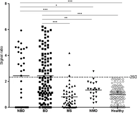
ELISA studies performed with the recombinant protein revealed high-titer autoantibodies in 18 of 32 (56.3%) NBD, 50 of 112 (44.6%) BD, 3 of 47 (6.4%) multiple sclerosis, 1 of 21 (4.8%) neuromyelitis optica patients and none of the healthy controls (Fig. 2). Overall, 68 of 144 (47.2%) BD patients with or without neurological involvement and 4 of 168 (2.4%) non-BD controls displayed Mtch1 antibody corresponding to a sensitivity of 47.2% at 97.6% specificity. NBD and BD patients had significantly higher signal ratio values as compared to control groups (pb 0.0001 by ANOVA and p b 0.05–0.001 by Tukey's post-hoc test,
Fig. 2). None of the sera reacted with the lysate of the E. coli strain used to express the proteins or irrelevant proteins (myc-associated zincfinger protein and zinc finger protein 553) expressed by the same E. coli strain (data not shown), supporting the specificity of the autoan-tibody measurements. There were no significant differences between average signal ratios of BD patients with (2.3 ± 1.9) or without (2.6 ± 1.7) immunosuppressive treatment (p = 0.148 by Student's
Fig. 2. ELISA detection of IgG antibodies directed against mitochondrial carrier homolog 1 (Mtch1) in sera of Behçet's disease (NBD) patients, Behçet's disease patients with no neuro-logical involvement (BD), multiple sclerosis patients (MS), neuromyelitis optica patients (NMO) and healthy controls. The dashed lines represent 2 standard deviations (2SD) above the mean of the healthy control samples (cut-off values for positivity). Horizontal lines indicate the mean value of each group. *, pb 0.05; **, p b 0.01; ***, p b 0.001 by Tukey's post-hoc test.
t-test). Mtch1 antibody seropositivity rates were also comparable among both groups (23/58 patients with immunosuppression vs 27/54 patients without immunosuppression, p = 0.431 by Fisher's exact test).
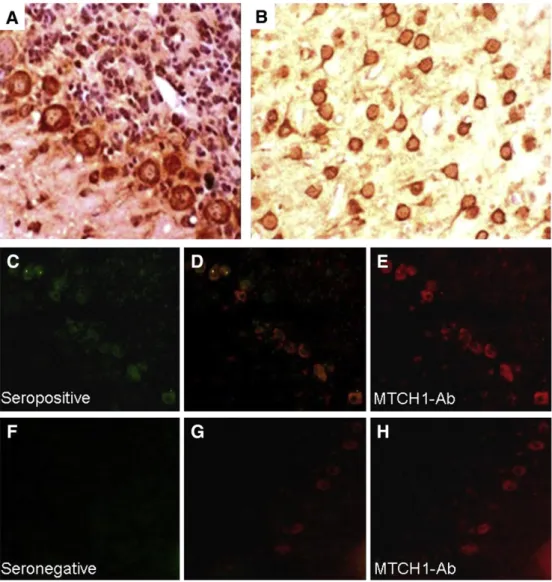
The antibody binding to Mtch1 was confirmed by Western blotting using purified Mtch1 protein. Both the commercially available antibody to Mtch1 and the seropositive NBD sera reacted with a band at the pre-dicted 40 kDa, whereas seronegative sera did not show any immunore-activity (Fig. 1). In immunohistochemistry studies, serum IgGs of Mtch1 antibody positive NBD patients showed strong cytoplasmic reactivity with neurons located throughout the whole brain section, including cer-ebellar Purkinje cells and cortical neurons (Fig. 3A,B), whereas those of Mtch1 antibody negative NBD patients did not show any appreciable immunoreactivity. Mtch1 is located at the mitochondrial membrane and is thus a cytoplasmic protein (Xu et al., 2002; Lamarca et al., 2007). Therefore, the cytoplasmic staining pattern obtained by NBD sera was considered to be due to immunoreactivity with neuronal Mtch1 protein. Immunofluorescence studies performed with sera of NBD patients and a commercially available Mtch1 antibody revealed a significant co-localization of reactivities with seropositive (Fig. 3C–E) but not seronegative (Fig. 3F–H) serum samples, further confirming the presence of Mtch1 antibodies.
3.2. Comparison of clinical features among Mtch1 antibody positive and negative NBD patients
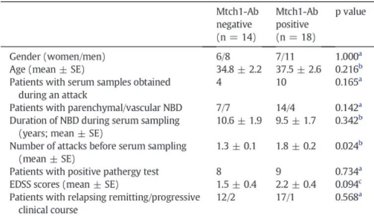
NBD patients with and without Mtch1 antibodies did not signi fi-cantly differ in terms of gender, age, clinical course, disease duration, neurological disability and pathergy test positivity. Although a higher antibody positivity rate was observed in samples obtained during a neurological attack, this difference did not attain statistical significance (p = 0.165 by Fisher's exact test). By contrast, Mtch1 antibody positive patients had significantly higher number of NBD attacks prior to blood sampling than Mtch1 antibody negative patients (p = 0.024 by Student's t-test). Also, Mtch1 antibody positive patients showed trends towards exhibiting higher Expanded Disability Status Scale (EDSS) scores (p = 0.094 by Mann–Whitney U) and parenchymal rather than vascular NBDfindings (p = 0.142 by Fisher's exact test) (Table 1).
3.3. Differential apoptotic cell death in Mtch1 antibody positive and negative patients
Mtch1 has been implicated to take part in apoptotic cell death mech-anisms (Xu et al., 2002; Lamarca et al., 2007). To investigate whether
Fig. 3. Immunolabeling of frozen rat brain sections with mitochondrial carrier homolog 1 antibody (Mtch1-Ab) positive and negative sera of neuro-Behçet's disease (NBD) patients. Both immunoperoxidase (A,B) and immunofluorescence (C–H) studies show intense reactivity with the cytoplasm of neurons throughout the brain including Purkinje cells (A) and cortical neurons (B). Double immunolabeling of cerebellar sections with Mtch1-Ab (C, green) and a commercially available antibody to Mtch1 (E, red) yield cytoplasmic staining; note the co-localization of reactivities (D, yellow). By contrast, a Mtch1-Ab seronegative NBD patient's serum IgGs fail to react with the same location as commercial Mtch1-Ab (F–H). Original mag-nification for panels A and B is ×100 and for panels C–H is ×40. Staining for panels A and B was performed with the avidin–biotin–peroxidase technique with hematoxylin counterstaining.
presence of Mtch1 antibodies alters apoptotic cell death rates, the inten-sity of apoptosis was estimated in sera of 32 NBD patients and an equal number of healthy controls (randomly selected from healthy control samples used in ELISA studies) using a cell death detection kit based on quantitation of circulating nucleosomes. NBD patients had signi fi-cantly higher serum nucleosome levels than healthy controls (p = 0.0001 by Student's t-test,Fig. 4A). Notably, Mtch1 antibody positive NBD patients had significantly lower nucleosome levels than Mtch1 antibody negative NBD patients (p = 0.034 by Student's t-test,Fig. 4B). In line with these results, Mtch1 antibody signal ratio values of NBD patients were negatively correlated with their circulating nucleosome levels (R =−0.474, p = 0.047 by Pearson's test).
4. Discussion
A number of autoantibodies have been described in serum and/or CSF samples of NBD patients. Most of these are directed against stress-related proteins, such as HSP-60, HSP-65, HSP-70,αB-crystallin and stress-induced-phosphoprotein 1. Antibodies to PINK1, α-enolase, cyclic citrullinated peptide and S. cerevisiae antigens have also been identified. However, all of these antibodies are found only in a small fraction (5–35%) of NBD patients and they can also be frequently detect-ed in patients with other neuroinflammatory diseases lowering their values as a diagnostic biomarker (Taşçi et al., 1998; Tanaka et al., 1999; Celet et al., 2000; Fresko et al., 2005; Koca et al., 2007; Birtas-Atesoglu et al., 2008; Lee et al., 2009; Vural et al., 2009; Vural et al., 2011). Annexin-V antibodies are highly prevalent in NBD and BD pa-tient cohorts (Gheita et al., 2012), but can be detected in a plethora of
rheumatological disorders (Iaccarino et al., 2011) and the prevalence in other neuroinflammatory disorders is currently unknown. The novel Mtch1 antibody described in this study does not only add a new member to the list of NBD-associated antibodies but also appears to be a potential diagnostic biomarker with its high frequency in NBD patients and low prevalence in other neuroinflammatory diseases that are in the differen-tial diagnosis list of NBD such as multiple sclerosis.
Presently, the only reliable diagnostic test for BD and NBD is the pathergy test, which also constitutes a part of the BD diagnostic criteria. However, problems with standardizing the induction method, needle size and type as well as the method of assessment of the response have limited the usefulness of this test in the clinical setting. Also, the prevalence of a positive pathergy test in BD varies among countries and test centers. Sensitivity of the pathergy test ranges between 30% and 45% at specificities of 87% to 100% (Dilşen et al., 1993; Chang and Cheon, 2002; Ozdemir et al., 2008; Davatchi et al., 2011). The sensitivity and specificity values of Mtch1 antibodies for NBD and BD patients in our study were easily comparable with those of pathergy test. As a matter of fact, the frequency of Mtch1 antibody seropositivity in our NBD cohort (56.3%) was higher than that of pathergy test positivity (53.1%). Therefore, ELISA-based Mtch1 antibody measurement might potentially be utilized as a reliable, easy-to-use, noninvasive and stan-dard diagnostic method.
NBD almost always develops several years after the onset of BD
(Akman-Demir et al., 1999) and thus NBD patients are generally
under immunosuppressive treatment. Although we managed to avoid the effects of steroid treatment on Mtch1 seropositivity, since all of our NBD patients were under long-term follow-up as BD patients, we could not compare Mtch1 antibody levels in naïve and immuno-suppressed NBD patients. Nevertheless, this comparison was made among BD patients. Although non-immunosuppressed BD patients showed trends towards exhibiting slightly higher Mtch1 antibody levels than immunosuppressed BD patients, this difference did not reach statistical significance, suggesting that measurement of Mtch1 antibody is not significantly affected from immunosuppression and lower Mtch1 antibody seropositivity rates in the BD group is not related with the dif-ferences in treatment status. However, for a better assessment of the specificity of Mtch1 antibody, patients with non-inflammatory CNS dis-orders as well as other vasculitic–rheumatological disorders need to be studied.
Based on the molecular mimicry between stress induced proteins and certain proteins expressed by microorganisms, it has recently been proposed that HSP antibodies develop as a result of the immune reaction against invading pathogens and coincidentally crossreact with human tissue thus causing BD symptoms (Ghasemi et al., 2012). How-ever, our extensive search in National Center for Biotechnology Informa-tion GenBank using BLAST and CLC Main Workbench software has failed tofind any considerable identity between Mtch1 and proteins of a wide range of microorganisms (data not shown). Moreover, Mtch1 antibody's association with NBD disease severity and apoptotic cell death rates
Table 1
Comparison of clinical and demographic features of neuro-Behçet's disease (NBD) patients with and without mitochondrial carrier homolog 1 antibodies (Mtch1-Ab).
Mtch1-Ab negative (n = 14) Mtch1-Ab positive (n = 18) p value Gender (women/men) 6/8 7/11 1.000a
Age (mean ± SE) 34.8 ± 2.2 37.5 ± 2.6 0.216b
Patients with serum samples obtained during an attack
4 10 0.165a
Patients with parenchymal/vascular NBD 7/7 14/4 0.142a
Duration of NBD during serum sampling (years; mean ± SE)
10.6 ± 1.9 9.5 ± 1.7 0.342b
Number of attacks before serum sampling (mean ± SE)
1.3 ± 0.1 1.8 ± 0.2 0.024b
Patients with positive pathergy test 8 9 0.734a
EDSS scores (mean ± SE) 1.5 ± 0.4 2.2 ± 0.4 0.094c
Patients with relapsing remitting/progressive clinical course
12/2 17/1 0.568a
SE; standard error, CSF; cerebrospinalfluid, EDSS; expanded disability status scale.
a
Fisher's exact test.
b
Student's t-test.
c Mann–Whitney U.
Fig. 4. Intensity of apoptosis in neuro-Behcet's disease (NBD) patients vs healthy controls (A) and NBD patients with vs without mitochondrial carrier homolog 1 antibodies (Mtch1-Ab) (B) estimated by serum levels (OD) of circulating nucleosomes measured using a cell-death detection ELISA kit. *; pb 0.05, ***; p b 0.001 by Student's t-test.
suggests that Mtch1 antibody is not a bystander side effect of auto-inflammation and might have certain pathogenic functions in NBD path-ogenesis. It is well known that, in BD, an increased occurrence of apoptotic cell death is observed in parenchymal lesions of CNS as well as other involved tissues (Hirohata, 2008). Notably, both Mtch1 and recently identified BD autoantigen annexin-V are associated with apo-ptosis and levels of antibodies to both antigens are correlated with dis-ease severity (Xu et al., 2002; Lamarca et al., 2007; Iaccarino et al., 2011; Gheita et al., 2012). Mtch1 is a proapoptotic protein, Mtch1 anti-bodies tend to occur in patients with a more severe disease course and patients with Mtch1 antibodies exhibit reduced intensity of apoptotic cell death, altogether suggesting that Mtch1 antibodies might plausibly be developing as a protective mechanism to reduce and neutralize the tissue damage afflicted by BD associated autoinflammation. Antibodies to progranulin, a protein associated with frontotemporal dementia, have recently been discovered in vasculitis patients (Thurner et al., 2012). Notably, Mtch1 is closely associated with presenilin, the dysfunc-tion of which might cause Alzheimer's disease (Xu et al., 2002; Lamarca et al., 2007). Whether these recent findings point to a possible link between neurodegeneration and neuroinflammation requires to be scru-tinized in future studies.
In conclusion, Mtch1 antibody seropositivity appears to have a con-siderable sensitivity and specificity for NBD and BD. The association be-tween Mtch1 antibody levels and clinical and apoptotic parameters of NBD suggests that Mtch1 antibody might have a pathogenic action. Therefore, further characterization of the functional role of Mtch1 anti-body is warranted.
References
Akman-Demir, G., Serdaroglu, P., Tasçi, B., 1999.Clinical patterns of neurological involve-ment in Behçet's disease: evaluation of 200 patients. The Neuro-Behçet Study Group. Brain 122, 2171–2182.
Birtas-Atesoglu, E., Inanc, N., Yavuz, S., Ergun, T., Direskeneli, H., 2008.Serum levels of free heat shock protein 70 and anti-HSP70 are elevated in Behçet's disease. Clin. Exp. Rheumatol. 26, S96–S98.
Celet, B., Akman-Demir, G., Serdaroğlu, P., Yentür, S.P., Taşci, B., et al., 2000.Anti-alpha B-crystallin immunoreactivity in inflammatory nervous system diseases. J. Neurol. 247, 935–939.
Chang, H.K., Cheon, K.S., 2002.The clinical significance of a pathergy reaction in patients with Behcet's disease. J. Korean Med. Sci. 17, 371–374.
Davatchi, F., Chams-Davatchi, C., Ghodsi, Z., Shahram, F., Nadji, A., et al., 2011.Diagnostic value of pathergy test in Behcet's disease according to the change of incidence over the time. Clin. Rheumatol. 30, 1151–1155.
Dilşen, N., Koniçe, M., Aral, O., Ocal, L., Inanç, M., et al., 1993.Comparative study of the skin pathergy test with blunt and sharp needles in Behçet's disease: confirmed specificity but decreased sensitivity with sharp needles. Ann. Rheum. Dis. 52, 823–825.
Dinc, A., Takafuta, T., Jiang, D., Melikoglu, M., Saruhan-Direskeneli, G., et al., 2003. Anti-endothelial cell antibodies in Behçet's disease. Clin. Exp. Rheumatol. 21, S27–S30.
Duygulu, F., Evereklioglu, C., Calis, M., Borlu, M., Cekmen, M., et al., 2005.Synovial nitric oxide concentrations are increased and correlated with serum levels in patients with active Behçet's disease: a pilot study. Clin. Rheumatol. 24, 324–330.
Fresko, I., Ugurlu, S., Ozbakir, F., Celik, A., Yurdakul, S., et al., 2005.Anti-Saccharomyces cerevisiae antibodies (ASCA) in Behçet's syndrome. Clin. Exp. Rheumatol. 23, S67–S70.
Ghasemi, Y., Dabbagh, F., Rasoul-Amini, S., Borhani Haghighi, A., Morowvat, M.H., 2012.
The possible role of HSPs on Behçet's disease: a bioinformatic approach. Comput. Biol. Med. 42, 1079–1085.
Gheita, T.A., Samir, H., Hussein, H., 2012.Anti-annexin V antibodies in neuro-Behçet patients: clinical significance and relation to disease activity. Int. J. Rheum. Dis. 15, e124–e126.
Gül, A., 2005.Behçet's disease as an autoinflammatory disorder. Curr. Drug Targets Inflamm. Allergy 4, 81–83.
Hirohata, S., 2008.Histopathology of central nervous system lesions in Behçet's disease. J. Neurol. Sci. 267, 41–47.
Iaccarino, L., Ghirardello, A., Canova, M., Zen, M., Bettio, S., et al., 2011.Anti-annexins autoantibodies: their role as biomarkers of autoimmune diseases. Autoimmun. Rev. 10, 553–558.
International Study Group for Behçet's Disease, 1990.Criteria for diagnosis of Behçet's disease. Lancet 335, 1078–1080.
Irani, S.R., Bera, K., Waters, P., Zuliani, L., Maxwell, S., et al., 2010.N-methyl-D-aspartate antibody encephalitis: temporal progression of clinical and paraclinical observations in a predominantly non-paraneoplastic disorder of both sexes. Brain 133, 1655–1667.
Koca, S.S., Akbulut, H., Dag, S., Artas, H., Isik, A., 2007.Anti-cyclic citrullinated peptide antibodies in rheumatoid arthritis and Behçet's disease. Tohoku J. Exp. Med. 213, 297–304.
Lamarca, V., Sanz-Clemente, A., Pérez-Pé, R., Martínez-Lorenzo, M.J., Halaihel, N., et al., 2007.Two isoforms of PSAP/MTCH1 share two proapoptotic domains and multiple internal signals for import into the mitochondrial outer membrane. Am. J. Physiol. Cell Physiol. 293, C1347–C1361.
Lee, J.H., Cho, S.B., Bang, D., Oh, S.H., Ahn, K.J., et al., 2009.Human anti-alpha-enolase antibody in sera from patients with Behçet's disease and rheumatologic disorders. Clin. Exp. Rheumatol. 27, S63–S66.
Ozdemir, M., Bodur, S., Engin, B., Baysal, I., 2008.Evaluation of application of multiple needle pricks on the pathergy reaction. Int. J. Dermatol. 47, 335–338.
Polman, C.H., Reingold, S.C., Edan, G., Filippi, M., Hartung, H.P., et al., 2005.Diagnostic criteria for multiple sclerosis: 2005 revisions to the“McDonald criteria”. Ann. Neurol. 58, 840–846.
Preuss, K.D., Pfreundschuh, M., Ahlgrimm, M., Fadle, N., Regitz, E., et al., 2009.A frequent target of paraproteins in the sera of patients with multiple myeloma and MGUS. Int. J. Cancer 125, 656–661.
Tanaka, T., Yamakawa, N., Koike, N., Suzuki, J., Mizuno, F., et al., 1999.Behçet's disease and antibody titers to various heat-shock protein 60s. Ocul. Immunol. Inflamm. 7, 69–74.
Taşçi, B., Direskeneli, H., Serdaroglu, P., Akman-Demir, G., Eraksoy, M., et al., 1998.Humoral immune response to mycobacterial heat shock protein (hsp)65 in the cerebrospinal fluid of neuro-Behçet patients. Clin. Exp. Immunol. 113, 100–104.
Thurner, L., Preuss, K.D., Fadle, N., Regitz, E., Klemm, P., et al., 2012. Progranulin antibodies in autoimmune diseases. J. Autoimmun.http://dx.doi.org/10.1016/j.jaut.2012.10.003. Vural, B., Demirkan, A., Ugurel, E., Kalaylioglu-Wheeler, Z., Esen, B.A., et al., 2009.
Seroreactivity against PTEN-induced putative kinase 1 (PINK1) in Turkish patients with Behçet's disease. Clin. Exp. Rheumatol. 27, S67–S72.
Vural, B., Uğurel, E., Tüzün, E., Kürtüncü, M., Zuliani, L., et al., 2011.Anti-neuronal and stress-induced-phosphoprotein 1 antibodies in neuro-Behçet's disease. J. Neuroimmunol. 239, 91–97.
Wingerchuk, D.M., Lennon, V.A., Pittock, S.J., Lucchinetti, C.F., Weinshenker, B.G., 2006.
Revised diagnostic criteria for neuromyelitis optica. Neurology 66, 1485–1489.
Xu, X., Shi, Y.C., Gao, W., Mao, G., Zhao, G., et al., 2002.The novel presenilin-1-associated protein is a proapoptotic mitochondrial protein. J. Biol. Chem. 277, 48913–48922.
Yurdakul, S., Yazici, H., 2008.Behçet's syndrome. Best Pract. Res. Clin. Rheumatol. 22, 793–809.