i
THE ROLE OF PROTEIN KINASE R IN
LIPOTOXICITY
A THESIS
SUBMITTED TO THE DEPARTMENT OF MOLECULAR
BIOLOGY AND GENETICS AND THE GRADUATE
SCHOOL OF ENGINEERING AND SCIENCE OF
BILKENT UNIVERSITY IN PARTIAL FULFILLMENT OF
THE REQUIREMENTS FOR THE DEGREE OF MASTER
OF SCIENCE
BY
BÜŞRA YAĞABASAN
AUGUST 2013
ii
I certify that I have read this thesis and that in my opinion it is fully adequate, in scope and in quality, as a thesis for the degree of Master of Science.
Assist.Prof.Dr.Ebru ERBAY (Advisor) I certify that I have read this thesis and that in my opinion it is fully adequate,
in scope and in quality, as a thesis for the degree of Master of Science.
Assist. Prof. Dr. Ali GÜRE
I certify that I have read this thesis and that in my opinion it is fully adequate, in scope and in quality, as a thesis for the degree of Master of Science.
Assoc. Prof. Dr. Sreeparna Banerjee
Approved for the Graduate School of Engineering and Science
Prof. Dr. Levent Onural Director of the Graduate School
iii
ABSTRACT
The Role of Protein Kinase R in Lipotoxicity
Büşra Yağabasan
M.Sc.in Molecular Biology and Genetics Supervisor Assist. Prof. Dr. Ebru Erbay
August 2013, 115 Pages
Endoplasmic reticulum (ER) is a central organelle for cellular homeostasis through its myriad of functions including protein and lipid biosynthesis, protein folding and secretion and calcium homeostasis. When protein folding or secretion is disrupted, ER elicits a unique signaling response initiated at its membranes called the unfolded protein response (UPR). UPR attempts to restore cellular homeostasis and survival via reducing unfolded protein levels, however, if this cannot be achieved or the stress is prolonged the UPR could lead to apoptosis. Three specific ER membrane proteins, inositol-requiring enzyme-1 (IRE1), Protein Kinase R-resemble like ER kinase (PERK) and activating transcription factor 6 (ATF6), act as ER stress sensors and initiate distinct but interlaced signaling pathways to restore ER homeostasis. Recently, studies demonstrated that over nutrition, especially high amount of saturated fatty acids or cholesterol in the circulation, leads to the induction of ER stress in metabolic tissues, resulting in the activation of UPR signaling pathways. Furthermore, ER stress was shown to play a causal role in the pathogenesis of metabolic diseases such as obesity, insulin resistance, type 2 diabetes and atherosclerosis.
Interferon inducible double strand RNA activated protein kinase R (PKR) is also known to be activated during ER stress. Recent studies showed it can be activated by lipids during ER stress in cells and in metabolic tissues of obese mice. Genetic ablation or inhbition of PKR
iv
enhances systemic glucose homeostasis and insulin sensitivity in obesity in rodent models. However, it is not known how PKR becomes activated by overnutrition or by ER stress. In fact, many of the specific cellular components and molecular mechanisms in lipid induced cellular stress or death, namely lipotoxicity, is not completely understood. PKR is one of the serine/threonine kinase that is known to be activated during lipid induced ER stress, but only a few specific downstream substrates are known and these fall short of explaining PKR’s role in lipotoxicity in chronic metabolic disease pathogenesis. PKR also plays a crucial role in activation of inflammasomes through interacting with the inflammasome components. There is a gap in our knowledge regarding PKR’s specific molecular actions in nutrient-induced inflammation and metabolism in chronic metabolic diseases.
In this thesis study, my major goal was to develop specific tools to modulate PKR’s activity and search for its specific substrates in lipotoxicity and its role in mediating lipid-induced ER stress response. For this purpose, I developed a novel chemical-genetic approach to specifically modify PKR’s kinase activity during ER stress. In this approach, the bulky side-chain of a gatekeeper amino acid (such as methionine) in the ATP binding cavity of PKR has been altered to a smaller side-chain amino acid (such as glycine) in order to slightly enlarge the cavity to accommodate bulky ATP analogs (activating or inhibiting). This mutant of the PKR has been named the analog sensitive kinase allele (ASKA) of PKR and was shown to utilize normal ATP as well as the bulky ATP analogs in kinase reactions. Furthermore, I demonstrated the specific inhibition of PKR kinase with the inhibitory, bulky ATP analogs such as1-tert-butyl-3-(1’-naphthyl)pyrazolo[3,4-d]pyrimidine (NAPP1) or 4-Amino-1-tert-butyl-3-(1’-naphthylmethyl)pyrazolo[3,4-d]pyrimidine (1-NMPP1). In order to move one step closer to identification of potential PKR substrates, I also optimized kinase reactions for immunoprecipitated PKR ASKA mutant and visualized several potential downstream substrates in my initial experiments.
v
Finally, I studied a unique relationship between two ER stress related kinases IRE1 and PKR in lipid induced ER stress conditions. I observed specific inhibition of IRE1’s endoribonuclease activity with an inhibitor, but not its kinase activity, completely blocks PKR activation by lipids. These findings strongly support hat IRE1’s RNAse activity is necessary for PKR kinase activation by lipids. This function of IRE1 RNAse domain is novel and unsuspected. The future goals of this research should be directed to discovering the RNA mediators of IRE1-PKR coupling and understanding their role in mediating the inflammatory and metabolic pathologies associated with chronic metabolic diseases.
In conclusion, in my thesis study, I developed novel chemical-genetic approach to specifically modify PKR kinase activity that could be useful in discovering novel PKR substrates. Based on the preliminary findings in this thesis, PKR appears to have many unidentified substrates regulated during lipid induced ER stress. Furthermore, using the chemical-genetic PKR as a tool as well as several other approaches I demonstrated the existence of a unique, functional relationship between IRE1 and PKR in lipotoxicity. In addition, the results in my thesis shows that IRE1’s endoribonuclease activity is required and sufficient for PKR kinase activation by lipids. These findings and tools developed during my studies can be further utilized for analyzing the specific role of PKR in lipotoxicity, which is important for the health consequences of metabolic diseases.
vi
ÖZET
Protein Kinaz R’nin Lipotoksisite’deki Rolü
Büşra Yağabasan
Moleküler Biyoloji ve Genetik Yüksek Lisansı Tez Yöneticisi: Yrd. Doç. Dr. Ebru Erbay
Ağustos 2013, 115 Sayfa
Protein ve yağ biosentezi, protein katlanması, kalsiyum salgılanması ve dengelenmesini de içeren sayısız fonksiyonuyla Endoplazmik Retikulum (ER) hücresel homeostaz için merkezi bir organeldir. Protein katlanması ya da salgılanmasıyla ilgili bir bozukluk olduğunda, ER zarlarından katlanmamış protein yanıtı (KPY) adı verilen özgün bir sinyal cevabı başlatır. KPY düzgün katlanmamış protein seviyesini azaltarak ER homeostazını eski haline getirmek için girişimde bulunur, fakat eğer ER homeostazı tekrar elde edilemezse ya da ER stresi uzun süreliyse KPY apoptoza yol açabilir. Üç özel ER zar proteini, inositol-gerektiren enzim-1 (IRE1), Protein Kinaz R’yi (PKR) anımsatan kinaz (PERK) ve aktifleştiren transkripsiyon faktör 6 bu yolakta stres sensörü olarak rol alır ve ER homeostazını tekrar sağlamak için ayrı fakat birbirinin içine geçmiş sinyal yolaklarını başlatır. Yakın zamanda, çalışmalar gösterdi ki aşırı beslenme, özellikle dolaşımda olan yüksek miktarda doymuş yağ asitleri ya da kolesterol, metabolik dokularda ER stresini indükler ve bu indüksiyon KPY sinyal yolaklarının aktifleşmesine yol açar. Ayrıca ER stresinin obezite, insülin direnci, tip 2 diyabet, ve aterosklerozis gibi metabolik hastalıklarının patogenezinde sebepsel bir rol oynadığı gösterilmiştir.
Interferon ile indüklenebilen ve çift sarmallı RNA ile aktifleşen protein kinaz R’nin (PKR) de ER stresi sırasında aktifleştiği bilinmektedir.Son çalışmalar ayrıca PKR’nin ER stresi boyunca
vii
lipidler ile ve obez farenin metabolik dokularında aktifleşebileceğini göstermiştir. PKR’nin genetik ablasyon ya da inhibe edilmesi kemirgen modellerdeki obezitede insülin duyarlılığını ve sistemik glukoz homeostazını arttırır. Fakat PKR’nin aşırı beslenme ile ya da ER stresiyle nasıl aktifleştiği bilinmemektedir. Aslında yağ ile indüklenmiş hücresel streste ya da ölümdeki, birçok özel hücresel bileşen ve moleküler mekanizma, yani lipotoksisite tam olarak anlaşılamamıştır. PKR yağ ile indüklenen ER stresinde aktifleştiği bilinen serin/treonin kinazlarından biridir, fakat sadece birkaç özel substratı bilinmektedir ve bunlar PKR’nin kronik metabolik hastalık patogenezindeki lipotoksisitede PKR’nin rolünü açıklamada yetersiz kalmaktadır. PKR ayrıca inflamazom bileşenleriyle etkileşime girerek, inflamazom aktivasyonunda önemli bir rol alır. PKR’nin kronik metabolik hastalıklarda besin ile indüklenen iltihaplanma ve metabolizmadaki özel moleküler eylemlerine ilişkin bilgimizde bir boşluk bulunmaktadır.
Bu tez çalışmasında, benim ana amacım PKR’nin aktivitesini düzenlemek için özel araçlar geliştirmek ve onun lipotoksisitedeki özel substratlarını ve yağ ile indüklenen ER stresi cevabında oynadığı rolü araştırmaktı. Bu amaçla, PKR’nin ER stresi sırasındaki kinaz aktivitesini özel olarak mofidiye etmek için özgün bir kimyasal-genetik yaklaşımı geliştirdim. Bu yaklaşımda, ATP analoglarının (engelleyen ya da aktifleştiren) PKR’nin ATP bağlanma boşluğuna uyum sağlayabilmesi için PKR’nin ATP bağlanma boşluğundaki iri hacimli yan-zincir bekçi aminoasit (metiyonin gibi) daha küçük yan-yan-zincir aminoasitine (glisin gibi) değiştirilir. PKR’nin bu mutantı ATP analoğuna duyarlı kinaz (AZKA) olarak adlandırılır ve bu mutantın kinaz reaksiyonlarında normal ATP’nin yanında iri hacimli ATP analoglarından da yararlandığı gösterilmiştir. Buna ek olarak, ben PKR kinazının inhibe eden iri hacimli-Amino-1-tert-butyl-3-(1’-naphthyl)pyrazolo[3,4-d]pyrimidine (NaPP1) or 4-Amino-1-tert-butyl-3-(1’-naphthylmethyl)pyrazolo[3,4-d]pyrimidine (1-NMPP1) gibi ATP analoglarıyla inhibe oluşunu gösterdim. Potansiyel PKR substratlarını tanımlamaya bir adım daha
viii
yaklaşmak için, ayrıca immünoçökeltilmiş PKR AZKA mutantı için kinaz reaksiyonlarını optimize ettim ve başlangıçtaki deneylerimde potansiyel aşağı sinyal yolaklarındaki substratlarını görüntüledim.
Son olarak, yağ ile indüklenen ER stresi koşullarında, iki ER stresi ile ilgili kinasın arasındaki özgün ilişkiyi çalıştım. IRE1 kinazının kinaz değil ama endoribonükleaz aktivitesini bir inhibitör ile inhibe ettiğimde, yağlara bağlı PKR aktivasyonunun tamamen kaybolduğunu gözlemledim. Bu bulgular IRE1 kinazının RNAse aktivitesinin PKR kinazının yağ ile aktifleşmesi için gerekli olduğu bilgisini güçlü bir şekilde destekler. IRE1 kinazının RNAse bölgesinin bu fonksiyonu özgündür ve umulmadıktır. Bu araştırmanın gelecekteki hedefi IRE1-PKR bağlantısındaki RNA aracılarını keşfetmeye ve bu bunların kronik metabolik hastalıklarla ilşkili inflamatuar ve metabolik patolojilerdeki rolünü araştırmaya yönlendirilmelidir.
Sonuç olarak, tez çalışmamda, PKR’nin kinaz aktivitesini özel olarak modifiye etmek ve bu şekilde özgün PKR substratlarını keşfetmek için özgün bir kimyasal-genetik yaklaşım geliştirdim. Bu tezde bulunan ön bulgulara göre, yağ ile indüklenen ER stresi esnasında PKR’nin birçok tanımlanmamış substratı var gibi görünmektedir. Buna ek olarak, kimyasal-genetik PKR’yi kimyasal kimyasal-genetik aracı olarak kullanarak ve başka diğer yaklaşımlarla, lipotoksisite esnasında IRE1 ve PKR kinazları arasında benzersiz, fonksiyonel bir ilişkinin varlığını gösterdim. Ayrıca, tezimdeki sonuçlar göstermekte ki; IRE1 kinazının endoribonükleaz aktivitesi PKR kinazının yağlar tarafından aktifleştirilmesi için gerekli ve yeterlidir. Çalışmalarım esnasında geliştirilen bu bulgular ve araçlarında, metabolik hastalıkların sağlıksal sonuçları için önemli olan PKR’nin lipotoksisitedeki özel rolünün analizi için daha fazla yararlanılabilir.
ix
ACKNOWLEDGEMENT
First, I would like to thank my thesis advisor Assist.Prof.Dr.Ebru Erbay for her guidance and help during my M.Sc thesis studies. She was not only a great advisor but also a great guide who shed light on my way, whenever I have personal or scientific troubles. In addition, I am really grateful to her as she always trusted and supported me even when I did not trust myself. I want to thank Assoc.Prof.Dr. Sreeparna Banerjee and Assist.Prof.Dr.Ali Güre for being members of my thesis committee and for their contribution in this thesis through their helpful suggestions.
I would like to give my special thanks to the former members of our group, Aylin Göke, Hakan Köksal and Arda Mızrak for helping the first step of this master thesis. Additionally, I cannot find words to express my gratitude to the current members of our group, Özlem Tufanlı, Seda Koyuncu, Erdem Murat Terzi and İnci Şimşek for their strong friendships, patience and support during my studies. I want to specially thank to Şeyma Demirsoy, our former lab member, for her support and valuable advices for my experiments in this thesis and also for being a sister for me. In addition, I would like to thank our former senior students Selin Kenet, Kübra Eren and our undergraduate students Elif Senem Kayalı and Hakan Köksal and Çağla Cömert for helping me during experiments and for their great friendship, patience and support. I am really proud and happy that I am a member of Erbay Lab and for future; I wish that I would study in an environment like this lab where lab feels like home. Furthermore, I would like to express my deep gratitudes to my friend Banu Bayyurt, who was always with me whenever I had a problem and was a valuable friend during my experiments and writing of this thesis. She not only gave great advices to me, but also supported me in each way. Many thanks to Gözde Güçlüler, Mehmet Şahin, İhsan Dereli, Verda Bitirim,
x
Defne Bayık, Ayşegül Örs, Derya Soner and Nilüfer Sayar for their friendships and their moral support. They became my second family during these two years. In addition, I would like to thank my former assistant, now a post-doc Ayça Arslan Ergül, for always being with me, with her moral support, friendship and her positive behavior.
I am very grateful to Assoc.Prof. İhsan Gürsel, Prof.Dr.Can Akçalı, Assist.Prof.Uygar Tazebay and Prof.Dr.Mehmet Öztürk and their groups for allowing me to use their instruments for my experiments and also for their valuable advices.
Most importantly, I want to thank my close friends Burak, Cemre, Gülhan, Burçak, Yunus and Aykut and my cousins Mehmet, Burak and Merve for their friendship, patience and support during my thesis studies.
I would like to thank Abdullah Ünnü and Füsun Elvan for being always there for us. They are the invisible people behind this thesis and I am sure this thesis would not have been completed if they had not helped me during my technical problems.
Finally and mostly, I am deeply indebted to my family for their continuous love, support and friendship. Without them, this thesis would not have been possible.
xi
xii
TABLE OF CONTENTS
1. INTRODUCTION ... 1
1.1. Endoplasmic Reticulum, Its Structure and Function ... 1
1.2. Endoplasmic Reticulum Stress ... 5
1.2.1. The Causes and the Consequences of Endoplasmic Reticulum Stress ... 5
1.2.2. The Unfolded Protein Response ... 7
1.2.3. The Unfolded Protein Response Outputs: Adaptive or Pro-Survival, Destructive or Pro-Apoptotic ... 15
1.3. The Relationship between Endoplasmic Reticulum Stress and Inflammation... 17
1.4. The role of Endoplasmic Reticulum Stress and Endoplasmic Reticulum Stress related Metabolic Diseases ... 19
1.5. Restoring ER function ... 22
1.6. Structure and Function of Protein Kinase R ... 24
1.7. Role of Protein Kinase R in Inflammation and Metabolic Regulation ... 30
1.8. The Relationship between Protein Kinase R and Endoplasmic Reticulum Stress ... 34
1.9. Overview of Chemical Genetics and Its Applications ... 35
2. OBJECTIVES AND RATIONALES ... 39
3. MATERIALS AND METHODS ... 41
3.1. MATERIALS ... 41
3.1.1. General Laboratory Agents ... 41
3.1.2. Tissue Culture Materials and Reagents ... 42
3.1.3. Bacterial Strains ... 42
3.1.4. Enzymes ... 42
3.1.5. Nucleic Acids: ... 43
xiii
3.1.7. Electrophoresis, Photograpy, Spectrophotometry and NanoDrop ... 44
3.1.8. Electroporation ... 45
3.1.9. Antibodies ... 45
3.2. SOLUTIONS AND MEDIA ... 46
3.2.1. General Solutions ... 46
3.2.2. Competent Cell Solutions ... 46
3.2.3. Bacteria Solutions ... 47
3.2.4. Tissue Culture Solutions ... 47
3.2.5. Sodium Dodecyl Sulfate Polyacrylamide Gel Electrophoresis (SDS-PAGE) and Western Blotting Solutions ... 49
3.2.6. In Vitro Kinase Assay Solutions ... 51
3.3. METHODS ... 51
3.3.1. The use of Software Programs ... 51
3.3.2. Molecular Cloning Methods ... 53
3.3.3. Tissue Culture Methods ... 60
3.3.4. Total Protein Isolation from Cultured Cells ... 65
3.3.5. Western Blot ... 66
3.3.6. In Vitro Kinase Assay ... 68
3.3.7. Total RNA isolation from Cultured Cells ... 69
3.3.8. First Strand c-DNA Synthesis ... 69
3.3.9. Semi Quantitative RT-PCR ... 70
4. RESULTS ... 71
4.1. Generation of Kinase Switch Mutant of PKR ... 71 4.2. Generation of Kinase Switch Mutants of Human PKR by Site Directed Mutagenesis
xiv
4.3. The expression of wild type and ASKA mutant of PKR in mammalian cells. ... 79
4.4. Confirming the Inhibition of PKR ASKA Mutant by the Bulky ATP Inhibitors in Mammalian Cells ... 80
4.5. Determining Substrates of Human PKR through In Vitro Kinase Assay ... 82
4.6. The Role of IRE1’s Endoribonuclease Function in the Activation of PKR by Lipid-induced ER Stress ... 83
5. DISCUSSION AND CONCLUSION ... 86
5.1. Creating an ATP Analog Sensitive Mutant of PKR ... 86
5.2. Searching Substrates of Human PKR Using the Chemical-Genetic Approach ... 88
5.3. Coupling of IRE1 Endoribonuclease Activity with PKR by Lipid-induced ER Stress 88 6. FUTURE PERSPECTIVES ... 91
xv
LIST OF FIGURES
Figure 1.1. ER Stress and UPR pathway. ... 9
Figure 1.2. IRE1 arm of the Unfolded Protein Response. ... 11
Figure 1.3. PERK arm of the Unfolded Protein Response. ... 13
Figure 1.4. ATF6 branch of unfolded protein response. ... 15
Figure 1.5. General structure of interferon inducible dsRNA activated Protein Kinase R. ... 27
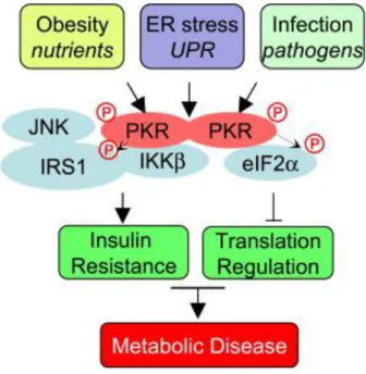
Figure 1.6. PKR induction, activation and its role in different cellular pathways. ... 30
Figure 1.7. PKR related inflammation pathways. ... 32
Figure 1.8. Hypothetical Metaflammasome complex and its components. ... 34
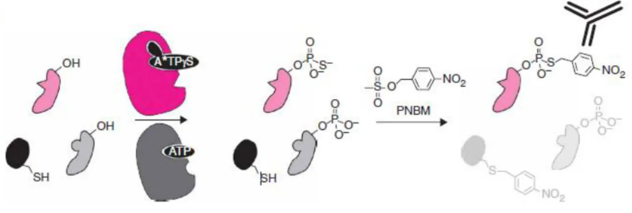
Figure 1.9. Strategy for labeling and recognizing individual kinase substrates through ASKA approach. ... 37
Figure 4.1. Genomic context and transcripts of PKR. ... 72
Figure 4.2. Chromosomal location and genomic context of PKR (EIF2AK2) gene in Homo Sapiens. ... 72
Figure 4.3. The conserved domains of PKR. ... 72
Figure 4.4. Multiple sequence alignment of kinases in order to identify specific gatekeeper residue in PKR via using Clustal OMEGA. ... 73
Figure 4.5. The hypothetical three dimensional (3D) structure of human PKR WT. ... 73
Figure 4.6. The hypothetical three dimensional (3D) model of PKR aligned with GCN2-ATP complex. ... 74
Figure 4.7. The hypothetical 3D model of PKR aligned with GCN2-1NM-PP1 complex. ... 75
Figure 4.8. Wild type human PKR in complex with ATP and 1NM-PP1. ... 76
Figure 4.9. Human PKR kinase-switch mutant in complex with 1NM-PP1 or ATP. ... 76
xvi
Figure 4.11. The sequencing result for M328A and mPKR M328G mutation. ... 78 Figure 4.12. PKR expression in HEK cells. ... 80 Figure 4.13. Bulky ATP analogs NaPP1 and 1NM-PP1 cannot inhibit wild type hPKR. ... 81 Figure 4.14. NaPP1 inhibits hPKR M366A ASKA mutant, whereas 1NM-PP1 cannot inhibit hPKR M366A mutant expressed in PKR-/- MEF cells. ... 81 Figure 4.15. NaPP1 inhibits hPKR M366G ASKA mutant, whereas 1NM-PP1 slightly reduces kinase activity of hPKR M366G mutant expressed in PKR-/- MEF cells. ... 82 Figure 4.16. Detection of PKR substrates with in vitro kinase assay. ... 83 Figure 4.17. The loss of IRE1 RNAse activity blocks PKR activation by lipids. ... 85
xvii
LIST OF TABLES
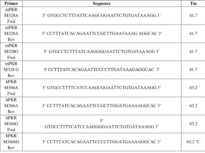
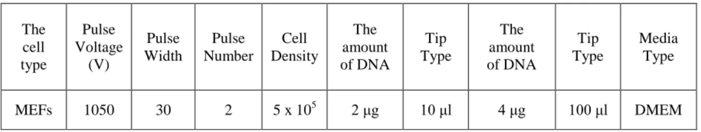
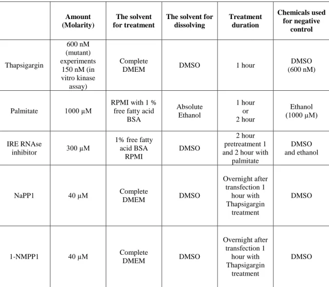
Table 3.1. The primer list (sequences and Tm) used during this research. ... 43 Table 3.2. The list of antibodies that were used during this research; including catalog number, working dilution and incubation times. ... 45 Table 3.3. Human PKR and Mouse PKR Primers. ... 57 Table 3.4. Optimal conditions for electroporation of plasmid DNA to MEFs with Neon Electroporation Kit. ... 63 Table 3.5. Optimized and used treatment conditions for cell lines used in this research... 64
1
1. INTRODUCTION
1.1. Endoplasmic Reticulum, Its Structure and Function
The endoplasmic reticulum (ER) is a central organelle for cellular homeostasis. ER, a membrane-bound organelle, is comprised of stacks of tubules and vesicles that connect it to a network of other membranous organelles2. Two types of ER are classified depending on association with or without ribosomes and called rough ER and smooth ER, respectively. The rough ER is important for protein synthesis. Secretory and transmembrane proteins are folded in the ER lumen and reach maturation before they leave the ER1. The smooth ER is the site for lipid and sterol synthesis as well as the major intracellular storage site for calcium (Ca2+)3. Furthermore, ER is the merging site for many extracellular and internal signals and orchestrates adaptive cellular responses to alternating cellular conditions. For example, via storing or releasing Ca2+, ER plays a role in muscle relaxation and contraction, learning and memory, metabolism and cell death2.
As the major reservoir for intracellular Ca2+, the ER can store up to three to four times more Ca2+ concentrations than in the cytosol. With the appropriate signal, this concentration gradient facilitates the exit of Ca+2 from the ER to the cytoplasm through one of the many ER membrane channels/receptors, such as Inositol triphosphate receptor (IP3R), leak channels and ryanodine receptor (RyR). In direct contrast to this, the sarcoplasmic-endoplasmic reticulum Ca2+ ATPase (SERCA) pumps take up calcium from cytosol into the ER lumen and as a result generate the high calcium gradient between ER and cytoplasm5.
Lots of enzymes and molecular chaperons function in the ER to facilitate its central role in protein folding, maturation and secretion. Collectively these are called the reticuloplasmins.
2
Molecular chaperones facilitate proteins folding by increasing the yield of structural maturation and folding rather than increasing the rate. One of the ER chaperones known as Binding immunoglobulin protein (BiP) also known as 78 kDa glucose-regulated protein (GRP-78), recognizes and binds to unfolded proteins through these proteins’ hydrophobic (specifically, aromatic) amino acids6. BiP also has an ATP hydrolysis function, acts as adenosinetriphopshatase (ATPase), it dissociates from its protein ligands when there is ATP. BiP/GRP78 protein and 94 kDa glucose-regulated protein (GRP94) bind to immunoglobulin heavy and light chains, and these ER stress proteins act as molecular chaperones and work together during folding and assembly of proteins8. Additionally, there are some ER-resident enzymes helping maturation and folding of proteins. For example, protein disulfide isomerase (PDI) acts as a catalyst in the formation of disulfide bridges. Another example is the calnexin/calreticulin/glycosyltransferase system, which helps the folding of proteins via interacting with nascent proteins and plays a role in ER-quality control system7. Newly synthesized unfolded proteins and proteins that reside in ER lumen are translocated into the lumen of the ER via one of the translocons called as Sec61 complex and are folded into mature forms with the assistance of molecular chaperones, helper folding enzymes and in the presence of high intralumenal Ca2+ concentrations. Then, these nascent polypeptides are N-glycosylated with N-glycosylating enzymes. Finally they mature into secondary and tertiary structures with the help of PDI. In addition, N-glycosylation enzymes and lectins also help the maturation of proteins7. After the mature forms of the proteins pass the ER quality control, they are then transported into the Golgi apparatus, whereas the unfolded or misfolded proteins are degraded by the highly conserved ER-associated degradation mechanism (ERAD).
In the ERAD pathway the unfolded proteins translocate into cytosol through the translocon channels for proteasomal degradation11. It was also showed that, Sec61 is needed for ERAD-L pathway which is a specific ERAD pathway that degrades integral unfolded ER luminal
3
proteins12,13. There are also other ERAD pathways such as ERAD-C, which is responsible for degrading membrane proteins with cytosolic lesions, and ERAD-M, which degrades membrane proteins with misfolded transmembrane domains143. To conclude, ER plays a multitude of important roles to maintain cellular homeostasis including but not limited to promoting protein folding, maturation and secretion.
Another fundamental but less well appreciated function of the ER is in lipid biosynthesis. It is the central site for synthesis of cholesterol, ceramides and phospholipids. When intracellular cholesterol levels are lowered this leads to activation of the ER resident sterol regulatory element-binding protein-1 and 2 (SREBP-1 and 2), which can transcriptionally upregulate the expression of key cholesterol synthesis enzymes15. When intracellular cholesterol is low, SREBP is released from Sterol regulatory element-binding protein cleavage-activating proteins (SCAP), which normally tethers SREBPs to the ER membranes. The SREBP can then translocate into Golgi to get cleaved and activated as a functional transcription factor. In particular SREBP1a and -2 upregulates synthesis of cholesterol, whereas SREBP1c upregulates the transcription of key enzymes in fatty acid synthesis pathways15. Insulin induced gene proteins (Insig-1 and 2), another ER membrane-bound proteins, negatively regulate fatty acid synthesis through controlling the activity of SREBP and 3-hydroxy-3-methylglutaryl coenzyme A reductase (HMGCoAR) degradation14, 144,145. In the presence of high amount of sterols, ER membrane proteins Insig-1 and Insig-2 interacts with HMGCoAR and recruits the tumor autocrine motility factor receptor protein gp78, a membrane-associated ubiquitin ligase, leading to ubiquitination of reductase. In result, the ubiquitinated reductase is degraded by the ERAD pathway14. Furthermore, when sterol levels are high, Insig-1 protein binds and changes the conformation of SCAP. Because of this conformational change, SCAP-SREBP-Insig-1 complex cannot be translocated to Golgi where SREBP is activated145. Another key lipid enzyme found the ER membrane is the Stearoyl-CoA desaturase (SCD), a
4
short lived enzyme which is needed for synthesizing monounsaturated fatty acids. SCD acts as a key component in hepatic synthesis of very-low-density proteins and triglycerides16. Also on the ER membrane, Diaclylycerol transferases (DGATs), act as catalysts in the final step of triacylglycerol (TAG) synthesis’17
. The ER-bound Acyl-coenzyme A: cholesterol acyltransferases (ACAT) catalyze the formation of cholesterol esters from cholesterol18. Some ER enzymes such as DGATs and acyl-CoA: sterol acyltransferases (ASATs) for steryl esters, act as catalysts in elongating fatty acid synthase synthesized palmitate product, which is beyond 16-C length. They also lengthen dietary polyunsaturated fatty acids synthesized by fatty acid synthase. Desaturases such as Delta6-desaturase, and some ER-resident proteins, NADH-cyt b5 Reductase and Cytochrome b5 take role in formation of double bond in fatty
acids20. To sum up, ER functions in lipid biosynthesis through these specific ER enzymes that regulate many cellular signaling pathways important in lipid biosynthesis.
Another essential role ER plays in lipid biology is contribution of the membrane components of the lipid droplets. Lipid droplets are major intracellular reservoir of mainly triacylglycerols (TAGs) and cholesterol esters (CEs). They consist of a hydrophobic neutral core containing these TAGs and CEs, surrounded by a phospholipid monolayer and cholesterol21. In addition, ER luminal proteins calnexin and BiP were detected in lipid droplet fractions22,147. Still there is limited information about lipid droplet assembly and expansion mechanisms and most models remain theoretical. Researchers strongly came up with two specific models for lipid droplet formation. These are the classical model and the hatching model. In the classical model, the budding lipid droplet is surrounded by the cytoplasmic leaflet of the ER membrane25. As ER luminal proteins calnexin and BiP association with lipid droplet was shown, Plough proposed another model for lipid droplet formation called Hatching model. In this model, lipid droplet is covered by phospholipid monolayer, with both cytosolic and luminal leaflets of ER membrane, this structure is bicellar structure22. This model also
5
supports the role of lipid droplet in misfolded protein degradation, as lipid droplet associates with luminal ER protein, BiP, bind to misfolded proteins and target them for degradation147. Recently, it was shown that several ER-resident lipid enzymes including the neutral lipid synthetic enzymes diacylglycerol acyltransferase 1 (DGAT) or 1-acylglycerol-3-phosphate acyltransferase (AGAT) also have role in lipid droplet formation24. Furthermore, it was shown that ER Glycerol-3-phosphate acyltransferase 4 (GPAT4) relocalizes from ER to lipid droplets and mediates their expansion2. Therefore, it can be concluded that ER functions in lipid droplet formation with specific ER enzymes and structural proteins which functions in formation of lipid droplets22,147.
Besides ER’s general functions that have been discussed, in the pancreatic beta cells ER assumes an especially important metabolic role in pro-insulin biosynthesis. The freshly translated insulin mRNA prepro-insulin gets translocated into the lumen of the ER, where ER proteases cleave off the signal peptide of turning preinsulin into insulin. Then pro-insulin is further folded by ER-enzymes via forming intramolecular double disulfide bonds. The properly folded pro-insulin is then translocated to Golgi and transferred into secretory granules. In secretory granules, pro-insulin is converted to mature insulin and released by exocytosis26. When the ER is stressed, however, insulin transcription, translation and secretion get blocked through several mechanisms involving the ER based stress response pathway26.
1.2. Endoplasmic Reticulum Stress
1.2.1. The Causes and the Consequences of Endoplasmic Reticulum Stress
ER is a central homeostatic organelle that functions in membrane protein and lipid biosynthesis, calcium homeostasis, secretion, folding and maturation of proteins41. If the ER cannot adapt to the ever-changing intracellular conditions, such as a dramatic increase in
6
unfolded or misfolded proteins, it undergoes an elaborative stress response called Unfolded Protein Response (UPR)41. ER stress can be induced by many other physiological and pathological stimuli such as environmental toxins, glucose deprivation, elevated saturated lipids or free cholesterol, viral infections, defects in protein trafficking, mutations in chaperone genes, aberrant calcium regulation, hypoxia and complex conditions associated with aging and tumorogenesis27,3,28. When ER stress is induced, unfolded proteins are identified through the Endoplasmic Reticulum Quality Control (ERQC) mechanism30. Specifically, calreticulin, calnexin and BiP chaperone proteins identify the unfolded proteins by the surface exposure of their hydrophobic sites, reactive thiols and immature glycans and bind these proteins to promote their proper folding30. Once these proteins are refolded they are released from the ER. Defects in the N-linked glycosylation pattern are also carefully monitored and such proteins become targets for ER α-mannosidases, following degradation-enhancing α-mannosidase-like protein (EDEM) binding to them30
. These are then translocated to cytoplasm and degraded via the ERAD pathway12,29,30.
ER stress can be viewed in three phases: acute, periodic and chronic32. The most widely studied form is the acute form, which can be experimentally induced via chemical treatments such as tunicamycin (protein glycosylation inhibitor), thapsigargin (SERCA pump inhibitor) and Dithiothreitol (DTT) (protein disulfide bond inhibitor)32. Exposure to toxic levels of saturated fatty acids or free cholesterol can also induce acute ER stress, whereas simultaneous treatments with unsaturated free fatty acids like oleate and palmitoleic acid can block this response32. Rhythmic or transient physiological activities such as feeding-fasting cycle, or transient glucose/lipid infusions induce the ―periodic‖ type of ER stress, and after each cycle, ER homeostasis is fully restored with help of UPR32. And the final form of ER stress is the ―chronic‖ form and in which ER homeostasis is never fully achieved in the continuous presence of ER-stress inducer leading to uninterrupted UPR signaling32. Chronic ER stress is
7
widely observed in metabolic diseases such as insulin resistance, obesity, dyslipidemias and atherosclerosis, in which the ER stressor being chronic nutrient excess32. Extended exposure to saturated fatty acids and cholesterol, lead to chronic, lipotoxic ER stress, which cannot be easily resolved32. A recent study in obese mice documenting reduction in ER protein synthesis and increase in lipid biosynthesis underscores the pivotal changes that occur around ER metabolism upon chronic, nutrient excess. Furthermore, the same study shows the alterations in ER lipid metabolism is coupled to the inhibition of SERCA activity and causally associated with chronic ER stress31.
1.2.2. The Unfolded Protein Response
The UPR is an elaborative, adaptive, intracellular signal transduction pathway initiated as a response to ER stress and first described in mammalian cells by Kozutsumi and colleagues in 198827, 33. In this study, investigators noted that expression of two ER-stress resident, glucose modulated proteins; GRP78/BiP and GRP94 are induced upon the accumulation of misfolded proteins at high temperatures33. In the same study, expression of wild type and mutant forms of influenza virus haemagglutinin (HA) protein in simian cells also lead to upregulation of the GRP78/BiP and GRP94 expression33. Unfolded protein response pathway was first described in yeast; a 22 base pair, cis-acting element (UPR element) was found sufficient for the induction of KAR2 (BiP) gene transcription by the accumulation of unfolded proteins34. Later it was shown that this UPR element is found in the promoter regions of five UPR target genes in yeast (karyogamy gene (KAR2) (homolog of BiP), protein disulfide isomerase gene (PDI1), protein disulfide isomerase EUG1 gene (EUG1), peptidylprolyl isomerase family gene (FKB2), and LHS1 Hsp70 family chaperone gene (LHS1)) and is required for their induction. Finally, the molecular UPR pathway was described upon the discovery of the yeast Hac1p, a UPR- activated transcription factor which binds to these UPR elements and activates the
8
transcription of ER chaperone proteins39,40. Hac1p is a basic-leucine transcription factor, which binds to UPR elements on genes that encode ER molecular chaperones38.
Peter Walter and his colleagues also identified IRE1, the key ER membrane protein that processes and activates Hac1p, in S.Cerevisae35. The human homolog of IRE1 gene, ERN1, encodes 1115 aminoacid length transmembrane kinase, Ire1p. Its glycosylated N-terminal side lies inside microsomes and its C-terminal cytosolic side has kinase activity36. Besides being kinase, Ire1p also has an endoribonuclease activity, which is responsible for the cleavage and activation of HAC1 mRNA37,38.
Historically, yeast IRE1 is the first discovered and conserved arm of the UPR that leads to the activation of XBP1 and production of molecular chaperoes50,28. In mammalian cells, in addition to the IRE-1, two additional UPR branches maintain ER homeostasis and are regulated by two ER membrane-bound proteins: PKR-like eukaryotic initiation factor 2 α kinase (PERK) and activating transcription factor 6 (ATF6)27. (Figure 1.1) A well-excepted model suggests that in homeostasis conditions, the molecular chaperone BiP/GRP78 interacts with PERK and IRE1 to keep them in an inactive state27. When ER homeostasis is disrupted, BiP/GRP78 dissociates from both PERK and IRE1 in order to bind to unfolded proteins. These kinases are thus activated and initiate the UPR41.
When ER homeostasis is disrupted, depending on the degree of disruption, adaptive or destructive downstream signaling pathways are activated upon the oligomerization and activating auto phosphorylation of IRE1 and PERK. In addition, ATF6 transcription factor translocates to Golgi, where it was cleaved by a serine protease site -1 protease (S1P), the metalloprotase site-2 protease (S2P) and it becomes fully activated. (Fig 1.1)
9
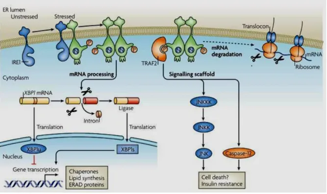
Figure 1.1. ER Stress and UPR pathway.
(reprinted with permission from Peter Walter and David Ron (2011) The Unfolded Protein Response: From Stress Pathway to Homeostatic Regulation. Science Reviews. Science 25 November 2011: Vol. 334 no. 6059 pp. 1081-1086 DOI: 10.1126/science.1209038.41) When there is ER stress, cell elicits a unique response called Unfolded Protein Response. During unfolded protein response three ER stress sensors PERK (PKR resemble ER kinase), activating transcription factor 6 (ATF 6) and Inositol requiring enzyme 1 (IRE1) are activated which represent three distinct arms of UPR. PERK and IRE1 are activated through auto phosphorylation and ATF6 is activated through cleavage by specific Golgi proteases S1P and S2P and when activated acts as transcription factor via upregulating UPR target genes’ expression49,76,81.When PERK is activated, it phosphorylates eIF2α,
leading to translational inhibition and specific ATF4 mRNA translation. ATF4 acts as transcription factor and up regulates chaperones’ genes expression such as XBP1 and CHOP. IRE1 splices XBP1 mRNA through its RNAse activity and leads to its translation and translated XBP1 protein acts as transcription factor via upregulating chaperones’ lipid synthesis’ ERAD proteins’ gene expression41
.
The first discovered arm of the UPR is regulated by IRE1 and conserved from yeast to mammals. There are two types of mammalian IRE1, which has differential tissue expression pattern. IRE1α is expressed in many tissues such as liver, skeletal muscle, lung, placenta and particularly in pancreas, whereas IRE1β is only expressed in intestinal epithelia51,52
. For activation, IRE1 first homo-oligomerizes and then auto-phosphorylates itself at serine 724 in its C-terminal kinase domain53. The only known substrate of IRE1 kinase is itself. Besides its kinase activity, IRE1 also has a unique endoribonuclease activity, which cleaves HAC1
10
mRNA in yeast or the homologous XBP1 (X-box binding protein) mRNA in metazoans53,54. The cleavage of the 26 nucleotide from the intron of XBP1 produces 41 kDA size, active XBP1 transcription factor. IRE1 also nonspecifically cleaves mRNAs on ER membrane in order to reduce protein synthesis and loading of protein synthesis to ER (IRE1 dependent mRNA decay pathway (RIDD). Recently, it was shown that IRE1α endoribonuclease (RNAse) domain also leads to fast decay of pre-miRNAs 17, 34, 96 and 125. These miRNAs all target caspase-2 mRNA translation. Therefore it can be concluded that, under ER stress conditions, IRE1α positively regulates expression of apoptotic Caspase-2 protein via inhibiting maturation of select miRNAs that have role in mediating the apoptotic outcome of UPR58.
Active XBP1 transcription factor up regulates the expression of UPR target genes including ER chaperones, ERAD proteins and ER membrane expansion related phospholipid synthesizing enzymes54,55. Furthermore XBP1 can work in coordination with ATF6 to induce the heat shock protein 40 member DnaJ (Hsp40) homolog, subfamily C, member 3 protein P58IPK. This protein assists in protein folding by behaving like a co-chaperone and
furthermore, can bind and inhibit PERK and PKR activities49, 142.
Additionally, IRE1 is known to interact with adaptor proteins like such as TNF receptor-associated factor 2 (TRAF2) in order to stimulate apoptosis signal regulating kinase (ASK1). ASK1 activates the pro-apoptotic cJUN NH2-terminal Kinase (JNK), p38 mitogen activated kinase (p38MAPK) and caspase 1257. Under normal conditions caspase 12 is bound and inactivated by TRAF2, but when there is ER stress caspase 12 dissociates from TRAF2 complex and this activated form leads to cell death 57 . IRE1 also through interaction with TRAF2 regulates JNK-AP1 and nuclear factor қB (NF-қB) pathways (Figure 1.2). Finally, by activating JNK, IRE1 can indirectly regulate insulin synthesis proteins Insulin receptor
11
substrate 1 (IRS1) and Insulin receptor substrate 2 (IRS2) via its JNK-induced inhibitory phosphorylation28.
Figure 1.2. IRE1 arm of the Unfolded Protein Response.
(reprinted with permission from Ron, D., and Walter, P. (2007). Signal integration in the endoplasmic reticulum unfolded protein response. Nat. Rev. Mol. Cell Biol. 8, 519–529. © Nature publishing Group, 200750.). In the presence of ER stress, IRE1 oligomerizes and autophosphorylates itself, leading to its activation50. After it is activated, through its RNAse domain, it splices XBP1 mRNA, which is then translated and acts as a transcription factor for expression of chaperones’, lipid synthesis’ and ERAD proteins’ genes50. It also interacts with TRAF2
through its kinase domain resulting in its activation and activated TRAF2 starts a signaling cascade, interacts and activates mitogen activated protein kinases leading to insulin resistance154. It also interacts with Caspase-12 leading to cell death50.
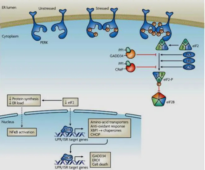
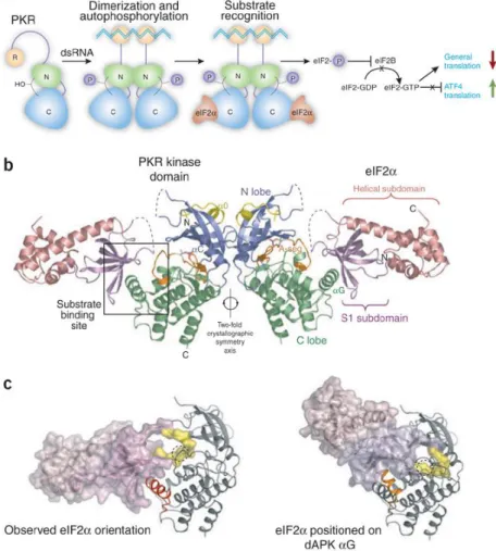
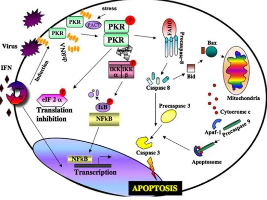
The second UPR arm is maintained by PERK. PERK is a type I transmembrane serine/threonine kinase which has N-terminal luminal ER stress sensor and a cytosolic kinase domain. Similar to IRE1 activation mechanism, PERK can oligomerize and auto phosphorylate (at threonine 981) upon dissociation of BiP42. PERK shares similarities with the three other eIF2α phosphorylating kinases at its C-terminal kinase domain43. The other eIF2α kinases are the interferon inducible viral infection derived double strand RNA activated Protein Kinase R (PKR), the amino acid deprivation activated general control
non-12
depressible kinase 2 (GCN2) and the heme regulated eif2α kinase (HRI)43 . Eif2α is a key protein that controls protein translation mechanism by transporting the initiator methionyl-transfer RNA (met-tRNA) to the ribosome in order to initiate protein translation44. When PERK is activated, it inactivates eukaryotic initiation factor 2 α (eIF2α) through phosphorylating it at Serine 51 site and thus, resulting in general protein translation inhibition. In this way, PERK reduces protein overload in the ER to alleviate ER stress. (Figure 1.3). PERK activity also induces activation of the nuclear factor kappa β (NF-қB)genes45. NF- қB has many functions especially as a key regulator of inflammation50. eIF2α phosphorylation leads to phosphorylation and translational suppression of inhibitory kappa B (IқB) resulting in release of inflammatory cytokines such as IL-6 and TNF-α46,47. Another protein PERK associates and phosphorylates is the Nuclear erytroid factor 228. Phosphorylation of the Nrf2/Keap1 (Kelch-like ECH-associated Protein 1) complex leads to Nrf2 dissociation and transport to nucleus28,. Nrf2 is a transcription factor that has role in eliciting antioxidant response146. In contrary with its role in general protein translation inhibition, PERK activates Activating Transcription Factor-4 (ATF-4) translation leading to the transcription of UPR target genes and chaperones (Figure 1.3)27,41. ATF4 stimulates transcriptional activation of apoptosis, inflammation, glucose metabolism, ER redox control, negative feedback eIF2α phosphorylation inhibitory genes28. Transcription factor C/EBP homologous protein also called CCAAT/enhancer binding proiculutein (CHOP) 41 and growth arrest- and DNA damage inducible- 34 (GADD34) 41 , ER redox control (ERO1) and activating transcription factor 3 (ATF3) are four gene targets of ATF428,44. GADD34 interacts with catalytic subunit of protein phosphatase (PP) and dephosphorylates eIF2α relieving its inhibition, leading to translational recovery44. Ero1 functions in balancing the redox potential in the ER191. CHOP is a transcription factor which regulates activation of apoptosis genes. ATF6 stimulated P58IPK
13
expressed several hours after PERK activation, this shows that in the end of UPR P58IPK
expression is induced and cells go apoptosis48,49. Therefore, it can be observed that besides its protective role and maintaining ER homeostasis through up regulation of UPR chaperones’ expression to relieve ER stress, PERK can also contribute to cell death pathways when ER stress is prolonged48,49,50.
Figure 1.3. PERK arm of the Unfolded Protein Response.
(reprinted with permission from Ron, D., and Walter, P. (2007). Signal integration in the endoplasmic reticulum unfolded protein response. Nat. Rev. Mol. Cell Biol. 8, 519–529. © Nature publishing Group, 200750.). In the presence of ER stress, PERK is activated through auto phosphorylation, leading to phosphorylation and inactivation of eIF2α. Besides leading to translational suppression, eIF2α results in ATF4 mRNA translation, therefore leading to UPR/ISR target genes expression such as XB1 and CHOP 62,45,149. CHOP then acts as transcription factor resulting in activation of GADD34, ERO1 genes and cell death genes expression 61,65,66,67.
ATF6 is another proximal sensor in the ER stress response and responsible for the initiation of the third branch of the UPR50. ATF6 is a 90 kDA Basic Leucine Zipper (bZIP) protein, which
14
is activated after cleavage by specific Golgi-resident proteases50. When ER stress is induced, ATF6 is reduced, and this monomeric, reduced ATF6 translocates to the Golgi, where it is cleaved by site-1 and site-2 protease (S1P and S2P)50. These proteases specifically cleave ATF6 from the luminal domain and its trans-membrane anchor. The active ATF6 (50 kDA after cleavage) translocates to the Golgi and binds to promoters that contain ER stress response element (ERSE; CCAAT(N)9CCACG), which are found in genes like ERAD pathway proteins, lipid biosynthesis, and ER chaperones59. There are two isoforms of ATF6, ATF6α and ATF6β which become activated with ER stress. Studies show that ATF6 also induces the expression of XBP159 and P58IPK, thus intercepting both the IRE-1 and PERK
branches49. On the other hand, the Wolfram Syndrome 1 (WSF1) protein can regulate ATF6 activity via targeting ATF6 for ubiquitylation and proteasomal degradation by HMG-CoA reductase degradation protein 1 (HRD1) and E3 ubiquitin ligase60. Recently, it was showed that ATF6 indirectly down regulates B-cell lymphoma 2 (BCL-2) family member myeloid cell leukemia sequence 1 protein (MCL-1) in myoblast cell line, pointing out the potential role of ATF6 in initiating apoptosis during ER stress61. It was also shown that, homologs of ATF6 like cAMP responsive element binding protein 3-like 3 (CREBH), old astrocyte specifically induced substance (OASIS), basic leucine zipper transcription factor (LUMAN also known as LZIP), cAMP responsive element binding protein 3-like 4 (CREB4) and box B-binding factor 2 human homolog on chromosome 7 (BBF2H7) are also similarly proteolytically cleaved in Golgi and give different responses under ER stress. (Figure 1.4)50.
15
Figure 1.4. ATF6 branch of unfolded protein response.
(reprinted with permission from Ron, D., and Walter, P. (2007). Signal integration in the endoplasmic reticulum unfolded protein response. Nat. Rev. Mol. Cell Biol. 8, 519–529. © Nature publishing Group, 200750). In the presence of ER stress, ATF6 dissociates from BiP and is transported to Golgi apparatus together with CREBH49,76,81. The lumenal site-1 protease and the intra-membrane site 2 protease cleave CREBH and ATF6 to activate these transcription factors76,81. These activated transcription factors then translocate to nucleus leading to ATF6 activation of UPR target genes and CREBH activation of acute phase genes’ expression.
1.2.3. The Unfolded Protein Response Outputs: Adaptive or Pro-Survival, Destructive or Pro-Apoptotic
The accumulation of unfolded proteins in the ER activates the UPR50. UPR first tries to restore folding process by inducing transcription of ER chaperones and ERAD proteins and reducing protein overload by inhibiting general translation61. It also promotes cell cycle arrest in G1 phase148. However if ER stress is prolonged, this response also results in apoptosis61. There are many UPR mediated mechanisms for adaptation to ER stress. For example, PERK phosphorylates eIF2α, resulting in translational inhibition and reduction of the protein overload in the ER50. In addition, PERK activation leads to cell cycle arrest through the loss of short-lived cell cycle regulatory proteins, such as CyclinD162. Even though general translation is inhibited as a result of PERK’s activity, certain select mRNAs’ translation continues in an increased manner due to their special regulation under the UPR149. One such
16
mRNA is ATF4 mRNA, whose translation increases in an eIF2α phosphorylation-dependent manner149.
ATF4 translocates to nucleus and activates expression of many genes which then help restore ER homeostasis and provide amino acid sufficiency149. PERK also activates NRF2 by phosphorylation and leads to its dissociation from KEAP145,63. Nrf2 then translocates into the nucleus and activates the expression of many antioxidant genes. This constitutes an important adaptive mechanism governed by PERK as NRF2 deficient cells showed increased rate of apoptosis under ER stress63, 45.
Other adaptive responses to ER stress are managed by the ATF6 arm. After its activation, ATF6 translocates into the nucleus and induces the expression of many genes that encode ER chaperone and ERAD proteins61. ATF6 also induces XBP1 transcription, which is another important factor for cell survival52.
The IRE1 arm also has a significant role in eliciting adaptive response. The main adaptive response of IRE1 is mediated by the spliced XBP1 mRNA, which generates the active XBP-1 transcription factor. XBP1 is a transcription factor that induces many genes that have role in protein folding and ERAD pathway64. XBP1 also regulates ER folding capacity itself64. Not all outcomes governed by UPR favor the cell’s survival. UPR can also promote apoptosis when ER stress is prolonged62,63,66,67. For example, PERK activates ATF4, which transcribes the CHOP gene, an activating transcription factor in apoptosis. CHOP down regulates transcription of anti-apoptotic BCL-2 and it up regulates pro-apoptotic BH3-only protein Bim65,66 and pro-apoptotic death receptor DR5 expression65,66,67. ERO1α, GADD34 and telomere repeat binding factor 3 (TRB3) are many other CHOP target genes have role in cell death mechanisms61. Recently, it was shown that the ATF6 arm also has an indirect role in triggering apoptosis via down regulation of anti-apoptotic BCL-2 family member MCL-1 in a myobast cell line61. Furthermore, studies also show that eventually IRE1 is turned off in
17
prolonged ER stress, whereas PERK arm remains on, suggesting the anti-survival role is dominated by PERK in continuous stress50. Nevertheless, IRE1 can also be pro-apoptotic by interacting with tumour necrosis factor receptor-associated factor 2 (TRAF2) and leading to Apoptosis signal-regulating kinase 1 (ASK1) and c-Jun N-terminal kinase (JNK) activation, which result in apoptotic death57. Under ER stress, JNK phosphorylates and suppresses BCL-2150 and B-cell lymphoma extra-large protein (BCL-XL)150 anti-apoptotic activity and it also
phosphorylates BH3 interacting-domain death agonist protein (BID) and proapoptotic BH3-only protein BIM, making these pro-apoptotic proteins active151. Recently Han et al., showed that IRE1 dependent RIDD pathway also have role in induction of cell death in the pancreatic beta cell line (INS-1)68.
1.3. The Relationship between Endoplasmic Reticulum Stress and Inflammation
Unfolded protein response is associated with inflammation through multiple mechanisms including interaction of the UPR branches with JNK and NF- қB pathways, by promoting ER ROS production and Nitric Oxide formation during ER stress and inflammation28.These pathways also play a major function in sterile inflammation observed metabolic diseases such as obesity, atherosclerosis, insulin resistance and diabetes69. Recent studies show that UPR signaling pathways result in production of many proinflammatory cytokines, through multiple mechanisms69. The historically first discovered and conserved branch of UPR, IRE1 pathway activates JNK leading to activation of the transcription factor activator protein 1 (AP1)69,70. AP1 translocates to the nucleus and activates transcription of inflammatory genes including tumor necrosis factor (TNF), keratinocyte growth factor (KGF), granulocyte macrophage colony (GM-CSF), interleukin-8 (IL-8) and some other cytokine receptors70. IRE1 also regulates inflammation related Mitogen activated protein kinase p38 and extracellular signal related kinase (ERK) via binding to non-catalytic region of tyrosine kinase adaptor protein
18
Nck71. Additionally, IRE1 signaling can interfere with NF-κB pathway by interacting with inhibitory IκB kinase (IKK) through TRAF2 complex50
. Recently it was shown that IRE1 kinase activity and not endoribonuclease activity activates NF-κB pathway through interacting with TRAF2/IKK complex and maintaining IKK activity72. Also shown was that in JNK deficient cells, tumor necrosis factor alpha (TNF-alpha), interleukin (IL6) and monocyte chemotactic protein-1 (MCP-1) proinflammatory cytokines’ expression are repressed28,152,153,154. As IRE1 can lead to the activation of JNK, it could induce the transcription of these pro-inflammatory cytokines28. Moreover, XBP1, downstream of IRE1, is necessary for B cells to secrete antibody, when they are induced with antigen73,74. XBP1 also has a role in production and release of antimicrobial peptides from Paneth cells73,74. NF- κB has also been shown to be regulated by the PERK arm through eif2α mediated inhibitor kappa B (IқB) translation suppression that results in NFқB activation46,50,160. Without IқB
present, NFқB translocates to the nucleus and induces expression of inflammatory cytokines69
. The third UPR arm, ATF6, has also been shown to activate NF-κB via Akt during subtilase cytotoxin induced ER stress75 ATF6 appears to play an additional role in initiation of acute phase response (APR), a highly complex inflammatory process observed at the onset of serious diseases such as infection, trauma and inflammation69. One study shows that in the liver, cyclic-AMP responsive element binding protein H (CREBH) cooperates with ATF6 in APR activation76.
Another link between ER stress and inflammation appears to be the ER-generated reactive oxygen species and oxidative stress45. Depending on UPR, Ero1p and endoplasmic reticulum flavin-linked sulfhydryl oxidase (Erv2p) enzymes up regulate chaperone proteins’ expressions through forming disulfide bonds in proteins77. These enzymes can also reduce molecular oxygen. This reduced molecular oxygen leads to oxidative stress and ER stress77. The activation of PERK then initiates anti-oxidant response with help of Nrf2 activation by
19
PERK45. Moreover, ER stress induces the transcription of inducible nitric oxide synthase (iNOS) via NF-κB activation, but which of the UPR arms is responsible remains unknown78. 1.4. The role of Endoplasmic Reticulum Stress and Endoplasmic Reticulum Stress
related Metabolic Diseases
Over the past decade we have reached an understanding on the importance of ER homeostasis for metabolic well-being and an important target to prevent chronic, inflammatory and metabolic diseases such as obesity, diabetes, insulin resistance, fatty liver and atherosclerosis32,155. Early studies showed that the UPR arms are activated in the liver and adipose tissue of rodents during obesity and its reduction by chemical chaperons can prevent insulin resistance and metabolic disease32,79,155. These landmark studies by Hotamisligil’ group were also extended to human obesity and has led to many groups to embark on research to understand the role of and the molecular mechanisms leading to ER Stress and UPR in chronic metabolic diseases157,158,159.
Several mechanisms have been demonstrated regarding how ER stress is tied to the development of insulin resistance and diabetes in obesity. For example, upon induction of UPR, JNK-AP1 pathway is activated in an IRE1-dependent manner. JNK has been shown to phosphorylate the insulin receptor substrate 1 (IRS1) at Serine 307 position, leading to the disruption of insulin signaling and promoting insulin resistance80. PERK also increases JNK activity, through unclear mechanisms28. Additionally, ATF675 and PERK46 activate NF-κB, which then transclocates into the nucleus and induces the expression of pro-inflammatory cytokines50,190. Other UPR-activated transcription factors can directly modulate expression of key lipogenic and gluconeogenic enzymes that contribute to the development of insulin resistance. For example, SREBP-1c161,162,163, XBP1160, and NRF2164,165 are such transcription factors that can lead to abnormal expression of lipogenic genes. On the other hand, CREBH
20
upregulates gluconeogenic genes while ATF6 and XBP1 downregulate their expression to promote insulin resistance81. UPR may also indirectly promote hepatic insulin resistance and hepatic steatosis via leading fat accumulation in the ER and inhibition of VLDL export through disrupting VLDL production by increasing apolipoprotein B100 (apoB100) degradation81,169,170. PERK- activated ATF4 is another factor that disrupts insulin signaling via activating the pseudokinase tribbles homolog 3 (TRB3) expression that negatively regulating protein kinase B (PKB), downstream component of the insulin signaling pathway82. ER stress can be caused by hepatic steatosis in ob/ob mice and this stress results in proteolytic cleavage of SREBP1c and SREBP2 transcription factors which upregulates sterol synthesis163. When BiP is overexpressed adenovirally in hepatocytes of ob/ob mice, SREBP1c activation decreases resulting in reduced hepatic triglycerides, and improved insulin sensitivity163. Additionally, it was showed that XBP1 deficient mice developed insulin resistance upon high fat diet induced obesity79.In hepatocytes, depending on insulin signaling, heterodimer structure formed by the regulatory subunits of phosphotidyl inositol 3-kinase (PI3K), p85α (encoded by Pik3r1) and p85β (encoded by Pik3r2) is altered leading to interaction of p85α and p85β with XBP1s. Then this p85-XBP1s complex translocates to nucleus, resulting in activation of expression of UPR target genes167. In insulin resistant, ob/ob mice, the XBP1s-p85 interaction is disrupted and when XBP1s-p85 is overexpressed in these mice, translocation of XBP1s to nucleus is retained, and glucose tolerance is improved167.According to a recent study, XBP1 was also shown to regulate gluconeogenic transcription factor Forkhead box protein O1 (FoxO1) resulting in affection of glucose homeostasis in obese mice166. Independent from its role in UPR, XBP1 improves glucose homeostasis through ubiquitination and degradation of FoxO1 in obese mice166. To sum up, XBP1 can improve insulin sensitivity through inducing UPR target genes’ expression such as Dnajb9168
21
also improve glucose tolerance in insulin resistant ob/ob mice by blocking gluconeogenesis as it interacts and degrades FoxO1166.
ER stress is also observed in adipose tissue of obese individuals. ATF6-regulated chaperones’ expression158, eif2α phosphorylation158 and expression of IRE1157 together with JNK157 and XBP1s157, increase in adipose tissue of obese individuals. In one research, it was shown that several ER-stress related proteins such as calreticulin, protein disulfide-isomerase A3, glutathione-S-transferase, calnexin, JNK-1 and XBP1s are upregulated in subcutaneous adipose tissue of obese individuals172. This result shows UPR activation in subcutaneous adipose tissue of humans172.
Even researchers showed the presence of ER stress in mouse skeletal muscle after high fat diet for 4 weeks173, the relationship between ER stress and insulin resistance is more complex compared to other tissues. For example, UPR elements’ expression levels remain unchanged in muscle tissue after HFD, however insulin resistance and lipogenesis increases174. Interestingly, researchers showed that single physical exercise causes ER stress in muscle whereas exercise training result in adaptive UPR through peroxisome proliferators-activator receptor gamma coactivator-1 alpha (PGC-1α) dependent coactivation of ATF6175. Interestingly, it was demonstrated that, in human myotube cells, when palmitate inducible Stearoyl-CoA Desaturase 1 (SCD1) expression increases. Depending on the high inducibility of SCD1 expression depending on palmitate treatment, insulin sensitivity also increases176. The increase in IRE1 and PERK phosphorylation together with JNK and BiP activity seen in the liver and adipose tissue of obese mice, suggested similar signs of ER stress may be evident in the brain. Indeed, a study showed ER stress leads to the activation of the NFκB pathway and inflammation in the hypothalamus, by engaging the Toll like receptor signaling83. Saturated free fatty acids, such as palmitate lead to activation of mostly TLR4 and modestly TLR2177. It is not clear; however, what is the direct ligand that stimulates hypothalamic Toll
22
like receptors177. After TLR4 and TLR2 are activated, they activate JNK and IκB kinase resulting in inflammation and leptin/insulin resistance177. Furthermore, chemical chaperone application inhibited NFκB signaling and reduced leptin- resistance84. Another recent research demonstrated that unsaturated fatty acids as well as chemical chaperones can revert high fat diet induced hypothalamic inflammation and can reduce body mass and adipocity178. In addition, reduction in body fat mass by dieting leads to decrease in the hypothalamic PERK acitivity, whereas ATF6, BiP and Xbp1 mRNA expression remain induced85 . Very recently, it was shown that during diet-induced obesity, post translational processing of proopiomelanocortin (POMC) mRNA is impaired by ER stress leading to reduced production of appetite-suppressing neuropeptide α-melanocyte-stimulating hormone (α-MSH) as this hormone’s synthesis is catalyzed by POMC protein179
. In this study, researchers demonstrated for the first time that in obesity, ER stress contributes in regulation of energy balance through altering neuropeptide processing179. From all these studies, it can be concluded that excess nutrients result in ER stress, which elicits an elaborative adaptive response UPR in hypothalamus, muscle, liver and adipose cells in obesity and diabetes. Even the mechanisms eliciting this response is not fully clarified, some results are common between these tissues. UPR causes inflammation and insulin/leptin resistance in the hypothalamus, liver and fat cells through some specific cellular pathways as explained above83.
1.5. Restoring ER function
After discovering the causality of ER stress in obesity induced metabolic diseases, a search to define novel approaches to restoring ER function has begun and the ER is viewed as a promising therapeutic target. For example, one of the first trials in rodents consisted of applying 4-phenylbutyric acid (PBA), a chemical chaperone which inhibits ER-stress related apoptosis, to mouse models of obesity and insulin resistance. PBA has been known to have a
23
protective function in cerebral ischemia and liver ischemia reperfusion injury86,87. Studies show it is also effective means to reduce ER stress and improve systemic metabolic homeostasis and insulin sensitivity in mouse models of obesity79. Another chemical chaperone, Tauroursodeoxycholic acid (TUDCA) also inhibits ER –stress and improves metabolism in mice79. Furthermore, TUDCA blocks ER stress-induced apoptosis in human liver cells through yet unknown mechanism88. Furthermore, chemical chaperone PBA also has been studied in another chronic, metabolic disease model, atherosclerosis, in mice. When PBA was chronically administered to Apolipoprotein E null (ApoE−/−) mouse model of atherosclerosis, ER stress was significantly reduced in atherosclerotic plaques88. Treatment with PBA reduced PERK phosphorylation and ATF3 expression in macrophages infiltrating the atherosclerotic plaques, as well as in isolated macrophages, resulting in protective effect against hypercholestrolemic stress88. Furthermore, a genetic-deficiency in a macrophage lipid chaperone also protected against ER stress and atherosclerosis. In addition, the lipid product of aP2 activity, a monounsaturated fatty acid known as palmitoleate, was also found to be protective effects against lipid-induced ER stress in macrophages82. Diakogiannaki et al., in their research also showed that when palmitoleate is given to β cells for 18 hours, Tunicamycin and Palmitate induced ER stress can be reduced89. Collectively, these data show that when palmitate (saturated and toxic) and palmitoleate (monounsaturated and non-toxic) are given in a combination treatment, ER stress and cell damage are prevented and meanwhile, evidence suggests ER membrane expands less in the combination treatment when compared to only palmitate treatment. This result shows that palmitoleate, also quite bioactive when given systemically, has protective effect on ER structure and function and a promising dietary approach to restoring ER stress in chronic metabolic diseases like atherosclerosis. These and other possibilities to reduce ER stress need to be vigilantly tested.