Research Article
Metin Yıldırımkaya*, Sedat Abusoglu, Setenay Arzu Yilmaz,
Beyza Saracligil, Esra Paydas Hataysal, Büsra Ecer and Ali Unlu
Serum and cord blood-methylated
arginine levels in gestational diabetic subjects
Gestasyonel Diyabetik Olgularda Serum ve Kord
Kan-Metillenmiş Arginin Seviyeleri
https://doi.org/10.1515/tjb-2018-0201
Received June 3, 2018; accepted August 2, 2018; previously published online December 25, 2018
Abstract
Objectives: Micro- and macrovascular endothelial
dete-rioration has been mentioned in diabetic pregnants with critical clinical outcome for the fetus. Our aim was to measure serum and cord blood concentrations of methyl-ated arginines in patients with gestational diabetes and find a relationship with endothelial dysfunction.
Materials and methods: Methylated arginines were
detected with high performance liquid chromatography mass spectrometry via electrospray ionization positive technique with a chromatographic C18 column.
Results: Although gestational diabetes mellitus (GDM)
groups’ asymmetric dimethylarginine (ADMA) levels were higher compared to control group, this difference was not significant. Control cord blood ADMA and N-monomethy-larginine levels were significantly higher than insulin-reg-ulated GDM cord blood ADMA and N-monomethylarginine levels (p = 0.001; p = 0.003, respectively). Diet-regulated GDM group’s cord blood N-monomethylarginine was sig-nificantly higher than insulin-regulated GDM group’s cord blood N-monomethylarginine (p = 0.045). A negative
correlation was found between cord blood symmetric dimethylarginine and oral glucose tolerance testing 0h glucose values (r = −0.453, p = 0.002).
Conclusions: According to this study’s results, methylated
arginine levels may not be associated with endothelial deterioration in GDM otherwise with preeclampsia risk.
Keywords: Methylated arginines; Gestational diabetes;
impaired glucose tolerance; Tandem mass spectrometry; Endothelial dysfunction.
Öz
Amaç: Diabetik gebelerde, fetüs için kritik klinik
sonuç-lara yol açacak mikro- ve makrovasküler endotelyal bozulma bildirilmiştir. Amacımız, gestasyonel diabeti olan hastalarda metilenmiş arjininlerin serum ve kord kanı konsantrasyonlarını ölçmek ve endotelyal disfonksi-yon ile ilişkisini ortaya koymaktır.
Gereç ve Yöntemler: Metillenmiş arjinin türevleri,
elekt-rosprey iyonizasyon pozitif mod tekniği ile kromatografik C18 kolon kullanılarak yüksek performanslı sıvı kroma-tografi kütle spektrometre yöntemi ile saptanmıştır.
Bulgular: Her ne kadar gestasyonel diabetes mellitus
grup-larının asimetrik dimetilarjinin seviyeleri kontrol grubuna kıyasla yüksek olsa da, bu fark önemli bulunamamıştır. Kontrol kord kanı asimetrik dimetilarjinin ve N-monome-tilarjinin seviyeleri, insülin-regüle gestasyonel diabetes mellitus grubunun kord kanı asimetrik dimetilarjinin ve N-monometilarjinin seviyelerinden anlamlı olarak yüksek idi (Sırasıyla, p = 0.001; p = 0.003). Diet-regüle gestasyo-nel diabetes mellitus grubunun kord kanı N-monometi-larjinin seviyeleri, insülin-regüle gestasyonel diabetes mellitus grubunun kord kanı N-monometilarjinin seviye-lerinden anlamlı olarak yüksek idi (p = 0.045). Kord kanı simetrik dimetilarjinin ve oral glukoz toleras testi 0.saat glukoz düzeyleri arasında negatif bir korelasyon saptandı (r = −0.453, p = 0.002).
*Corresponding author: Metin Yıldırımkaya, MD, Department of
Biochemistry, Lokman Hekim University Faculty of Medicine, 06311, Ankara, Turkey, Mobile: +90 5323752771, Phone: +90 3124449911, Fax: +90 3122760818, e-mail: [email protected]. https://orcid.org/0000-0002-4588-3797
Sedat Abusoglu, Esra Paydas Hataysal, Büsra Ecer and Ali Unlu:
Department of Biochemistry, Selcuk University Faculty of Medicine, Konya, Turkey
Setenay Arzu Yilmaz: Department of Obstetrics and Gynecology,
Selcuk University Faculty of Medicine, Konya, Turkey
Beyza Saracligil: Department of Biochemistry, Karatay University
Sonuçlar: Bu çalışmanın sonuçlarına göre metilenmiş
arjinin seviyeleri, preeklempsi riski olmadığı durumda gestasyonel diabetes mellitus hastalığında endotelyal bozulma ile birliktelik göstermeyebilir.
Anahtar kelimeler: Metilenmiş arjininler; Gestasyonel
diabet; Bozulmuş glukoz toleransı; Sıralı kütle spektro-metresi; Endotelyal disfonksiyon.
Introduction
Gestational diabetes mellitus (GDM), identified as glucose intolerance with the period of pregnancy, is one of widely acquired obstetrical complications, affect-ing 5–8% of all pregnancies. Duraffect-ing last decade, the incidence of GDM was notified to rise up [1]. GDM rep-resents a major risk for fetal development and outcome and is of importance for development of such metabolic and cardiovascular consequences [2]. Hyperglycaemia becomes evident after birth. These individuals are at high risk of developing type 2 diabetes and distinct car-diovascular disease (CVD). Therefore, early diagnosis of GDM might be useful for clinicians to identify a pro-portionally young individuals at high risk for cardiovas-cular outcomes and to respond promptly to reduce this possible consequences [3]. Vascular reactivity might be explained by many pathways such as locally generated vasoactive chemicals from the endothelium. Several pathological consitions are related with attenuated potential of endothelium to produce the efficient vasodi-lator nitric oxide (NO) [4]. NO is a potent vasodivasodi-lator, and has an important role for the function of endothelial pro-genitor cells, which can immigrate to and fractionate at damage area to support the formation of new vessels in healthy subjects [5]. Lack of endothelial properties such as organization of vascular tonus means that deficiency of cellular functions, mentioned as endothelial dysfunc-tion [6]. Methylated arginine derivaties are compounds synthesized via the action of enzymes called protein methyltransferases (PRMT) types I and II (PRMT I and II). Methylated arginines including asymmetric dimethy-larginine (ADMA), symmetric dimethydimethy-larginine (SDMA) and N-monomethylarginine (L-NMMA) has a capacity of acting as inhibitor molecules for NO production by nitric oxide synthase (NOS). Whereas the structure of ADMA is similar to arginine, it competes with arginine for NOS binding, hereby blocking the formation of NO from arginine by NOS directly. NOS is mainly localized in the cell, thus the intracellular ADMA and arginine levels regulate NOS activity. In addition, extracellular ADMA is
an antagonist to extracellular arginine on cell membrane transporter level, whereas they are both transported into the cell via the cell membrane by the cationic amino acid transporter (CAT). Since ADMA competes with arginine for NOS and for cell transport via CAT-2, the bioavail-ability of NO depends on the balance between the two, the so-called arginine/ADMA ratio. The arginine/ADMA ratio is an important indicator of NO bioavailability and therefore of the risk of formation of atherosclerotic plaques [7].
Even though L-NMMA and ADMA has been con-cluded to have potential fundamental inhibitory effects on NO synthesis, SDMA is also known to prevent the uptake of basic NO substrate, arginine. Due to this prop-erty, SDMA has also effects to diminish NO production [8]. NO synthase activity is regulated by both negative feedback and availability of L-arginine. L-Arginine is a semi-essential amino acid obtained either from the diet or synthesized endogenously from L-citrulline. L-citrul-line, in turn, can be derived from ornithine in the catab-olism of proline or glutamine and glutamate, or from L-arginine via arginine–citrulline pathway. L-Citrulline can also be obtained during the degradation of ADMA, the process catalyzed by dimethylarginine dimethylami-nohydrolase (DDAH), yielding dimethylamine (DMA) as a coproduct [9].
In this study, our aim was to measure cord blood and serum concentrations of methylated arginine levels in gestational diabetic subjects and find a relationship with endothelial dysfunction.
Materials and methods
Study design
Pregnant women admitting to the pregnancy outpatient units of Selcuk University Faculty of Medicine hospital between July 2017 and March 2018 were included in the present study. Participiants were classified into 36 diet-reg-ulated GDM (mean age 33 ± 5 years), 38 insulin- regdiet-reg-ulated GDM (mean age 33 ± 5 years) and 31 control subjects (mean age 28 ± 5 years). GDM was diagnosed via confirmation of at least two elevated serum glucose levels with 100 g 3 h oral glucose tolerance testing (OGTT) according to Car-penter and Cousten criteria (fasting serum glucose value: 95 mg/dL, first hour serum glucose value: 180 mg/dL, second hour serum glucose value: 155 mg/dL, third hour serum glucose value: 140 mg/dL). For 50 g glucose-loading test, a cut-off value of ≥140 mg/dL serum glucose levels were
accepted as positive. According to The International Asso-ciation of Diabetes in Pregnancy Study Groups (IADPSG) consensus paper, a 75 g OGTT in additon to the Carpenter and Coustan criteria should be performed to all pregnant subjects without a previous diagnosis of diabetes mellitus [10]. However, present study has been planned accord-ing to the routine follow up procedure of our hospital and Turkish Endocrinology Organization recommendations.
All participants was given written informed consent and this study was approved by Ethics Committe. The exclusion criteria of this study were to suffer from diabe-tes mellitus, chronic kidney disease, hypertension, and several chronic inflammatory diseases. There were three preeclampsia, four early delivery abortion and five intrau-terine growth restriction (IUGR) patients. Demographic and clinical properties such as patient names, birth dates, week of gestation, the number of participiants, body mass index, weights on the onset and after birth, smoking status, family history of diabetes and weights of babies were collected for all subjects.
Biochemical measurements
Eight milliliters of venous blood samples were collected into plain tubes (BD Vacutainer, Franklin Lakes, NJ, USA) by phlebotomy from each participiants and analyzed according to stability procedures. After the centrifugation at 3500 × g for 10 min at 4°C, sera were collected and stored at −80°C until the biochemical measurements. Methylated arginine derivatives such as SDMA, ADMA, L-NMMA and also arginine and citrulline (Sigma, Karlsruhe, Germany) of serum and cord blood were determined via high perfor-mance liquid chromatography (Shimadzu LC-20AD system (Tokyo, Japan) tandem mass spectrometry [Applied Bio-systems MDS SCIEX (Foster City, CA, USA) API 3200]. This electrospray ionization (ESI) technique was operated in positive mode with a high resolution chromatography column [Phenomenex (Torrance, CA, USA) Luna C18] [11].
According to this prodecure, 100 μL of internal stand-ard [(deuterated7-ADMA (Cambridge Isotopes, Tewksbury, MA, USA)] dissolved in methanol (Merck, Darmstadt, Germany) were added to 200 μL of sample and proteins were seperated after centrifugation via 6000 g for 10 min. The clear supernatant was taken and evaporated under a nitrogen gas flow at 60°C. Two hundred microliters of fresh butanol (Merck, Darmstadt, Germany) solution including 5% (vv−1) acetyl chloride (Merck, Darmstadt, Germany)
were used for derivatizion of samples. The mixture was incubated at 60°C for 20 min. The mixture was dried under nitrogen flow at 60°C. The bulks were dissolved in
100 μL of water (Merck, Darmstadt, Germany) – methanol (Merck, Darmstadt, Germany) (90:10, vv−1) containing
0.1% (vv−1) formic acid (Sigma, Karlsruhe, Germany) and
40 μL were used for injection to chromatographic column. Mobile phase A and B consist of high performance liquid chromatography grade water containing 0.1% (vv−1) formic
acid and methanol containing 0.1% (vv−1), respectively
with a total binary flow of 0.8 mL. Chromatographic sep-eration was performed on Phenomenex Luna C18 column (Torrance, CA, USA) (250 × 4.6 mm, 5 μm, 100 Å) in 5 min analysis time. Mass spectrometric parameters were such as: ion source gas 1: 60; ion source gas 2: 60; entrance potential: 7.5; collision cell exit potential: 4; ion sprey voltage: 5500 V; declustering potential: 40; collision gas: 5; collision energy: 24; temperature: 550°C. Methylated arginine derivatives’ analysis was assessed by an optimi-sation procedure with an infusion of a 50 μM solution of each molecule. According to this method, either intra-day coefficient variation (CV) or inter-day CV values for meth-lated arginine molecules were both under 20%. The limit of detection and the limit of quantification were below or equal than the lowest calibration point: LOD was 0.01 μM for all compounds in serum. The LOQ was 0.05 μM for ADMA, SDMA, L-NMMA, citrulline and arginine. The observed bias for all added concentrations was <± 17% and recoveries were between 80 and 92% (80% for L-NMMA).
Statistical analysis
Statistical analyses were assessed by Statistical Package for the Social Sciences (SPSS) v 15.0. Distrubition of para-meters were controlled via Shapiro–Wilk test.
The descriptive properties of methlated arginine and demographic data were given as mean ± standard deviation or median (minimum-maximum). Student t-test and Mann-Whitney U-tests were used for parametric and non-para-metric distrubitions to identify the statistical difference of groups, respectively. Spearman correlation test was per-formed. p < 0.05 was accepted as statistically significant.
Results
In this study, no significant difference was found for preg-nants’ serum methylated arginine values (for all groups p > 0.05). The demographic data and methylated arginine levels were expressed in Table 1. No significant difference was found between diet-regulated GDM cord blood SDMA [2.47 (1.25–7.49) μmol/L] and insulin-regulated GDM cord blood SDMA levels [2.03 (1.22–6.63) μmol/L] (p = 0.200).
0.32 Control (n = 31) p = 0.001 p < 0.001 Diet regulated GDM (n = 36) Cord bl ood ADMA (µ mol/L) Cord bl ood SDMA (µ mol/L)
Insulin regulated GDM (n = 38) Control (n = 31) Diet regulated GDM (n = 36) Insulin regulated GDM (n = 38)
0.82 1.32 1.82 2.32 2.82 3.32 3.82 0.078 Control (n = 31) p = 0.045 Diet regulated GDM (n = 36) Cord b lood L-NMMA (µ mol/L) Cord b lood citr ulline (µ mol/L)
Insulin regulated GDM (n = 38) Control (n = 31) Diet regulated GDM (n = 36) Insulin regulated GDM (n = 38)
0.128 0.178 0.228 0.278 0.328 0.378 0.428 0.478 148.91 p = 0.016 128.91 108.91 88.91 68.91 48.91 28.91 8.91 7.22 6.22 5.22 4.22 3.22 2.22 1.22
Figure 1: Cord blood ADMA, SDMA, L-NMMA, citrulline levels of control and GDM groups. Table 1: Maternal and cord blood methylated arginine levels and demographic data of all groups.
Control (n = 31) Diet regulated GDM (n = 36) Insulin regulated GDM (n = 38) p-Value
Maternal serum ADMA (μmol/L) 0.37 ± 0.13 0.39 ± 0.16 0.37 ± 0.11 0.808
Maternal serum SDMA (μmol/L) 0.44 (0.28–1.02) 0.46 (0.19–1.01) 0.47 (0.15–0.87) 0.647 Maternal serum L-NMMA (μmol/L) 0.05 (0.02–0.4) 0.05 (0.006–0.19) 0.04 (0.01–0.2) 0.501 Maternal serum arginine (μmol/L) 149 (30–272) 145 (14–270) 164 (22–898) 0.233
Maternal serum citrulline (μmol/L) 29 (6–62) 35 (4–76) 34 (4–59) 0.165
Maternal arginine/ADMA ratio 422 (73–776) 404 (82–836) 422 (73–1448) 0.311
Cord blood arginine (μmol/L) 174 ± 120 170 ± 101 154 ± 72 0.673
Cord blood arginine/ADMA ratio 79 ± 51 94 ± 44 107 ± 54 0.084
Cord blood ADMA (μmol/L) 2.28 ± 0.80 1.93 ± 0.86 1.61 ± 0.62 0.001a
Cord blood SDMA (μmol/L) 3.68 (1.65–6.39) 2.47 (1.25–7.49) 2.03 (1.22–6.63) <0.001a
Cord blood L-NMMA (μmol/L) 0.23 ± 0.09 0.19 ± 0.06 0.17 ± 0.03 0.045a
Cord blood citrulline (μmol/L) 41 (8–125) 44 (23–149) 37 (9–103) 0.016a
Maternal age (years) 28 ± 5 33 ± 5 33 ± 5 <0.001a
Maternal height (cm) 160 ± 6 160 ± 8 159 ± 4 0.745 Maternal weight (kg) 77 ± 10 81 ± 11 81 ± 11 0.205 Weight of birth (g) 3419 ± 408 3397 ± 591 3240 ± 515 0.237 BPD (mm) 38 (35–41) 38 (32–84) 38 (34–41) 0.189 AC (mm) 38.2 ± 1.41 38.0 ± 2.0 37.7 ± 1.68 0.309 FL (mm) 38 (36–41) 38 (33–78) 38 (33–41) 0.017a
Parameters were expressed as median (minimum-maximum) and mean ± SD, respectively. n = number of participiants. aConsidered
as significant. GDM, gestational diabetes mellitus; BPD, biparietal diameter; AC, abdominal circumference; FL, femur length; ADMA, asymmetric dimethylarginine; SDMA, symmetric dimethylarginine; L-NMMA, N-monomethylarginine.
(Figure 1). Maternal serum L-NMMA positively correlated with 3 h OGTT serum glucose level (r = 0.294, p = 0.043).
Subjects with preeclampsia (n = 3) were found to have fold increase in ADMA (0.75 vs. 0.36 μmol/L) and two-fold decrease in Arginine/ADMA ratios (429 vs. 207).
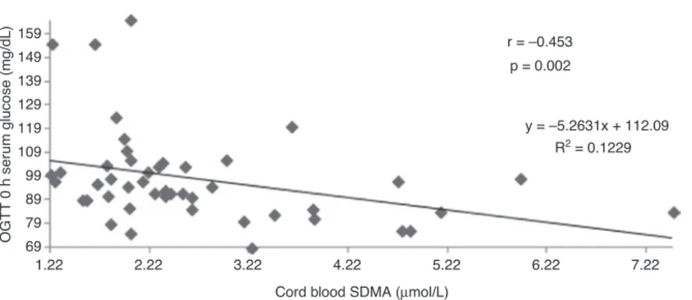
Cord blood SDMA negatively correlated with 0 h OGTT serum glucose levels (r = −0.453, p = 0.002) (Figure 2).
There was a negative correlation between cord blood L-NMMA and OGTT 1 h serum glucose levels (r = −0.401, p = 0.006) (Figure 3). There was no significant correla-tion between other maternal or cord blood methylated arginines and OGTT glucose levels.
Discussion
Gestational diabetes is a syndrome that occurs during preg-nancy and associated with impaired glucose metabolism
leading to endothelial deterioration and improper organi-zation of vascular tonus. One important local mechanism involved in the control of vascular tonus is endothelial NO synthesis, a process that could require membrane trans-port of L-arginine, the substrate for eNOS [12].
In recent years, alterations in methylated arginine metabolites have been suggested as a early onset predic-tor of endothelial deterioration and proved to be a unique, distinct risk factor of several cardiovascular and meta-bolic conditions.
There are contradictious ADMA and SDMA results in patients with GDM in the literature. Telejko et al. reported the levels of ADMA in 56 patients with gestational diabe-tes (GDM), 68 pregnant subjects with normal glucose tol-erance (NGT) and 36 healthy non-pregnant participiants. ADMA concentrations were found to be substantially lower in NGT (0.48 μmol/L) compared to GDM (0.50 μmol/L) and healthy non-pregnant (0.57 μmol/L) groups and reported that a decrease in circulating ADMA concentrations might
69
1.22 2.22 3.22 4.22
Cord blood SDMA vs. OGTT 0 h glucose
Cord blood SDMA (µmol/L)
OGTT 0 h ser um glucose (mg/dL) 5.22 6.22 r = –0.453 p = 0.002 y = –5.2631x + 112.09 R2 = 0.1229 7.22 79 89 99 109 119 129 139 149 159
Figure 2: Correlation between cord blood SDMA and OGTT 0 h glucose.
0.08 132 152 172 192 212 232 0.13 0.18 0.23
Cord blood L-NMMA (µmol/L)
OGTT 1 h ser
um glucose (mg/dL)
Cord blood L-NMMA vs. OGTT 1 h glucose
0.28 0.33 0.38
r = –0.401 p = 0.006 y = –187.18x + 232.85
R2 = 0.1849
be associated with physiological adaptation process [13]. In their study an enzyme linked immunosorbent assay commercial kit was used for the detection of ADMA and Arginine/ADMA ratio was not calculated. A multimarker approach by determining serum methylated arginines might provide useful information about the all parameters related to NO synthesis. Thus, a single step and more sensi-tive measurement of these molecules by liquid chromato-graphy tandem mass spectrometry offers us to have more accurate results of this NO-related pathway. In this study, accurate and precise measurement of ADMA and arginine was performed with tandem mass spectrometry and there was no difference for serum ADMA levels and Arginine/ ADMA ratios for all groups (Table 1).
Mittermayer et al. conducted a study with 46 obese, 31 non-obese GDM patients and 17 healthy women. ADMA concentrations were similiar for obese and non-obese GDM subjects (0.58 ± 0.02 and 0.57 ± 0.02 μmol/L, respectively), and the levels were found to be elevated compared to control group (0.47 ± 0.03 μmol/L). Although SDMA levels and ADMA/SDMA ratios were found to be higher in obese GDM, SDMA concentrations were higher in GDM groups than controls [14]. In another study with 77 subjects with GDM who carried out a 75 g OGTT and follow-up, they reported a correlation between serum ADMA levels and blood pressure without a correlation with metabolic para-meters (body-mass index, fasting glucose levels and lipid levels). In their study, Arginine and symmetrical dimethyl arginine were not associated with glucose values. During the follow-up period, Arginine and ADMA were found to diminish. Glucose tolerance was affected in 36% of GDM patients over 0.56 μmol/L median ADMA values [15].
These results are in consistency with previous data. Such that Pettersson et al. analyzed serum concentrations of arginine and ADMA that were collected at 32–39 weeks of gestation, 3–5 days and 3 months after birth from 12 pregnant subjects with advanced preeclampsia and from pregnant participiants without hypertension (n = 12). In their study, it was found that at the course of third tri-mester, serum ADMA levels tended to be higher (p < 0.05) in the preeclampsia patients (0.55 ± 0.02 μmol/L) com-pared to the control group (0.36 ± 0.01 μmol/L). There was no stastistically significant difference between both groups for arginine levels (80.7 ± 5.8 μmol/L) and 74.5 ± 3.8 μmol/L, respectively), arginine/ADMA ratio was found to be decreased (p < 0.05) in the preeclamptic group (145.6 ± 10.5) compared to controls (211.0 ± 14.3) [16]. These findings suggest a link between high blood pressure and circulating methlated arginine levels. Also, in this study there were three preeclampsia, four early delivery abor-tion and five IUGR patients. Patients with preeclampsia
were found to have two-fold increase in ADMA levels (0.75 vs. 0.36 μmol/L) and two-fold decrease in Arginine/ADMA ratios (429 vs. 207). In this study, although not statistically significant (p = 0.084), the cord blood of patients with GDM regulated by diet or insulin has a trend to increase the ratio of Arginine/ADMA. This may be a condition that develops in response to decreased NO synthesis due to gestational diabetes (Table 1).
Pleiner et al. performed a research with seven over-weight and five non-overover-weight women with GDM. They found that there was no significant difference between non-overweight participiants and healthy sub-jects for serum ADMA levels. SDMA and ADMA levels were found to be elevated in obese GDM than non-obese group (0.58 ± 0.04 and 0.55 ± 0.03 vs. 0.46 ± 0.07 and 0.40 ± 0.07 μmol/L, respectively) [17].
Gumus et al. performed a study with 30 subjects with GDM and 40 healthy controls participiants with simil-iar age and body-mass index values. According to their study’s results, serum ADMA levels were elevated in patients with GDM rather than control group (0.45 ± 0.11 vs. 0.31 ± 0.13 μmol/L, respectively; p = 0.01) [18].
Sertkaya et al. compared plasma concentrations of ADMA in 58 GDM, 30 pregnant subjects with positive glucose challange test but negative OGTT (AGCT) and 50 healthy pregnant individuals. ADMA values were signif-icantly elevated compared to control subjects (3.60 ± 1.21; 4.00 ± 1.70; 2.65 ± 0.82 μmol/L, respectively, p = 0.001). They concluded that the rise of ADMA values in pregnant subjects with glucose intolerance might probably be by virtue of higher insulin values [19]. Interestingly, there was a 10-fold increase in ADMA levels in this study’s control group compared to other studies at all. In this study, while the methylated arginine derivatives that indirectly block NO synthase, play an important role in vascular patholo-gies due to hyperglycaemia and endothelial dysfunction, especially in the chronic basis, the negative correlation between OGTT 0 and 3-h glucose levels and concentrations of these molecules might not be considered as clinically significant. The decrease in the levels of methylated argi-nine derivatives that inhibit the uptake of argiargi-nine, such as SDMA and L-NMMA, can be considered as a dilution effect since the sudden increase in glucose values does not reflect exactly whether glucose’s endotoxic effects occur.
Akturk et al. analyzed plasma ADMA levels in 54 gestational diabetic subjects and 69 participiants with normal glucose tolerance (NGT) between 32 and 39 weeks. They reported the elevation of serum ADMA levels in GDM subjects compared to NGT controls (p = 0.03). The ADMA concentrations in the third trimester were positively cor-related with the glucose levels the 50-g glucose challenge
test (GCT) during 24–28 weeks in the whole group (r = 0.21, p = 0.02) [20]. In our study, ADMA, SDMA and L-NMMA levels, which are antagonists of NO synthesis, were found to be significantly lower in cord blood of insulin- or diet-regulated gestational diabetic patients compared to control group. This can be explained by the fact that there is a decrease in the level of inhibitory molecules in the patient group regulated with insulin or diet. However, due to the low molecular stability and difficulty of measuring NO, NO levels could not be analyzed as a limitation of this study. The net effect of insulin or dietary therapy on NO production could be observed if NO levels were measured and could be assessed in all three groups.
Conclusion
According to this study’s results, methylated arginine levels may not be associated with endothelial deteriora-tion in GDM otherwise with preeclampsia risk.
Acknowledgements: No funding from any pharmaceutical
firm was received for this project, and the authors’ time on this project was supported by their respective employers.
Authors’ conflict of interest disclosure: The authors stated
that there are no conflicts of interest regarding the publi-cation of this article.
Employment or leadership: None declared. Honorarium: None declared.
References
1. Stanley JL, Cheung CC, Rueda-Clausen CF, Sankaralingam S, Baker PN, Davidge ST. Effect of gestational diabetes on maternal artery function. Reprod Sci 2011;18:342–52.
2. Capobianco E, Martínez N, Fornes D, Higa R, Di Marco I, Basualdo MN, et al. PPAR activation as a regulator of lipid metabolism, nitric oxide production and lipid peroxidation in the placenta from type 2 diabetic patients. Mol Cell Endocrinol 2013;377:7–15.
3. Brewster S, Zinman B, Retnakaran R, Floras JS. Cardiometabolic consequences of gestational dysglycemia. J Am Coll Cardiol 2013;62:677–84.
4. Guzmán-Gutiérrez E, Arroyo P, Salsoso R, Fuenzalida B, Sáez T, Leiva A, et al. Role of insulin and adenosine in the human placenta microvascular and macrovascular endothelial cell dysfunction in gestational diabetes mellitus. Microcirculation 2014;21:26–37.
5. Mordwinkin NM, Ouzounian JG, Yedigarova L, Montoro MN, Louie SG, Rodgers KE. Alteration of endothelial function markers in
women with gestational diabetes and their fetuses. J Matern Fetal Neonatal Med 2013;26:507–12.
6. Sobrevia L, Abarzúa F, Nien JK, Salomón C, Westermeier F, Puebla C, et al. Differential placental macrovascular and microvascular endothelial dysfunction in gestational diabetes. Placenta 2011;32:159–64.
7. Brinkmann SJ, Wörner EA, Buijs N, Richir M, Cynober L, van Leeuwen PA, et al. The Arginine/ADMA ratio ıs related to the prevention of atherosclerotic plaques in hypercholesterolemic rabbits when giving a combined therapy with atorvastatine and arginine. Int J Mol Sci 2015;16:12230–42.
8. Di Gangi IM, Pirillo P, Carraro S, Gucciardi A, Naturale M, Baraldi E, et al. Online trapping and enrichment ultra performance liquid chromatography-tandem mass spectrometry method for sensitive measurement of “arginine-asymmetric dimethylargi-nine cycle” biomarkers in human exhaled breath condensate. Anal Chim Acta 2012;754:67–74.
9. Fleszar MG, Wiśniewski J, Krzystek-Korpacka M, Misiak B, Frydecka D, Piechowicz J, et al. Quantitative analysis of l-arginine, dimethylated arginine derivatives, L-citrulline, and dimethylamine in human serum using liquid chromatography-mass spectrometric method. Chromatographia 2018;81:911–21. 10. International Association of Diabetes and Pregnancy Study
Groups Consensus Panel, Metzger BE, Gabbe SG, Persson B, Buchanan TA, Catalano PA, et al. International association of diabetes and pregnancy study groups recommendations on the diagnosis and classification of hyperglycemia in pregnancy. Diab Care 2010;33:676–82.
11. Di Gangi IM, Chiandetti L, Gucciardi A, Moret V, Naturale M, Giordano G. Simultaneous quantitative determination of N(G),N(G)-dimethyl-L-arginine or asymmetric dimethylargi-nine and related pathway’s metabolites in biological fluids by ultrahigh-performance liquid chromatography/electro-spray ionization-tandem mass spectrometry. Anal Chim Acta 2010;677:140–8.
12. Vásquez G, Sanhueza F, Vásquez R, González M, San Martín R, Casanello P, et al. Role of adenosine transport in gestational diabetes-induced L-arginine transport and nitric oxide synthesis in human umbilical vein endothelium. J Physiol 2004;560(Pt 1): 111–22.
13. Telejko B, Zonenberg A, Kuzmicki M, Modzelewska A, Niedziolko-Bagniuk K, Ponurkiewicz A, et al. Circulating asymmetric dimethylarginine, endothelin-1 and cell adhesion molecules in women with gestational diabetes. Acta Diabetol 2009;46:303–8.
14. Mittermayer F, Mayer BX, Meyer A, Winzer C, Pacini G, Wagner OF, et al. Circulating concentrations of asymmetrical dimethyl-L-arginine are increased in women with previous ges-tational diabetes. Diabetologia 2002;45:1372–8.
15. Mittermayer F, Kautzky-Willer A, Winzer C, Krzyzanowska K, Prikoszovich T, Demehri S, et al. Elevated concentrations of asymmetric dimethylarginine are associated with deterioration of glucose tolerance in women with previous gestational diabe-tes mellitus. J Intern Med 2007;261:392–8.
16. Pettersson A, Hedner T, Milsom I. Increased circulating concen-trations of asymmetric dimethyl arginine (ADMA), an endog-enous inhibitor of nitric oxide synthesis, in preeclampsia. Acta Obstet Gynecol Scand 1998;77:808–13.
17. Pleiner J, Mittermayer F, Langenberger H, Winzer C, Schaller G, Pacini G, et al. Impaired vascular nitric oxide bioactivity in
women with previous gestational diabetes. Wien Klin Wochen-schr 2007;119:483–9.
18. Gumus II, Kargili A, Kaygusuz I, Derbent A, Karakurt F, Kasapoglu B, et al. The association between serum asymmetric dimethyl arginine levels and a history of gestational diabetes among healthy women. Blood Coagul Fibrinolysis 2012;23: 391–5.
19. Sertkaya AC, Kafkasli A, Turkcuoglu I, Karabulut AB. Asymmetric dimethylarginine level in hyperglycemic gestation. Endocrine 2011;40:237–42.
20. Akturk M, Altinova A, Mert I, Dincel A, Sargin A, Buyukkagnici U, et al. Asymmetric dimethylarginine concentrations are elevated in women with gestational diabetes. Endocrine. 2010;38: 134–41.