RESEARCH TJH-2020-0682.R2
doi: 10.4274/tjh.galenos.2021.2020.0682
Generation of Induced Pluripotent Stem Cells from Patients with Multiple Myeloma
Yılmaz Başaran and Karaöz. iPS cells-Multiple myeloma İrem Yılmaz Başaran1, Erdal Karaöz2,3,4,5
1Eskişehir Osmangazi University, Cellular Therapy and Stem Cell Production Application and
Research Center, Eskisehir, Turkey
2İstinye University, Faculty of Medicine, Department of Histology and Embryology, İstanbul,
Turkey
3İstinye University, Center for Stem Cell and Tissue Engineering Research and Practice,
İstanbul, Turkey
4İstinye University, 3D Bioprinting Design and Prototyping R&D Center, İstanbul/Turkey. 5Liv Hospital, Center for Regenerative Medicine and Stem Cell Manufacturing (LivMedCell),
İstanbul, Turkey Erdal Karaoz, Ph.D.
Department of Histology & Embryology, Faculty of Medicine, İstinye University, İstanbul, Turkey
+90-212-481-36-55 [email protected] November 15, 2020 March 16, 2021
Keywords: iPS, Multiple myeloma, MSC, Bone Marrow Mesenchymal Stem Cells, Sendai virus Financial disclosure statement: This study was supported by grants (112S296) from the Scientific and Research Council of Turkey (TUBITAK).
Conflict of interest statement: The authors declare no conflict of interest.
Introduction
uncorrected
Yamanaka and Takahashi made a discovery in the world of life science by transforming mouse somatic fibroblasts into pluripotent cells as a result of transferring 4 gene sets (Sox2-Oct4-Klf4&c-Myc) in 2006 (1). Since that day, induced pluripotent stem cells (iPSCs) are considered to be one of the main sources for regenerative medicine; like embryonic stem cells (ESCs). Because of their pluripotent features, both cell types are building blocks of regenerative medicine. There are no ethical limitations and immunological problems for iPSCs when compared to ESCs (2). Additionally, iPSCs with disease genotypes have been used for human disease modeling (3).
To date, iPSCs have been generated from many different sources (1,4-9), including
mesenchymal stem cells (MSCs). MSCs were shown to be more efficient in reprogramming compared to other somatic cells (10,11).
In the last decade, iPSCs have proven to be a powerful in vitro system for studying diseases (12,13), especially genetic disorders (1,14). Patient specific iPSCs have powerful potential in regenerative medicine, and notably in human disease modeling (15). The in vitro phenotype of disease-specific iPS-derived cells can enable us to comprehend the differences and/or similarities between the molecular/cellular pathophysiology and clinical phenotype. This technology can also facilitate and improve the understanding of the disease pathology. To date, many disease models have been established with iPSCs. There are efforts for drug screening tests and genetic modifications of cells for the treatment of diseases (15). On the other hand, in many diseases, patient specific iPSCs have been shown to exhibit the characteristics of these diseases (13,16). In addition, use of iPSCs is very important in the research of cancer microenvironment (17-19). Multiple myeloma (MM) progresses with uncontrolled increase and accumulation of malignant plasma cells in the bone marrow (BM) (20). MM bone disease is observed due to the increase of osteoclastic activity through the factors synthesized from malignant plasma cells and the
decrease in the differentiation of osteoblasts originating from MSCs. Imbalance in this process leads to overproduction of the many responsible chemokines and cytokines and besides, various signaling cascades are involved in this complex process. (21-23). Advanced lesions and fractures occur as a result of this imbalance in bone formation and destruction.
The construction and differentiation of osteoblasts from MSCs is controlled by many factors and pathways in the BM microenvironment. Various inhibitory substances released by plasma cells in the BM microenvironment in MM, stop bone formation as a result of disruption or disruption of different stages of osteoblastogenesis (24-26). Furthermore, there are many factors in MM disease that disrupt osteoblastogenesis with different pathways. Many of these factors may be indirectly secreted or secreted by MM cells (27,28). In studies carried out, osteogenic
differentiation defects were detected in BM derived MSCs (BM-MSCs) obtained from MM patients, even in vitro, where MM cells did not have all the inhibitory factors secreted (29-33). In recent years, various approaches have been developed in the treatment of MM bone disease, especially regarding the use of MSC (34,35). However, the limited proliferation of BM-MSCs obtained from MM patients and the low capacity of osteoblastic differentiation in vivo and in
vitro conditions will prevent possible autologous MSCs treatments in the future. In addition to
the purpose of revealing the molecular development stages of diseases and designing
disease/personalized drugs, iPSCs technology is expected to serve its potential for future use in cell therapy or tissue engineering in many disease models. With the development of iPSC technology, it will be possible in the future to obtain genetically repaired autologous stem cells from patients or reproduce & replace the missing tissue.
uncorrected
Recently, different types of cells that are gathered from patients with various diseases have been used for generating iPSCs, except for patients with MM. Based on all this information, we aimed to obtain the MM disease model for the first time by reprogramming BM-MSCs obtained from MM patients in our study. Initially, iPSCs have a big potential to start with, because of their MM patient cell origin and the inclusion of disease genotype in a stem cell. MM-iPSCs would
undeniably contain the genotype that causes the disease. With this study, patient-specific cells enable patient-specific disease modeling possible, and defects in MSCs can be studied by programming them into the pluripotent stage. This topic will lead to other studies to be carried out for the first time in the literature.
Material and Methods
Selection of Patients and Control Groups
In this study, MSCs were isolated from BM obtained from newly diagnosed MM patients’ iliac crest (n=3). Biopsies were performed for diagnosis, staging and evaluation of ongoing treatment. Control groups to generate IPSCs were derived from newborn babies’ foreskin fibroblasts after informed consent approved under protocols.
MSC Isolation from MM Patients and Cell Culture
The isolation and culture of hBM-MSCs were performed as previously described by Karaoz et al (37). Briefly, bone marrow aspirates were obtained from the iliac crest of MM patients. And then, samples were diluted to 1:3 with Phosphate-buffered saline (PBS). Histopaque-1077 (1.0777 g/ml, Sigma-Aldrich, St. Louis, MO) was used for gradient centrifugation. The low-density mononuclear cells were collected and plated in tissue culture flask.
iPSCs Generation
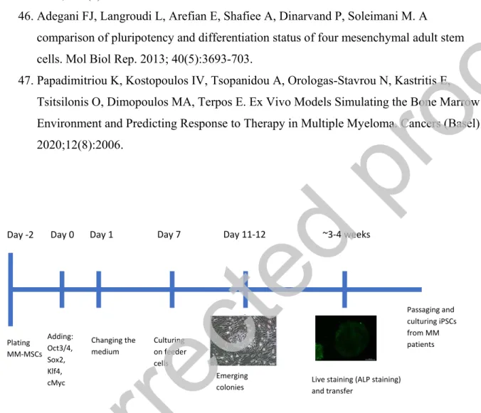
For the generation of iPSCs, CytoTune-iPS Reprogramming Kit (Thermo Fisher Scientific, USA) was used. The manufacturer’s instructions were followed for setting up the generation procedure (Figure 1). 2 days before transduction, the cells were plated into 2 wells of a 6-well plate (Day -2). On the day of transduction (Day 0), cell medium was aspired, and Yamanaka factors were added to cells, which then incubated overnight. The cells then cultured with their specific culture media for 6 days. When the colonies were grown to an appropriate size for transferring; live staining was done by using Alkaline Phosphatase (ALP Live Stain, Thermo Fisher) for selecting reprogrammed colonies. The selected colonies were then harvested. Manually picked colonies were transferred onto fresh MEF plates. Next day, the medium was changed to iPSC medium (DMEM-F12+ 20% KnockOut Serum Replacement, 100μM MEM Non-Essential Amino Acids, 1X GlutaMAX, 100 μM β-mercaptoethanol, 0,2% Primocin, 4 ng/mL FGF) and replaced everyday thereafter. Colony formation was monitored and photographed every day.
iPSCs Culture
iPSCs were passaged in order to avoid overgrowth and to maintain them in undifferentiated state. Before splitting the colonies, differentiated colonies were removed under a microscope in sterile conditions. Differentiated areas were excised and discarded before bulk passaging. Colonies were mechanically cut into pieces using a needle for passaging. The colony was usually ready to passage in 2–3 days.
For feeder free culture, picked colonies were added to a freshly prepared Geltrex™ (Invitrogen, Life Tech.) coated plate. The medium was gradually changed to StemPro® hESC Serum Free
Medium (Invitrogen, Life Tech.) as explained in Table 1. StemPro® was used every day thereafter. The colonies were passaged at a 1:3 ratio. Continued passaging was done by StemPro® EZPassage™ Disposable Stem Cell Passaging Tool (Invitrogen, Life Tech.).
uncorrected
Characterization of iPSCs Cell staining
The same method used for IF staining of MSCs (Supplemental Material and Methods) was done. Following primary antibodies were used for staining: Oct4, NANOG, TRA1-60, TRA1-81 and Sox2 (Table 2). DAPI was used for nuclear staining.
Flow Cytometry
Pluripotency associated markers’ expressions were analyzed by flow cytometry. Feeder free cultured iPSCs were passaged by TrypLE (Life Technologies) to be prepared as a suspension. The cells were stained with antibodies for SSEA4, Tra1-81 and Oct3/4 (BD Biosciences Pharmingen, USA).
Gene Expression Analysis
The cell specific gene expressions (Lin28, Nr6A, Klf4, FoxD3, Myc, Utf1, Msx1, Gata6, endogenous Oct4, endogenous Sox2, Nanog, and Rex1) in the undifferentiated cells were determined by PCR as previously described (39). The gene expression level detection was done with LightCycler 480 DNA SYBR Green I Master (Roche) with specific primers on LightCycler 480 real-time PCR instrument (Roche). Firstly, the PCR reactions that were performed for GAPDH (reference gene) were as follows: 45 cycles, denaturation: 10s at 95°C, annealing and extension: 30s at 60°C. Analysis for the results was performed using Roche LightCycler 480 software. In vitro embryonic body formation
Spontaneous embryonic body (EB) generation was used for testing of in vitro differentiation capacity of iPSCs. Cells were cultured with medium without bFGF2 in bacteriological culture dishes for 21 days. Formation was monitored daily.
Results
iPSCs Generation & Culture
First colonies were obtained on the 6th day of culture after transduction. The structure of these
colonies had a scattered appearance compared to ESC colonies, but the boundaries have become more apparent in the following days. After the colonies reached a certain size, they were
transferred to new feeder cell layers by mechanical passaging. These new colonies have been observed to form tight cell assemblies with clearly defined boundaries observed as ESCs.
Colony-like structures photographed under the microscope in which they grew over days (Figure 2). When differentiated parts were identified, these parts were cleaned, and the culture was continued. During the culture, the colonies kept their borders.
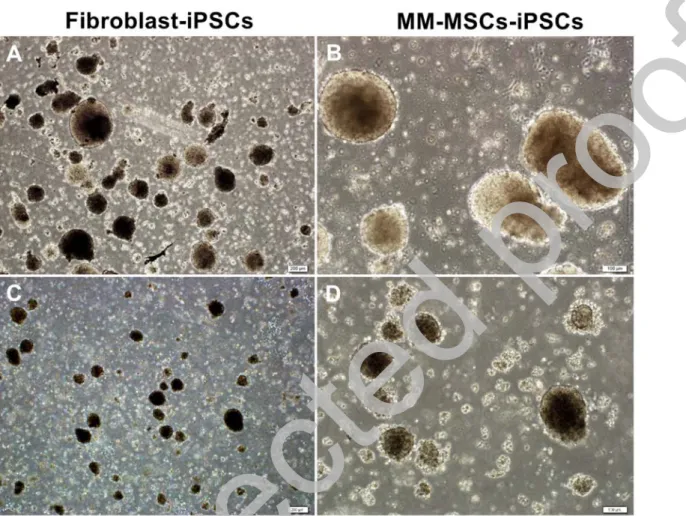
Following the mechanical passaging of colonies which were cultured on MEF, colonies were successfully performed growing in Geltrex-coated culture dishes. It was observed that the colonies retain their classical morphology (Figure 3).
Characterization
The resulting colonies were stained against ALP while on the feeder layer in the culture dish. Colonies were marked with ALP-FITC dye, which was performed without loss of viability. With this labeling, cells in colonies with ESC character were stained (Figure 4). Green colonies were selected under fluorescence microscopy and first cell lines were formed by physical passaging. ESC markers such as SSEA-4, TRA-1-81 and Oct3/4 were positive for cells in flow cytometry analysis (Figure 5). iPSCs cultured on feeder layers were further characterized by IF methods. The colonies were positive for the pluripotent cell markers Oct4, TRA1-60, Nanog, TRA1-81 and Sox2 (Figure 6).
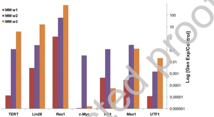
According to the expression analysis, a significant increase was observed between 1st and 3rd
weeks in iPSCs cultures. The significant increase in c-Myc and Klf4 gene expressions in the 2nd
uncorrected
week, decreased in the 3rd week. Since these genes were transmitted by viruses, the initial
expression was ectopic and turned into internal expression of the cells at the 3rd week (Figure 7).
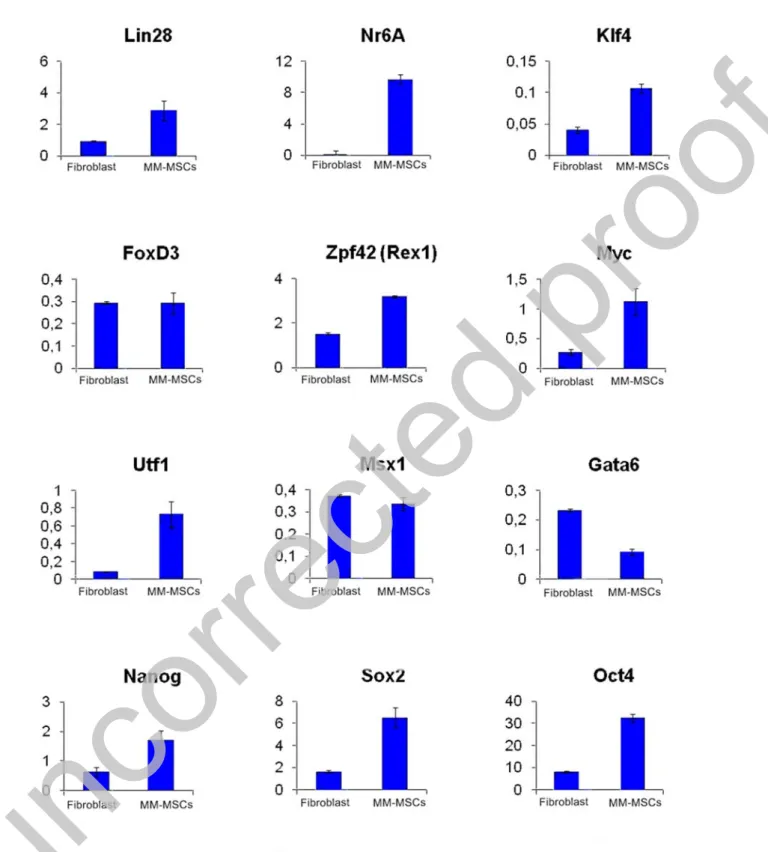
Expressions of pluripotent genes were shown in all iPSCs obtained. Significant increases have been observed in Oct4, Nanog, Sox2, Rex1, Utf1 and Lin28 genes. Using Sendai virus, Oct4, Sox2, c-Myc and Klf4 genes were transferred and their expressions were provided with the help of ectopic vectors. According to these transferred vectors, Oct4, Sox2, c-Myc and Klf4
expressions may not be endogenous gene expressions of the cells. However, the increased expression of highly specific pluripotent genes such as Nanog, Lin28 and Utf1 constitutes the most serious evidence that cells acquire pluripotent cell character (Figure 8). EB formation was obtained after 4th day of suspended culture (Figure 9).
Discussion
iPSCs carry an immense potential for future cellular therapies. However, they were shown to carry characteristics of the cells they were originated from and their niche (40) thorough their epigenetic memory (41,42). The first type of reprogrammed cells was found to be fibroblasts. Other types of human cells have also been tried for reprogramming, which might be potentially easier (42).
In this study, we attempted the reprogramming of MSCs obtained from MM patients’ BM. Our study uses a standardized reprogramming approach to evaluate reprogramming of two cell lines on various stages of differentiation; terminally differentiated fibroblasts (as control) and
multipotent MSCs that were obtained from MM patients. Both cells were reprogrammed by CytoTune-iPS 2.0 Sendai Reprogramming Kit that contains Yamanaka factors. Yamanaka factors have been reported many times in literature as adequate for effective reprogramming (4,43-45). The efficiency of iPSCs generation using Sendai virus, is much higher than the conventional vectors (45). The elimination of Sendai virus is easier than the conventional vectors, which allows obtaining transgene-free iPSCs.
Firstly, microscopic analysis revealed that the generated iPSCs possessed classical ESC-like morphological characteristics. Secondly, reprogramming experiments demonstrated that both cell lines can be reprogrammed up to the pluripotent stage, which was confirmed by flow cytometry, IF staining and gene expression analyses. To confirm spontaneous differentiation potential, in
vitro EB formation assay was performed.
iPSCs have been successfully obtained from MM patients for the first time. One of the major findings of our study is the rapid reprogramming of MSCs, which started as early as 6th day with
the appearance of first colony-forming cell accumulations. Considering the results of the previous studies, this rapid reprogramming can be attributed to the multipotent nature of MSCs, which implies that the effectiveness of reprogramming is related to the differentiation stage of the cell line. Adegani et al. demonstrated that human MSCs of various sources such as adipose tissue and BM-MSCs expressed core pluripotency factors such as Lin28, Klf4 and Sox2 at higher levels, Nanog at moderate levels and Oct4 low levels, intrinsically, which allows them to
reprogram easily (46).
Our data shows that human iPSC can be derived from MSCs more rapidly than fibroblasts. Obtaining MSCs from patients does not require a great effort, because BM aspirates are taken almost daily for diagnostic purposes in the hematology clinics. MSCs can be isolated from these samples.
As a result, we generated iPSCs form MM-MSCs for the first time. As we know from the
previous studies, osteogenic differentiation of MM-MSCs is weak. Our next goal is to discuss the differences between healthy donors MSCs-iPSCs’ and MM-MSCs-iPSCs’ osteogenic
uncorrected
differentiation potential. We are planning further studies to understand the pathogenesis of this disease, because MM-iPSCs could clarify molecular mechanisms behind this disease. Therefore, further studies can be developed to understand the molecular mechanisms of this disease.
Understanding the pathogenetic mechanisms underlying this disease is crucial for effective management and improving the quality of MM patients’ life (22). Based on our knowledge, the investigation of novel targeted drugs and understanding the role of novel targeted therapies in this disease are great interests (23). For more successful results, researchers develop the complex 3D environments by using MM patients’ cells (47). iPSCs offer unprecedented opportunities for drug discovery and screening with their ability to differentiate into all kinds of cells found in the body.
Conclusion
The data obtained from this study expose that iPSCs can be derived from MSCs more rapidly than fibroblasts and iPSCs have been successfully obtained from MM patients for the first time. iPSCs generated from MM-MSCs could clarify molecular mechanisms behind this disease. Thus, further studies can be developed to understand the molecular mechanisms of this disease. Our next goal is to discuss the differences between healthy donors MSCs-iPSCs and MM-MSCs-iPSCs osteogenic differentiation potential.
Acknowledgments
This study was supported by grants (112S296) from the Scientific and Research Council of Turkey. The authors would like to thank Dr. Ozgur Mehtap for providing BM aspirates from patients and Cansu Demir, Dr. Ayca Dikmen and Dr. Gokhan Duruksu for their technical assistance.
Ethics
The study received ethical approval from Kocaeli University, Faculty of Medicine Ethical Committee (KAEK 2012/38) for collection of human samples.
References
1. Takahashi K, Yamanaka S. Induction of pluripotent stem cells from mouse embryonic and adult fibroblast cultures by defined factors. Cell 2006; 126(4):663-76.
2. Moradi S, Mahdizadeh H, Šarić T, Kim J, Harati J, Shahsavarani H, Greber B, Moore JB. Research and therapy with induced pluripotent stem cells (iPSCs): social, legal, and ethical considerations. Stem Cell Res Ther. 2019; 10: 341.
3. Maehr R, Chen S, Snitow M, Ludwig T, Yagasaki L, Goland R, Leibel RL, Melton DA. Generation of pluripotent stem cells from patients with type 1 diabetes. Proc Natl Acad Sci USA 2009;106(37):15768-73.
4. Takahashi K, Tanabe K, Ohnuki M,Narita M, Ichisaka T, Tomoda K, Yamanaka S. Induction of pluripotent stem cells from adult human fibroblasts by defined factors. Cell 2007; 30;131(5):861-72.
uncorrected
5. Re S, Dogan AA, Ben-Shachar D, Berger G, Werling AM, Walitza S, Grünblatt E. Improved generation of induced pluripotent stem cells from hair derived keratinocytes – A tool to study neurodevelopmental disorders as ADHD. Front. Cell. Neurosci. 2018; 12: 321.
6. Wang J, Gu Q, Hao J, bai D, Liu L, Zhao X, Liu Z, Wang L, Zhou Q. Generation of induced pluripotent stem cells with high efficiency from human umbilical cord blood mononuclear cells. Genomics Proteomics Bioinformatics. 2013; 11(5): 304–311. 7. Brown ME, Rondon E, Rajesh D, Mack A, Lewis R, Feng X, Zitur LJ, Learish RD,
Nuwaysir FE. Derivation of induced pluripotent stem cells from human peripheral blood t lymphocytes. PLoS ONE 2010; 29;5(6):e11373.
8. AoiI T, Yae K, Nakagawa M, Ichisaka T, Okita K, et al., Generation of pluripotent stem cells from adult mouse liver and stomach cells. Science 2008; 321: 699–702.
9. Stadtfeld M, Brennand K, Hochedlinger K. Reprogramming of pancreatic beta cells into induced pluripotent stem cells. Curr Biol. 2008; 18: 890– 894.
10. Niibe K, Kawamura Y, Araki D, Morikawa S, Miura K, Suzuki S, Shimmura S, Sunabori T, Mabuchi Y, Nagari Y, Nakagawa T, Okana H, Matsuzaki Y. Purified mesenchymal stem cells are an efficient source for iPS cell induction. PLoS One 2011; 6(3):e17610. 11. Cai J, Li W, Su H, Qin D, Yang J, Zhu F, Xu J, He W, Guo X, Labuda K, Peterbauer A,
Wolbank S, Zhong M, Li Z, Wu W, So KF, Redl H, Zeng L, Esteban MA, Pei D. Generation of human induced pluripotent stem cells from umbilical cord matrix and amniotic membrane mesenchymal cells. J Biol Chem 2010; 285(15):11227-34. 12. Jiang Z, Han Y, Cao X. Induced pluripotent stem cell (iPSCs) and their application in
immunotherapy. Cell Mol Immunol. 2014;11(1):17-24.
13. Doss MX, Sachinidis A. Current challenges of iPSCs -based disease modeling and therapeutic implications. Cells 2019; 8(5): 403.
14. Shi Y, Inoue H, Wu JC, Yamanaka S. Induced pluripotent stem cell technology: A decade of progress. Nat Rev Drug Discov. 2016; 16:115‐130.
15. Ebert AD, Liang P, Wu JC. Induced pluripotent stem cells as a disease modeling and drug screening platform. J Cardiovasc Pharmacol. 2012; 60(4): 408–416.
16. Ohnuki M, Takahaski K. Present and future challenges of induced pluripotent stem cells. Philos Trans R Soc Lond B Biol Sci. 2015; 370(1680): 20140367.
uncorrected
17. Papapetrou E. Patient-derived induced pluripotent stem cells in cancer research and precision oncology. Nat Med. 2016; 22(12): 1392–1401.
18. Rami F, Mollainezhad H, Salehi M. Induced pluripotent stem cell as a new source for cancer immunotherapy. Genet Res Int. 2016; 2016: 3451807.
19. Saito S, Lin YC, Nakamura Y, Eckner R, Kuo KK, Lin CS, Yokoyama K. Potential application of cell reprogramming techniques for cancer research. Cell Mol Life Sci 2019; 76(1):45-65.
20. Chaidos A, Barnes CP, Cowan G, May PC, Melo V, Hatjiharissi E, Papaioannou M, Harrington H, Doolittle H, Terpos E, Dimopoulos M, Abdalla S, Yarranton H, Naresh K, Foroni L, Reid A, Rahemtulla A, Stumpf M, Roberts I, Karadimitris A. Clinical drug resistance linked to interconvertible phenotypic and functional states of tumor-propagating cells in multiple myeloma. Blood 2013; 121 (2): 318–328.
21. Terpos E, Christoulas D, Kastritis E et al. High levels of periostin correlate with increased fracture rate, diffuse MRI pattern, abnormal bone remodeling and advanced disease stage in patients with newly diagnosed symptomatic multiple myeloma. Blood Cancer Journal 2016; 6, e482.
22. Terpos E, Ntanasis-Stathopoulos I, Gavriatopoulou M et al. Pathogenesis of bone disease in multiple myeloma: from bench to bedside. Blood Cancer Journal 2018; 8, 7.
23. Terpos E, Ntanasis-Stathopoulos I, Christoulas D et al. Semaphorin 4D correlates with increased bone resorption, hypercalcemia, and disease stage in newly diagnosed patients with multiple myeloma. Blood Cancer Journal 2018; 8, 42.
24. Edwards CM, Zhuang J, Mundy GR. The pathogenesis of the bone disease of multiple myeloma. Bone 2008; 42, 1007–13.
25. Mundy GR, Luben RA, Raisz LG, Oppenheim JJ, Buell DN. Bone-resorbing activity in supernatants from lymphoid cell lines. N Engl J Med 1974; 290, 867–71.
26. Mundy GR, Raisz LG, Cooper RA, Schechter GP, Salmon S. Evidence for the secretion of an osteoclast stimulating factor in myeloma. N Engl J Med 1974; 291, 1041–6. 27. Fowler JA, Edwards CM, Croucher PI. Tumor-host cell interactions in the bone disease
of myeloma. Bone 2011; 48(1), 121-8.
28. Roodman GD. Osteoblast function in myeloma. Bone 2011; 48(1), 135-40.
uncorrected
29. Todoerti K, Lisignoli G, Storti P, Agnelli L, Novara F,Manferdini C, Codeluppi K, Colla S, CrugnolaM, AbeltinoM, BolzoniM, Sgobba V, Facchini A, Lambertenghi-Deliliers G, Zuffardi O, Rizzoli V, Neri A, Giuliani N. Distinct transcriptional profiles characterize bone microenvironment mesenchymal cells rather than osteoblasts in relationship with multiple myeloma bone disease. Exp Hematol 2009; 38: 141-53.
30. Tian E, Zhan F, Walker R, Rasmussen E, Ma Y, Barlogie B, et al. The role of the Wnt-signaling antagonist DKK1 in the development of osteolytic lesions in multiple myeloma. N Engl J Med 2003; 349:2483–94.
31. Wang Y, Nishida S, Elalieh HZ, Long RK, Halloran BP, Bikle DD. Role of IGF-I signaling in regulating osteoclastogenesis. J Bone 2006; Miner Res, 21:1350–8. 32. De Bruyne E, Bos TJ, Schuit F, Van Valckenborgh E, Menu E, Thorrez L, Atadja P,
Jernberg-Wiklund H, Vanderkerken K. et al. IGF-1 suppresses Bim expression in multiple myeloma via epigenetic and posttranslational mechanisms. Blood 2010, 115(12):2430-40.
33. Abe M, Hiura K, Wilde J, Shioyasono A, Moriyama K, Hashimoto T, et al., Osteoclasts enhance myeloma cell growth and survival via cell–cell contact: a vicious cycle between bone destruction and myeloma expansion. Blood 2004; 104:2484–91.
34. Rabin N, Kyriakou C, Coulton L, Gallagher OM, Buckle C, Benjamin R, et al. A new xenograft model of myeloma bone disease demonstrating the efficacy of human
mesenchymal stem cells expressing osteoprotegerin by lentiviral gene transfer. Leukemia 2007; 21:2181–91.
35. Li X, Ling W, Pennisi A, Wang Y, Khan S, Heidaran M, Pal A, Zhang X, He S, Zeitlin A, Abbot S, Faleck H, Hariri R, Shaughnessy JD Jr, van Rhee F, Nair B, Barlogie B, Epstein J, Yaccoby S, et al. Human placenta-derived adherent cells prevent bone loss, stimulate bone formation, and suppress growth of multiple myeloma in bone. Stem Cells 2011; 29(2):263-73.
36. Meng G, Liu S, Krawetz R, Chan M, Chernos J, Rancourt DE. A novel method for generating xeno-free human feeder cells for human embryonic stem cell culture. Stem Cells Dev 2008; 17:413–422.
uncorrected
37. Karaöz E, Okçu A, Gacar G, Sağlam O, Yürüker S, Kenar H. A comprehensive
characterization study of human bone marrow MSCs with an emphasis on molecular and ultrastructural properties. J Cell Physiol 2011; 226(5):1367-82.
38. Karaoz E, Aksoy A, Ayhan S,Sarıboyacı AE, Kaymaz F, Kasap M. Characterization of mesenchymal stem cells from rat bone marrow: ultrastructural properties, differentiation potential and immunophenotypic markers. Histochem Cell Biol 2009; 132:533–546. 39. Yilmaz I, Sariboyaci AE, Subasi C, Karaoz E. Differentiation Potential of Mouse
Embryonic Stem Cells into Insulin Producing Cells in Pancreatic Islet Microenvironment. Exp Clin Endocrinol Diabetes. 2016; 124: 120–129.
40. Polo JM, Liu S, Figueroa ME, Kulalert W, Eminli S, Tan KY, Apostolou E, Stadtfeld M, Li Y, Shioda T, Natesan S, Wagers AJ, Melnick A, Evans T, Hochedlinger K. Cell type of origin influences the molecular and functional properties of mouse induced pluripotent stem cells.Nat Biotechnol. 2010; 28(8): 848–855.
41. Kim K, Doi A, Wen B, Ng K, Zhao R, Cahan P, Kim J, Aryee MJ, Ji H, Ehrlich L, Yabuuchi A, Takeuchi A, Cunniff KC, Hongguang H, Mckinney-Freeman S, Naveiras O, Yoon TJ, Irizarry RA, Jung N, Seita J, Hanna J, Murakami P, Jaenisch R, Weissleder R, Orkin SH, Weissman IL, Feinberg AP, Daley GQ. Epigenetic memory in induced pluripotent stem cells. Nature 2010; 16; 467(7313): 285–290.
42. Yan X, Qin H, Qu C, Tuan RS, Shi S, Huang GTJ. iPS cells reprogrammed from human mesenchymal-like stem/progenitor cells of dental tissue origin. Stem Cells Dev. 2010; 19(4): 469–480.
43. Sochackia J, Devallea S, Reisa M, Maciel RM, Paulsen BS, Brentanic H, Abreue PSB, Rehen S. Generation of iPS cell lines from schizophrenia patients using a non-integrative method. Stem Cell Res 2016; 17(1):97-101.
44. Sampaio GLA, Martins GLS, Paredes BD, Nonaka CKV, Silva KN, Rossi SEA, Santos RRD, Soares MBP, Souza BSF. Generation of an induced pluripotent stem cell line from a patient with autism spectrum disorder and SCN2A haploinsufficiency. Stem Cell Res 2019; 39:101488.
45. Tan X, Dai Q, Guo T, Xu J, Dai Q. Efficient generation of transgene- and feeder-free induced pluripotent stem cells from human dental mesenchymal stem cells and their
uncorrected
chemically defined differentiation into cardiomyocytes. Biochem Biophys Res Commun 2018; 495(4):2490-2497.
46. Adegani FJ, Langroudi L, Arefian E, Shafiee A, Dinarvand P, Soleimani M. A
comparison of pluripotency and differentiation status of four mesenchymal adult stem cells. Mol Biol Rep. 2013; 40(5):3693-703.
47. Papadimitriou K, Kostopoulos IV, Tsopanidou A, Orologas-Stavrou N, Kastritis E, Tsitsilonis O, Dimopoulos MA, Terpos E. Ex Vivo Models Simulating the Bone Marrow Environment and Predicting Response to Therapy in Multiple Myeloma. Cancers (Basel) 2020;12(8):2006.
Figure 1: Experiment timeline for reprogramming experiment for MM-MSCs.
Day -2 Day 0 Plating MM-MSCs Adding: Oct3/4, Sox2, Klf4, cMyc Day 1 Changing the medium Day 7 Culturing on feeder cells Day 11-12 Emerging
colonies Live staining (ALP staining) and transfer
~3-4 weeks Passaging and culturing iPSCs from MM patients
uncorrected
proof
Figure 2: The development of the first iPSCs colonies produced after Sendai virus transfection is monitored. A: 6th day after transfection, B: 7th day after transfection, C: 8th day after transfection
and D: 9th day after transfection. Scale bars: 50 µm (A, B, C and D).
uncorrected
uncorrected
Figure 3: The development of new iPSCs colonies obtained by mechanical passaging method is observed in the culture plate. (A, B, C, D, E, F, G and H) After feeder layer cultured iPS colonies picked up and cultured under feeder free conditions. Microscopic views of iPSCs colonies in the culture plate without feeder free are monitored (I and J). A: 2nd day of P0, B: 14th day of P0, C:
3rd day of P1, D: 4th of P1, E: 2nd day of P2, F: 5th day of P2, G: 5th day of P3, H: 7th day of
P3, I: 2nd day of P0 on Geltrex and I: 5th day of P0 on Geltrex. Scale bars: 20 µm (A). 100 µm
(B, D), 200 µm (C, F, G, H, I and J), 50 µm (E).
Figure 4: Combined images of light and fluorescent microscopes in which produced iPS colonies react positively with ALP are observed. A and D: Brightfield; B and E: FITC; C and F: Overlay. Scale bars: 200 µm.
uncorrected
Figure 5: Flow cytometric analysis of pluripotency marker antigens (SSEA-4 and Tra-1-81 and Oct 3/4) in normal and MM-MSCs- iPSCs.
uncorrected
Figure 6: Immunofluorescence staining of pluripotency marker antigens Oct4 (A, B; green), TRA1-60 (C, D; red), Nanog (E,F; green), TRA1-81 (G, H; red) and Sox2 (I, J; red) in fibroblasts
uncorrected
and MM-MSCs-iPSCs. All markers were positive for colonies. Scale bars: 20 µm (A). 100 µm (B, D), 200 µm (C, F, G, H, I and J), 50 µm (E).
Figure 7: Pluripotent gene expression analysis of colonies formed after virus infection. Gene expressions were monitored for 3 weeks (w1, w2, w3). As a result, it was seen that iPSCs express pluripotent markers.
uncorrected
Figure 8: Measuring the expression of pluripotent genes by Real-Time PCR. HPRT gene was used as reference gene. Gene expression values are expressed according to fold values relative to the HPRT gene. iPSCs obtained from fibroblasts were used as control in gene expression
analysis.
uncorrected
Figure 9: Embryoid body formation of iPSCs (A and B; EBs generated from fibroblasts, C and D; EBs generated from MM-MSCs. Scale bars: 200 µm (A and C). 100 µm (B and D).
Table 1. Media percentages of MEF Conditioned Medium and StemPro® hESC Serum Free Medium in the first days of feeder free culture
Days in
Geltrex MEF-Conditioned Medium
StemPro® hESC Serum Free Medium
1st Day: 75% 25%
2nd Day: 50% 50%
3rd Day: 25% 75%
4th Day: - 100%
Table 2. Primary antibodies used for characterization of iPSCs
Antibody/Marker Dilution Source Cat.
Oct4 1:50 Abcam ab18976
uncorrected
NANOG 1:50 Santa Cruz Biotechnology
SC-293121
TRA1-81 1:50 Santa Cruz
Biotechnology
SC-21706
Sox2 1:100 Santa Cruz
Biotechnology
SC-17320