Joint Diseases and
Related Surgery Experimental Study / Deneysel Çalışma 2009;20(1):52-58
Evaluation of morphometric characteristics of the
fibular incisura on dry bones
Kuru kemikler üzerinde insisura fibularis’in morfometrik özelliklerinin değerlendirilmesi
Figen Taşer, M.D.,1 Serdar Toker, M.D.,2 Volkan Kılınçoğlu, M.D.3
Departments of 1Anatomy and 2Orthopedics and Traumatology, Medicine Faculty of Dumlupınar University, Kütahya, Turkey; 3Department of Orthopedics and Traumatology, Fatih Sultan Mehmet Research and Training Hospital, İstanbul, Turkey
Objectives: The purpose of this study was to determine the anatomical characteristics of the fibular incisura of the tibia and the distal end of the fibula that form together the tibiofibular syndesmosis joint and to obtain the mor-phometric data in both genders.
Materials and methods: Current study has been per-formed on 35 dry adult tibia-fibula sets (22 males and 13 females).
Results: 35% of cases presented a significantly concave shape (≥4 mm) and 65% had shallow concave fibular incisura (<4 mm) in both genders. The posterior tubercle was bigger on 62% of cases, anterior and posterior tubercles were approxi-mately equal on 32% and the anterior tubercle was bigger only in 6% of the cases. The angle between the anterior and the posterior facets was approximately 126° in both genders.
Conclusion: These morphometric data may help to eas-ily understand and interpret plain radiographs, computed tomography and magnetic rezonans images. It could also be important to perform surgical reconstructions after dislocation fractures.
Key words: Fibular incisura; fibular notch; morphometry;
tibia.
Amaç: Bu çalışmada syndesmosis tibiofibularis’i şekil-lendiren tibianın insisura fibularis’inin ve fibula’nın distal ucunun anatomik karakteristikleri değerlendi-rildi ve her iki cinsiyette morfometrik verileri elde edildi.
Gereç ve yöntem: Bu çalışma 35 adet yetişkin kuru tibia-fibula kemik seti (22 erkek ve 13 kadın) üzerinde gerçekleştirildi.
Bulgular: İnsisura fibularis, her iki cinsiyette de olguların %35’inde derin konkav (≥4 mm) ve %65’inde ise sığ bir şekil (<4 mm) gösteriyordu. Olguların %62’sinde tuber-culum posterior büyük, %32’sinde ön ve arka faset’ler yaklaşık eşit ve %6’sında ise tuberculum anterior büyüktü. Ön ve arka faset arasındaki açı her iki cinssiyette yaklaşık 126° idi.
Çıkarımlar: Bu morfometrik veriler radyografilerin, manyetik rezonans ve bilgisayarlı tomografi görüntüle-rinin daha kolay anlaşılmasına yardımcı olabilir. Ayrıca parçalı kırıkların cerrahi rekonstrüksiyonunun yapılma-sında önem taşıyabilir.
Anahtar sözcükler: İnsisura fibularis; fibular çentik;
morfo-metri; tibia.
• Received: May 07, 2008 Accepted: October 15, 2008
• Correspondence: Serdar Toker, M.D. Dumlupınar Üniversitesi Tıp Fakültesi Ortopedi ve Travmatoloji Anabilim Dalı, 43820 Kütahya, Turkey. Tel: +90 274 - 212 53 43 Fax: +90 274 - 265 22 70 e-mail: [email protected]
The lower ends of the fibula and tibia form an ana-tomic and functional unit. The distal tibiofibular joint takes on a shape between the medial convex surface on the distal end of the fibula and the lat-eral concave surface of the fibular incisura of the tibia. This joint is a syndesmosis type of the fibrous joint. The ligaments of the syndesmosis keep the
fibula closely approximated in the fibular notch, thus forming an articulation.[1-3] The lateral surface
of the distal tibia is the triangular fibular notch: its anterior and posterior edges, which are called the anterior and posterior tubercles, project and converge proximally to the interosseous border. The floor of the notch is roughened proximally by
a substantial interosseous ligament but is smooth distally. The fibular incisura of the tibia is called the incisural notch, fibular notch of the tibia, peroneal groove of the tibia or syndesmotic notch.[2-5]
The relationship between the tibia and the fibu-la at the level of the ankle is of primary importance for the proper function of the distal tibiofibular joint. Considering treatment and prognosis, it is important to know whether the syndesmosis is disrupted in cases of ankle fractures. Syndesmotic injury may be difficult to appreciate by radio-graphic criteria because of variations in the amount of rotation, the wide anatomic variability in the depth of the fibular incisura and the shape of the tibial tubercle.
The comprehensive anatomical knowledge of the bone structure and morphometrics of the distal tibia and fibula may help to easily understand and interpret the plain radiographs, CT and MR images. These data are also important in order to perform surgical reconstructions after dislocation fractures. The purpose of this study was to determine the anatomical characteristics of the fibular incisura of the tibia and the distal end of the fibula, forming together the tibiofibular syndesmosis joint, and to obtain the morphometric data comparing the gen-ders and right-left sides.
MATERIALS AND METHODS
The current study was performed on thirty five dry adult tibia-fibula sets (22 males, 13 females). The bone collection consisted of 17 right and 18 left crural bone sets (tibia and fibula). The genders of the skeletons in our collection were known from their records. The measurements were taken by the Mitutoyo Caliper (accuracy value 0.01 mm) and performed by the same author. Goniometer was used for angular measurements. All data including mean, minimum and maximum range and stan-dard deviation were recorded and calculated using the SPSS 11.0 software. The Mann-Whitney U-test was used for the comparison between male and female subjects and the comparison between left and right sides. The confidence level for signifi-cance was p<0.05. Pearson correlation test was used for correlations.
The following parameters were measured at 1 cm proximal level from the tibial plafond: a, width of the tibia, which was measured between the
medial and lateral borders of the tibia; b, width of the incisura fibularis measured between the anterior and the posterior tubercles; c, depth of the incisura fibularis measured from the deepest point of the fibular incisura to a line between the tips of the anterior and posterior tubercles; d, the length of the anterior facet measured between the tip of the anterior tubercle and the deepest point of the fibular incisura; e, length of the posterior facet measured between the tip of the posterior tubercle and the deepest point of the fibular incisura; f, the angle between the anterior and posterior facets; g, the angle between the anterior surface of the tibia and intertuberculer line which was drawn between the tips of the both tubercles; h, the width of the fibula measured between the medial and lateral borders of the fibula (Figure 1).
Additionally, the following parameters were measured (Figure 2): 1, height of the fibular incisu-ra measured between the tibial plafond and top of the fibular notch; 2, the height of the most promi-nent point of the anterior tubercle from the tibial plafond; 3, the width of the incisura fibularis at the level of the tibial plafond; 4, the length of the tibia measured between the tip of the medial malleolus and the medial plateau of the tibia. Parameters and measurement points which were measured at 1 cm proximal level from the tibial plafond are presented in Table I and Figure 1. Other parameters are presented in Table II and Figure 2.
RESULTS
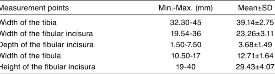
The fibular incisura is situated at the distal epiphy-sis of the tibia on the lateral side, bordered by the tibial tubercles. The tibiofibular syndesmosis has a pyramidal shape. The mean height, the width and the depth of the fibular incisura are given at Table III. Pearson correlation test was used to find any possible correlation between fibular incisura and the other parameters. There was a statistically significant (at the level of 0.05), strong positive correlation (coefficient of correlation (r)=0.432) between the width of the tibia and the depth of the fibular incisura at the 1 cm proximal level from the tibial plafond. There was no correlation with the tibial length.
The difference of the anterior and posterior facet lengths is statistically significant (p=0.000), (Table IV). The posterior facet was bigger on 62% of cases, anterior and posterior facets were
approxi-mately equal on 32% (difference less than 1.5 mm), and the anterior facet was bigger only on 6% of cases. There was no significant difference between genders.
Having said that there was no significant dif-ference between genders regarding the angle of anterior and posterior facets (126.6. in males, 126.9. in females, p>0.05), it must be stated that the fre-quency of the distribution was different.
100°-120° interval and 120°-150° interval were equal (42%) in females, but 120°-150° interval was significantly more prevalent among male subjects (64%), (Figure 3). The angle between the anterior and the posterior facets was statistically different between the right and the left sides (p=0.02).
The angle between the anterior surface of the tibia and the intertuberculer line, which may effect superposition of tubercles on AP radiographs was changing between 70° and 82° (mean 74.63°±3.13). There was no significant difference between male and female subjects. Also there was no correlation between this angle and the angle of the anterior and posterior tubercles, the depth of the fibular incisura or the length of the anterior facet.
We measured the height of the most prominent point of the anterior tubercle from the tibial pla-fond. We thought that this measurement can be helpful to understand the tibiofibular overlap and shed light on bone structures on the plain radio-graphs. This dimension was 15.1±2.4 mm (range between 11.4-22.5 mm).
The difference in the width of the tibia measure-ment between genders is statistically significant (p=0.006). Our observations clearly demonstrated that the width of the tibia, the width of the fibula and the width of the fibular incisura at the 1 cm proximal level from the tibial plafond were sig-nificantly different between male and female bone subjects (p<0.05). But there was no significant dif-ference regarding the depth of the fibular incisura (Table V). 35% of cases had shown significantly concave shape (depth of the fibular incisura ≥4 mm) and 65% had shallow concave fibular incisura (<4 mm) in both genders.
DISCUSSION
An intact tibiofibular syndesmosis joint is impor-tant in maintaining normal ankle function. The function of the syndesmosis is to maintain the nor-mal articulation of the ankle joint by determining
Figure 1. The measurement points of 1 cm proximal level from the tibial plafond. a: Width of the tibia; b: Width of the incisura fibularis; c: Depth of the incisura fibularis; d: Length of the anterior facet; e: Length of the posterior facet; f: Angle between the anterior and posterior facets; g: Angle between the anterior surface of the tibia and intertuberculer line; h: Width of the fibula; ITL: Intertuberculer line; AS: Anterior surface of tibia.
a h g f d b c e Medial Lateral ITL AS Anterior Posterior
the precise relationship between the distal tibia and the fibula.[1,5,6] The knowledge of the
tibiofibu-lar syndesmosis is essential for the preoperative
and postoperative assessment of ankle sprains and ankle fractures. The tibiofibular syndesmosis may be injured in ankle sprains and fractures with or without dislocation. The fibula could be displaced posteriorly from the tibia in severe injuries.[5,7-12] It is
clear that the relationship of the distal tibia, espe-cially the fibular incisura, and the fibula is of prime importance in maintaining the integrity of ankle mortise.[7,11] This is the morphometric study of the
fibular incisura of the tibia. There are only few ana-tomic studies in the literature concerning this area on dry human bones. The anatomy of the tibio-fibular syndesmosis and the tibio-fibular incisura has been described by CT and MRI previously.[4,5,11-13]
Yıldırım et al.[12] evaluated the fibular incisura using
MRI and concluded that a syndesmotic injury may be observed much more frequently in women than men because of anatomic differences. Mavi et al.[5]
performed a study on MRI to investigate the fibu-lar incisura characteristics on recurrent sprained ankle subjects and determined that in this group the anterior and posterior tubercles were lengthier, the depth of fibular incisura was deeper and fibula was more anterior than the control group. The depth of the fibular incisura in men was greater than those of the women in both groups. Höcker and Pachucki[14] performed a study on cadavers and
typified the bony structures of the syndesmosis into three groups. They reported that 75% of the fibular incisura were concave, 16% were convex, and 8% were irregular forms. Mavi et al.[5] observed in their
TABLE I
Measurement points and parameters at 1 cm proximal level from the tibial plafond
Parameter Name of parameter Measurement points
a Width of the tibia Between the medial and lateral borders of the tibia
b Width of fibular incisura Between the anterior and posterior tubercles
c Depth of incisura fibularis From the deepest point of the fibular incisura to a
line between tips of the anterior and posterior tubercles
d Length of the anterior facet The tip of the anterior tubercle and the between deepest
point of the fibular incisura
e Length of the posterior facet Between the tip of the posterior tubercle and the deepest
point of the fibular incisura
f Angle between the anterior and posterior Angle between the anterior and posterior facets facets
g Angle between the anterior surface of the Angle between the anterior surface of the tibia and tibia and intertuberculer line intertuberculer line which was drawn tangent of tip of the
both tubercles
h Width of the fibula Between the medial and lateral borders of the fibula
1 cm
3
2 1
Figure 2. The measurement points of the fibular incisura
of the tibia. 1: Height of the fibular incisura; 2: Height of the most prominent point of the anterior tubercle from the tibial plafond; 3: Width of the incisura fibularis at the level of the tibial plafond. (The dotted line represents 1 cm proximal level from the tibial plafond which cross section was used for most of measurements).
series that 90% of the specimens in men and 50% in women were significantly concave (4 mm and more of the depth of the fibular incisura) and others were shallowly concave in the patient group. In the control group, the ratio of the significantly concave fibular incisura were 54.65% in men and 29.6% in women. Ebraheim et al. performed a CT study on cadaveric specimens and demonstrated that 60% of the fibular incisura were significantly concave and 40% were shallowly concave (1.72-6.78 mm)[4] Sora
et al.[13] found that the depth of the fibular incisura
was 5.07 mm (range 3.6-6.1 mm). In our study, 35% of cases have shown significantly concave shape (depth of the fibular incisura ≥4 mm) and 65% had shallow concave fibular incisura (<4 mm) in both genders. Shallow concave fibular incisura might be a predisposing factor for the displacement of the fibula, associated with fracture dislocation. While the depth of fibular incisura decreases, the angle between the anterior and posterior facets increases and therefore the vertical distance of tibiofibular overlapping decreases.[4,12] We measured the
ante-rior and posteante-rior facets and found a statistically significant difference between them. Our findings showed that the posterior tubercle was bigger in 62% of cases, anterior and posterior facets were approximately equal in 32% (difference less than 1.5 mm), and the anterior facet was bigger only in 6% of the cases. We also measured the angle between
the anterior and the posterior facets as 126° (mean value), together with the findings for the different distribution between male and female subjects. Yıldırım et al.[12] found this angle as approximately
139° with no gender difference.
In our study, the depth of the fibular incisura were compared with the other parameters and a statistically significant, strong positive correlation was found with the width of the tibia at the 1 cm proximal level from the tibial plafond. McDermott et al.[15] performed a study to evaluate the
relation-ship between the position of the fibula and a recur-rent ankle instability and concluded that an ankle mortise with a more posteriorly positioned fibula has less structural stability and is more susceptible to sprains. Although it is clear that the disruption of the distal tibiofibular syndesmosis represents a serious injury, there is controversy regarding the appropriate criteria that should be used to establish the diagnosis.[7] Syndesmotic injury may be
diffi-cult to appreciate by radiographic criteria because of the variations in the amount of rotation, the wide anatomic variability in the depth of the fibu-lar incisura, and the shape of the tibial tubercle.[6]
This study intended to define the anatomic vari-ability and the morphometric characteristics of the fibular incisura; tibial tubercles and distal fibula to facilitate the understanding of the plain radio-graphic films widely used in routine procedures.
TABLE II
Other measurement parameters of the tibia and fibular incisura
Parameter Name of parameter Measurement points
1 Height of the fibular incisura Between the tibial plafond and top of the fibular notch
2 Height of anterior tubercle Height of the most prominent point of the anterior tubercle from the
tibial plafond
3 Width of the fibular incisura Between the anterior and posterior tubercles at the level of the tibial plafond
4 Length of the tibia Between the tip of the medial malleolus and medial plateau of the tibia
TABLE III
Measurements at 1 cm proximal level from the tibial plafond and corresponding level of the fibula (n= 35)
Measurement points Min.-Max. (mm) Mean±SD
Width of the tibia 32.30-45 39.14±2.75
Width of the fibular incisura 19.54-36 23.26±3.11
Depth of the fibular incisura 1.50-7.50 3.68±1.49
Width of the fibula 10.50-17 12.71±1.64
TABLE IV
Measurements of the anterior and posterior facet of the tibia (n=35)
Measurement points Min.-Max. (mm) Mean±SD
Length of the anterior facet 7.00-16.60 10.89±2.08
Length of the posterior facet 11.00-17.00 13.28±1.49
Angle between anterior and posterior facet 100-160 126.74±17.78
TABLE V
Comparison of the bone measurements at 1 cm proximal level from the tibial plafond and corresponding level of the fibula between male and female subjects
Measurement points Male (n=22) Female (n=13) p
Width of the tibia 40.18±2.50 37.38±2.26 0.006*
Width of the fibular incisura 24.13±3.44 21.78±1.75 0.015*
Depth of the fibular incisura 3.67±1.55 3.70±1.44 0.814
Width of the fibula 13.32±1.77 11.77±0.79 0.028*
Angle of anterior and posterior facets 126.64±17.20 126.92±19.59 0.885
*: p<0.05 Statistically significant difference.
Identification of the distal tubercles is very impor-tant for the radiographic assessment of the ankle and the tibiofibular syndesmosis. The anterior tubercle has a sharp structure unlike the posterior tubercle which has a smooth curvature. The ante-rior and posteante-rior tubercles have unique features that can be recognized in different positions of rotation. To evaluate the tibiofibular syndesmosis on ankle radiographs, two parameters are com-monly used. These are the tibiofibular clear space (TFCS) and the tibiofibular overlap (TFO).[6,7,16-20]
The configuration and the projection of the distal tibial tubercles and the tibiofibular syn-desmosis in different positions of ankle rotation has been described radiologically by different investigators.[16,20]
Beumer and Swiersta[16] noticed that the anterior
tubercle has an angular shape with its maximum dimension approximately 1 cm above the joint line. According to our observations, the height of the most prominent point of the anterior tubercle from the tibial plafond was approximately 1.5 cm. Since the measurements of the tibiofibular syndesmosis taken from a position 1 cm above the joint line have been accepted widely in routine procedures, we also used measurements at this line. Sora et al.[13]
investi-gated the position of the fibula on plastination slices of cadaver ankles and concluded that the fibula is situated posteriorly to tibia, seen from the front.
There is a relationship between the position of the fibula and recurrent ankle instability. Ankle mortise with a more posteriorly positioned fibula has less structural stability and is more susceptible to sprains.[15] The position of the tibial tubercles,
which depends on their dimensions and angle between them, directly shows the location and position of the fibula that is located within the fibular incisura of the tibia.
In our study, we found that the angle between the anterior surface of the tibia and intertuberculer
Figure 3. Comparison of the angle between anterior and
posterior facets in male and female subjects. 64 % 32 0 100-120 121-150 Angle Male Female 151-and over 48 16 27 42 64 42 9 16
line varies between 70° and 82°. This angle shows that the position of the fibula depends on the tibia. This angular measurement could be important for the interpretation of the mortise radiograph.
These data could be useful for X-ray and CT scan examinations of the ankle and are therefore of practical clinical significance.
In conclusion, this study has shown the morpho-metric characteristics and the anatomic variability of the fibular incisura; the tibial tubercles and the distal fibula in order to facilitate our understand-ing of the plain radiographic films widely used in routine procedures, since the identification of the distal tubercles is very important for the radio-graphic assessment of the ankle and the tibiofibu-lar syndesmosis.
REFERENCES
1. Ebraheim NA, Taser F, Shafiq Q, Yeasting RA. Anatomical evaluation and clinical importance of the tibiofibular syndesmosis ligaments. Surg Radiol Anat 2006;28:142-9.
2. Sarrafian SK. Osteology, syndesmology. In: Sarrafian. SK, editor. Anatomy of the foot and ankle. Philadelphia: Lippincott; 1983. p. 35-143.
3. Standring S. Gray’s anatomy. In: Williams A, Davies MS, editors. Ankle and foot. Chapter 115. 39th ed. London: Churchill Livingstone; 2005. p. 1525-30. 4. Ebraheim NA, Lu J, Yang H, Rollins J. The fibular
incisure of the tibia on CT scan: a cadaver study. Foot Ankle Int 1998;19:318-21.
5. Mavi A, Yildirim H, Gunes H, Pestamalci T, Gumusburun E. The fibular incisura of the tibia with recurrent sprained ankle on magnetic resonance imaging. Saudi Med J 2002;23:845-9.
6. Ebraheim NA, Lu J, Yang H, Mekhail AO, Yeasting RA. Radiographic and CT evaluation of tibiofibular syndesmotic diastasis: a cadaver study. Foot Ankle Int 1997;18:693-8.
7. Bozic KJ, Jaramillo D, DiCanzio J, Zurakowski D, Kasser JR. Radiographic appearance of the normal
distal tibiofibular syndesmosis in children. J Pediatr Orthop 1999;19:14-21.
8. Pankovich AM. Fractures of the fibula at the dis-tal tibiofibular syndesmosis. Clin Orthop Relat Res 1979;143:138-47.
9. Scurran BL. Foot and ankle trauma. In: Gumann G., editor. Ankle fractures. Chapter 28. New York: Churchill Livingstone 1990. p. 579-625.
10. Snedden MH, Shea JP. Diastasis with low distal fibula fractures: an anatomic rationale. Clin Orthop Relat Res 2001;382:197-205.
11. Taser F, Shafiq Q, Ebraheim NA. Three-dimensional volume rendering of tibiofibular joint space and quan-titative analysis of change in volume due to tibiofibular syndesmosis diastases. Skeletal Radiol 2006;35:935-41. 12. Yildirim H, Mavi A, Büyükbebeci O, Gümüflburun
E. Evaluation of the fibular incisura of the tibia with magnetic resonance imaging. Foot Ankle Int 2003;24:387-91.
13. Sora MC, Strobl B, Staykov D, Förster-Streffleur S. Evaluation of the ankle syndesmosis: a plastination slices study. Clin Anat 2004;17:513-7.
14. Höcker K, Pachucki A. The fibular incisure of the tibia. The cross-sectional position of the fibula in distal syn-desmosis. Unfallchirurg 1989;92:401-6. [Abstract] 15. McDermott JE, Scranton PE Jr, Rogers JV. Variations in
fibular position, talar length, and anterior talofibular ligament length. Foot Ankle Int 2004;25:625-9.
16. Beumer A, Swierstra BA. The influence of ankle positioning on the radiography of the distal tibial tubercles. Surg Radiol Anat 2003;25:446-50.
17. Harper MC, Keller TS. A radiographic evaluation of the tibiofibular syndesmosis. Foot Ankle 1989;10:156-60. 18. Leeds HC, Ehrlich MG. Instability of the distal
tibio-fibular syndesmosis after bimalleolar and trimalleolar ankle fractures. J Bone Joint Surg [Am] 1984;66:490-503. 19. Ostrum RF, De Meo P, Subramanian R. A critical anal-ysis of the anterior-posterior radiographic anatomy of the ankle syndesmosis. Foot Ankle Int 1995;16:128-31. 20. Pneumaticos SG, Noble PC, Chatziioannou SN,
Trevino SG. The effects of rotation on radiographic evaluation of the tibiofibular syndesmosis. Foot Ankle Int 2002;23:107-11.