Akash Chandawarkar and
P. Hande Özdinler*
MGH-HMS Center for Nervous System Repair, Department of Neurosurgery , Harvard Medical School, Massachusetts General Hospital, Boston, MA, 02114 USA
Abstract
ALS (Amyotrophic lateral sclerosis) is a complex neurodegenerative disorder that affects corticospinal motor neurons in the cortex, cortico-brainstem neurons and the spinal motor neurons in the spinal cord. ALS can develop both due to genetic or sporatic causes. The genetic causes of the disease are largerly unknown, but the cellular interactions between motor neurons and their environment are beginning to elicudate insights into the cellular pathogenesis of the disease. Even though cognition, memory and other brain functions are left intact in patient, the severed motor neuron circuitry leaves them paralyzed. To develop therapeutic approaches in ALS, there is a growing need to understand each and every component of the motor neuron circuitry, and their interaction in detail. In this rewiew, we will introduce cellular components of the complex neurocircuitry and discuss their possible contribution for therapeutic approaches in ALS.
Introduction to ALS
Amyotrophic lateral sclerosis (ALS) is a complex neurodegenerative disorder that is defined by specific degeneration of both corticospinal motor neurons (CSMN) in the cortex, cortico-brainstem neurons, and
spinal motor neurons in the spinal cord (Cleveland et al., 1996; Brown, 1997; Martin, 1999; Brown and Robberecht, 2001; Eisen and Weber, 2001; Carri et al., 2003; Bruijn et al., 2004). To date the molecular, and cellular mechanisms of cell death is poorly understood.
The complexity of ALS stems from various aspects of the disease: i) ALS could be due to genetic (familial ALS, FALS) factors or it could develop sporadically, without having any known genetic prevalence in the family (Pasinelli and Brown, 2006); ii) disease may initiate, develop and progress differently in each patient, making it very hard to diagnose and follow. The overall effect of the disease varies with the time of initiation, pace of development, and pace of progression in each patient (Brown and Robberecht, 2001); iii) disease degenerates both corticospinal motor neurons in the cortex and the spinal motor neurons in the spinal cord, severly limiting the motor neuron circuitry that is involved in controlling voluntery movements. (Brown, 1997; Brown, 1998; Brown and Robberecht, 2001; Bruijn et al., 2004). This speaks to the complexity of the disease as a systems degeneration.
i) Even though most incidences of ALS are sporadic about 10% of patients have a familial history (familial ALS, FALS). A remarkable emphasis has been focused on ALS caused by mutations in Cu/Zn superoxide dismutase (SOD1), since the mutations in this gene has are the most common form of inherited ALS (Beckman et al., 2001; Bendotti and Carri, 2004; Brown, 1998; Bruijn et al., 2004; Cudkowicz and Brown, 1996; Kunst et al., 1997; Trotti et al., 1999; Valentine and Hart, 2003). To date there have been more than 114 mutations found in this one gene which leads to the specific degeneration of motor neurons, with an unknown mechanism (Bruijn et al., 2004). Different mutations within the same gene show some predominence in some regions of the world. For example, the alanine-to-valine subtitution at postion 4 of SOD1 (SOD1A4V) is responsible for about 50% of familial ALS cases in North America, and this information has been very helpful in prediagnosis of ALS (Broom et al., 2006; Juneja et al., 1997).
*Correspondence author:
MGH-HMS Center for Nervous System Repair, Department of Neurosurgery, Harward Medical School, Massachusetts General Hospital, Boston MA, 02114 USA
E-mail: [email protected]
Received: August 22, 2007; Accepted: August 29, 2007.
Review
Effective treatment in amyotrophic lateral sclerosis?
Invest in each player
The mutations in the SOD1 gene has been the focus in the field, but even to date the SOD1 gene carries its mysteries within. Animal models that lack SOD1 gene did not develop any motor neuron degeneration phenotype or the over expression of SOD1 did not have any deletirious effects on motor neuron survival. An ALS phenotype is observed only when this gene is mutated. Therefore the effect is the “toxic gain of function of SOD1” and its mechanism is not well-understood. Even though there have been other genes that were found to be linked to ALS, the animal models generated either by knocking down the gene or by overexpression did not lead to any motor neuron defect (Wang et al., 2003). It is not clearly understood, why only motor neurons are effected when mutant SOD1 is expressed in all the cells of the body. Never the less, “toxic gain of function of SOD1” has been an important tool for the investigation of the cellular, molecular, pyhsiological perspectives of the disease. The presence of various animals models that express mutant SOD1, and construction and analysis of mice carrying a deletable “floxed” mutant SOD1 gene that can be excised by the activation of the Cre recombinase, enabled scientists to directly investigate the role of different cell types in the initiation and progression of the disease.
ii) ALS patients come in different ages, different genders, different socio-economic bacgrounds, different ethnic groups, different locations from all round the world. Thus it is very hard to pin point the causes of the diease (Baek and Desai, 2007). The initial diagnosis is also very hard to perform because the diease does not present one specific area of degeneration. Degeneation in some patients begins in the cortex, while in other patients the spinal motor neurons of cervical region or the lumbar region of the spinal cord maybe the initial targets. Thus, it is important to approach each patient differently and develop different individualized therapies, if possible. In addition to the variations at the initiation phase of the disease, patients show differences in the progression of the disease. In some patients the disease spreads like a hay-fire, where the devastating effects are seen within a year, and in some patients the disease progress slowly (Rowland, 1998; Shaw et al., 1997; Stewart et al., 2006; ten Donkelaar et al., 2004; Veldink et al., 2004). This indicates complexity of the disease, multi-factorial aspect of the disease with the involvement of multiple cellular components, and the dynamic interaction between these components. At this point, it is important to emphasize that motor neurons in the cortex and the spinal cord do not share
exact same biology, or the same developmental paradigm, but they are both named “motor neurons” because they are both involved in the execution of motor function. Thus if we focus only to one aspect of the disease, we may easily miss target since there seems to be multiple targets for an effective therapy in this devastating neurodegenerative disorder.
iii) ALS is not the only motor neuron degeneration disease, but it differs from others by one important aspect. In spinal muscular atrophy (SMA) (Harding, 1992; Hanemann and Ludolph, 2002; Figlewicz and Orrell, 2003; Krivickas, 2003), the spinal motor neurons are affected and show progressive degeneration. In hereditary spastic paraplegia (HSP) (Bruyn, 1992; Fink, 2002), neuronal degeneration is restricted to the corticospinal motor neurons in the cortex. But in ALS, both motor neurons in the cortex and the spinal motor neurons in the spinal cortex degenerate. This tells us something very important about the disease: this is a systems degeneration disorder, and it is the breakdown of the whole system that controls our voluntery movements. ALS thus can not be understood fully if one focuses on spinal cord motor neurons or corticospinal motor neurons; the whole system must be put under investigation, with all components.
Motor neuron circuity
Neurons get input from other neurons, relay information to other neurons, form networks of communication and circuitries. This is the most simplistic explaination of how neurons work in the complex cortex. There are various neuronal networks and circuitries in our bodies that control various different aspects of our functions, the circuitry that control voluntary movements is the “motor neuron circuitry”. The motor neuron circurity has both upper and lower components, which means they have players both in the cortex and the spinal cord. The motor pathways originate in the brain and descend down the spinal cord and spinal motor neurons extend their axons to the muscle (Gammie, 2005; Kern et al., 2005; Selverston, 2005; Molnar and Cheung, 2006) (Figure 1). Therefore the communication of neurons in the cortex is not restricted to cortex. Corticospinal motor neurons extend their axons within the corticospinal tract, in the dorsal funiculus of the spinal cord. CSMN exit the corticospinal tract at different locations of the spinal cord, reach and recognize their targets and form connections mainly with spinal motor neurons (Kaufmann and Mitsumoto, 2002; Martin,
2005; Winhammar et al., 2005). These axonal extensions could be more than a meter long. Spinal motor neurons extend their axons to the periphery and reach, recognize and innervate their muscular targets.
For proper execution of motor neuron function, the cellular environment of the motor neuron is also important. Astrocytes and microglia is present in the environment and we will discuss their contribution separately in this review. The motor neuron circuitry thus involves a cortical component and a spinal component of motor neurons, and the non-neuronal cells that surround the motor neurons both in the cortex and the spinal cord as well as the muscle that is innervated by spinal neurons (Figure 1).
To date much of the emphasis has been focused on spinal motor neurons in the spinal cord, but the upper motor neurons have not been investigated in great detail in mouse models of ALS. Thus in this review, we will mainly highlight findings that focus on spinal motor neurons, but would like you to keep in mind that most of the parts of the puzzle is still missing.
Motor neurons
Motor neurons are very large, excitatory neurons that have a remarkable role in the control of voluntery motor function. Motor neurons have one of the longest axonal projections and they require very high levels of energy for their survival, and for proper functioning. The corticospinal motor neurons, that are sometimes refered to as the “upper motor neurons” reside in layerV of motor cortex. They have a large cell body, a very prominent apical dendrite and an extensively long axon.
The “lower motor neurons” , also called α-motor neurons, are the large neurons in the ventral horns of the spinal cord that send their axons out via spinal roots and directly control muscles. At this point it is important to remember that spinal cord is in fact a column, with continuous tracts and cell columns. The spinal cord can be divided into segments by the nerve roots that come off of it such as 8 cervical, 12 thoracic, 5 lumbar, 5 sacral, and 1 coccygeal. At each segment, rootlets appear to come out of both the dorsal and ventral halves of the spinal cord. At segments that control a limb, the motor neurons are large and numerous. This causes enlarged ventral horns in two places: the lower cervical sections (C5-C8) and the lumbar/sacral sections, and these areas are the major sites of degeneration in ALS patients.
In ALS a specific degeneration is observed in motor neurons both in the cortex and in the spinal cord. In mouse models of ALS, the precise timing of the disease is well established where spinal motor neuron loss is documented at each key point of disease progression, such as the pre-symhomatic stage, symtomatic stage, and end-stage. In SOD1 mice model, having mutant SOD1 in motor neurons induces their degeneration, but other neurons such as the peripheral neurons (e.g. DRG) are not effected. Detailed investigation of motor neurons that degenerate in ALS, revealed that large caliber (>5αm) mylinated axons are more selectively vulnerable in ALS and that different sets of motor neurons that innervate different muscle fibers have different susceptibilities to mutant SOD1 toxicity. Fast-fatiguable motor neurons were shown to be effected first, followed by fast-fatigue-resistant motor neurons (Frey and Gerry, 2006) and the slow type neurons were shown to degenerate the last (Pun et al., 2006).
Even though the progressive paralysis results due to the loss of motor neurons in ALS, evidence from various directions point to the possibility that toxicity is not cell-autonomous and that other cells that are not neurons are involved in the pathogenesis of ALS (Hall et al., 1998; Skene and Cleveland, 2001; Newbery and Abbott, 2002; Clement et al., 2003; Harrington et al., 2004; Tanaka et al., 2006; Di Giorgio et al., 2007; Johansson et al., 2007). Construction and analysis of chimeric mice where different cell types expressed mutant SOD1 in a WT background, enabled researchers to inverstigate the direct controls of each cell type to the pathogenesis of ALS (Clement et al., 2003). Studies where only motor neurons expressed SOD1 mutations did not develop the well-defined ALS phenotype in mice, suggesting that having the mutant SOD1 only in the motor neurons was not enough for the disease to progress. So what are the other components of the disease and at which stage of the disease are they involved the most? Do they play a role in the initiation of the disease, or in the progression of the disease?
Microglia
Microglia are one of the immune cells of the central nervous system, whose primary role is to clean up CNS debris (McGeer et al., 1993; Sargsyan et al., 2005; Streit et al., 2005). Microglia, the smallest of the glial cells, are derived from myeloid progenitor cells that come from bone marrow. During embryonic development they migrate to CNS and differentiate into microglia. Microglial activation is referred to the
state of these cells where their cellular metabolism is increased and cellular products either fully processed or non-processed are secreted outside of the cell (Boillee et al., 2006). Microglial activation has been reported in various neurodegenerative diseases such as Alzheimer’s and Parkinson’s (Giulian, 1999; Gonzalez-Scarano and Baltuch, 1999; Stoll and Jander, 1999; Streit, 2002; Schenk and Yednock, 2002; Streit, 2005; Kim and Joh, 2006). Do microglia play a role in ALS? That question gave rise to various investigations using ALS mouse models and taking advantage of Cre/Lox trangenic system. For example lowering mutant SOD1 expression in microglia and peripheral macrophages (Using a Cre transgene with a CD11b promoter that is specifically expressed in microglia and peripheral macrophages) slowed disease progression, and extended overall survival, but did not affect the initiation of the disease. A similar study where WT microglia was allowed to grow and develop in mutant SOD1 mice model by the complete replacement of entire myeloid lineage by transplantation of normal bone marrow cells into SOD1 mutant mice that had deletion of PU.1 transcription factor and could not synthesize their own myeloid cells yielded interesting results. The replacement of WT microglia into mutant SOD1 mice model did not affect the disease onset, but slowed disease progression (Beers et al., 2006). These results suggested that mutant SOD1 within macrophages do not effect the initiation of the disease, but accelerate disease progression. Studies where introduction of mutant SOD1 expressing microglia to control animals did not lead motor neuron degeneration, suggesting that mutant SOD1-expressing microglia is not enough by itself to cause motor neuron death.
One approach to treat motor neuon diseases has been to decrease the activation of microglia, which occurs during progression of the disease. Minocyline, a tetracyline derivative which was shown to inhibit microglial activation has been tested in mutant SOD1 mice (Kriz et al., 2002), and reported to increase their survival by slowing disease progression when administered at late stages of disease (Zhu et al., 2002). Minocycline has thus been proposed for clinical trials in ALS. An other target in microglial activation has been the cyclooxygenase-2 (Cox-2), which plays a role in the production of proinflamatory cytokines (McGeer, 2001; Consilvio et al., 2004; Okuno et al., 2004; Yiangou et al., 2006; Almer et al., 2006; Benatar, 2007). Use of Cox-2 in ALS mice models prolonged survival by slowing disease onset, but did not provide any benefit to the overall survival in the disease.
Astrocytes
Astrocytes have long been thought to support neurons by secreting growth factors and by removing excitory molecules such as glutamate from synaptic clefts via the action of glial glutamate transporter EAAT2. Neuron are vulnerable to glutame induced damage if not removed from the synaptic clefts, as glutamate triggers repetitive firing and drives abundant calcium entry through AMPA receptors inside the neuron (Holden, 2007; Rothstein et al., 1995; Vandenberghe et al., 2000; Tortarolo et al., 2004; Vermeiren et al., 2006). Motor neurons, different from other neurons, are more succeptible to excitotoxicity. Astrocytes are key cells in regulating the glutamate levels and keeping the neurons from uncontrolled firing. The disturbed balance between the motor neuron and the astrocyte may be an important aspect of motor neuron degeneration in ALS (Van Damme et al., 2005).
Astrocytes respond to damage in many ways: by the varried assembly of their intermediate filaments, by GFAP expression, by the increase in the number and size of processes. This process is called astrocyte activation. Astrocyte activation is seen in ALS patients spinal cords and also the spinal cords of ALS mouse models (Figure 1B). However, activation of astrocyte is not specific to ALS disease; it is also observed in Alzheimer’s as well as Parkinson’s patients. So the specific contribution of astrocyte to ALS has not been fully described, but its effective role has been indicated in various investigations.
Astrocytes are the major sources of growth factors and cytokines that motor neurons require for survival (Acsadi et al., 2002; Ekestern, 2004; Cassina et al., 2005; Vande Velde and Cleveland, 2005; Wilczak and de Keyser, 2005; Zhang and Huang, 2006). Even though the complete list of growth factors needed for motor neuron survival and the complete list of factors secreted from astrocytes is currently unknown, recent investigations showed that activated astrocytes secrete factors that bind to p75NTR receptor and induce apoptosis of motor neurons (Pehar et al., 2004). Most recent studies using cultured motor neurons derived from mouse embyonic spinal cord or differentiated from stem cells, reported that co-cultured motor neurons survived less when they were cultured on astrocytes that expressed mutant SOD1, than on WT astrocytes (Di Giorgio et al., 2007). These investigations, in line with the previous investigations suggest the importance of a functional motor neuron/astrocyte interaction for the health of the motor neuron. Using conditioned medium isolated from
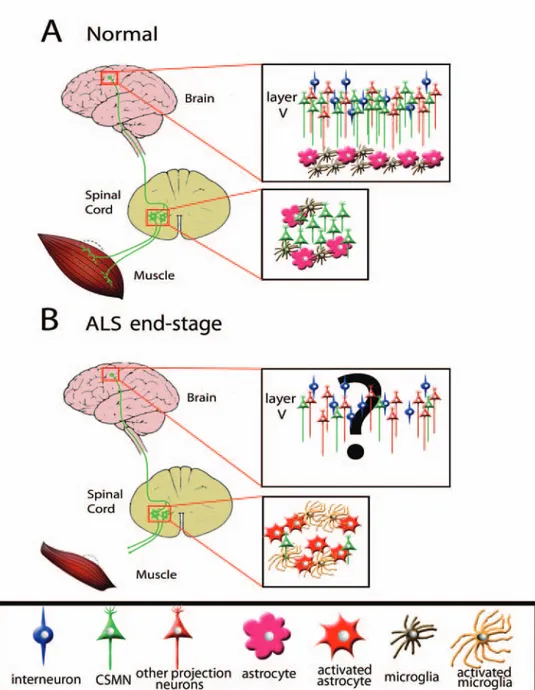
Figure 1: A Schematic representation of motor neuron circuitry that degenerates in ALS. (A) Cortical input is carried to spinal cord
via corticospinal motor neurons (CSMN) , and spinal motor neurons extend their axons to the peripheral muscle. CSMN are in layer V of the cortex, together with other projection neurons (e.g. callosal projection neurons) and interneurons. For simplicity only layer V of the cortex has been shown in the figure. Astrocyte and microglia are present in the cortex, but they are not activated. (B) In diseased patients, the cortiospinal motor neurons are lost, but other projection neurons or interneurons are not affected. Even though the astrocytes and microglia has been reported to be active in ALS patient cortex, their location with respect to CSMN in layer V is not clear and this aspect of the disease is not well-charecterized in ALS mouse model. In the spinal cord the spinal motor neurons are lost and the astrocyte and microglia are activated. This finding has been confirmed both in ALS patients and ALS-mouse models.
mutant SOD1-expressing astrocytes also decreased the survival of motor neurons suggesting the presence of a secrteted molecule from astrocytes that specifically effect motor neurons(Nagai et al., 2007). On the other hand, when motor neurons were cultured with other cells such as fibroblasts which express the mutant SOD1, their survival was not altered. In addition, the astrocytes which expressed the mutant SOD1 did not have a toxic effect on other neurons such as the dorsal root ganglion neurons or GABAergic neurons. These findings suggest a close relationship between astrocytes and motor neurons, the toxic effect of astrocytes are specific to motor neurons in this system and that astrocytes may be important in motor neuron degeneration. All put together, these studies suggest that astrocyte support of motor neuron survival decreases during disease progression, and that SOD1-expressing astrocytes become toxic to motor neurons.
One approach to treat ALS has been to restore the impaired glutamate induced toxicity. The only drug to that has been approved by FDA (Federation of Drug Administration in USA) to be used by ALS patients, Riluzole, has been effective in reducing the glutamate induced toxicity. Its mechanism of action is mediated via blocking the sodium channel, high-voltage calcium channel and by acting as an NMDA/glutame receptor antagonist (Neatherlin, 1998; Meininger et al., 2000). Even though Riluzole increases the life-span of patients very modestly, and is associated with various side effects, it is currently the only FDA-approved drug available to patients. This by itself is a reason why we should focus on better treatment possibilities in ALS.
Future therapies in ALS
Even though ALS is a motor neuron disease, there is now ample evidence to suggest that interaction of motor neurons with their microenvironment is very important for the health and survival of the motor neuron. Having said that, one must again keep in mind that the motor neurons studied in these investigations are spinal motor neurons and the cortical component of the disease has been kept in the dark for those investigations. Thus as is, the picture is not very clear and it is missing pieces, but it has a potential to give us a direction for future investigations.
It is now obvious that there is no single way to success. There must be ways to bring therapeutic approaches that aim different targets together. The chimeric mouse studies as well as the Cre/Lox
recombination studies suggested that the disease is initiated with the death of motor neurons but the progression of the disease is influenced by non-neuronal cells in the environment. So there are key questions to be answered: Why does the motor neuron death begin? How can one support motor neuron survival? Can one enhance motor neuron survival by controlling the possible damage imposed by microglia and astrocyte activation? Can microglia and astrocyte be targets of therapy? Would it make sense to target astrocyte or microglia at later stages of desease, to reduce their “killing” effect on motor neurons? Are cell therapies realistic approaches? Does it make sense to transplant mix of uncharacterized “stem cells” that we have no control over their directed differentiation? Does it make more sense to transplant healthy motor neuons or even healthy non-neuronal cells towards the end stage of disease? How should different therapies be combined, and how could this be tailored for each patient?
Even though there is a race to find a cure for ALS, there is no one single route. Scientists and investigators that take different routes should at times get together to discuss not only the progress they have in their field, but also to put the information they have in a cumulative pot for a better perception of the disease. Afterall, everyone is right depending on the angle they are looking from, but we have to be able to view ALS from a birds-eye view to take a realistic approach for the development of effective therapies in the near future.
References
Acsadi G, Anguelov RA, Yang H, Toth G, Thomas R, Jani A, Wang Y, Ianakova E, Mohammad S, Lewis R A, and Shy M E. Increased survival and function of SOD1 mice after glial cell-derived neurotrophic factor gene therapy. Hum Gene Ther. 13: 1047-1059, 2002.
Almer G, Kikuchi H, Teismann P, and Przedborski S. Is prostaglandin E(2) a pathogenic factor in amyotrophic lateral sclerosis? Ann Neurol. 59: 980-983, 2006.
Baek WS and Desai NP. ALS: pitfalls in the diagnosis. Pract Neurol. 7: 74-81, 2007.
Beckman JS, Estevez AG, Crow JP, and Barbeito L. Superoxide dismutase and the death of motoneurons in ALS. Trends Neurosci 24: S15-20, 2001.
Beers DR, Henkel JS, Xiao Q, Zhao W, Wang J, Yen AA, Siklos L, McKercher SR, and Appel SH. Wild-type microglia extend survival in PU.1 knockout mice with familial amyotrophic lateral sclerosis. Proc Natl Acad Sci. USA 103: 16021-16026, 2006. Benatar M. Lost in translation: treatment trials in the
SOD1 mouse and in human ALS. Neurobiol Dis. 26: 1-13, 2007.
Bendotti C and Carri MT. Lessons from models of SOD1-linked familial ALS. Trends Mol Med. 10: 393-400, 2004.
Boillee S, Yamanaka K, Lobsiger CS, Copeland NG, Jenkins NA, Kassiotis G, Kollias G and Cleveland D W. Onset and progression in inherited ALS determined by motor neurons and microglia. Science 312: 1389-1392, 2006.
Broom WJ, Russ C, Sapp PC, McKenna-Yasek D, Hosler BA, Andersen PM and Brown RH Jr. Variants in candidate ALS modifier genes linked to Cu/Zn superoxide dismutase do not explain divergent survival phenotypes. Neurosci Lett. 392: 52-57, 2006.
Brown RH Jr. Amyotrophic lateral sclerosis. Insights from genetics. Arch Neurol. 54: 1246-1250, 1997. Brown RH Jr. SOD1 aggregates in ALS: cause,
correlate or consequence? Nat Med. 4: 1362-1364, 1998.
Brown RH Jr and Robberecht W. Amyotrophic lateral sclerosis: pathogenesis. Semin Neurol. 21: 131-139, 2001.
Bruijn LI, Miller TM and Cleveland DW. Unraveling the mechanisms involved in motor neuron degeneration in ALS. Annu Rev Neurosci. 27: 723-749, 2004.
Bruyn RP. The neuropathology of hereditary spastic paraparesis. Clin Neurol Neurosurg. 94 Suppl, S16-18, 1992.
Carri MT, Ferri A, Cozzolino M, Calabrese L and Rotilio G. Neurodegeneration in amyotrophic lateral sclerosis: the role of oxidative stress and altered homeostasis of metals. Brain Res Bull. 61: 365-374, 2003.
Cassina P, Pehar M, Vargas MR, Castellanos R, Barbeito AG, Estevez AG, Thompson JA, Beckman JS and Barbeito L. Astrocyte activation by fibroblast growth factor-1 and motor neuron
apoptosis: implications for amyotrophic lateral sclerosis. J Neurochem. 93: 38-46, 2005.
Clement AM, Nguyen MD, Roberts EA, Garcia ML, Boillee S, Rule M, McMahon AP, Doucette W, Siwek D, Ferrante RJ, Brown RH Jr, Julien JP, Goldstein LS and Cleveland DW. Wild-type nonneuronal cells extend survival of SOD1 mutant motor neurons in ALS mice. Science 302: 113-117, 2003.
Cleveland DW, Bruijn LI, Wong PC, Marszalek JR, Vechio JD, Lee MK, Xu XS, Borchelt DR, Sisodia S S and Price, DL Mechanisms of selective motor neuron death in transgenic mouse models of motor neuron disease. Neurology. 47: S54-61; discussion S61-52, 1996.
Consilvio C, Vincent AM and Feldman EL. Neuroinflammation, COX-2, and ALS--a dual role? Exp Neurol. 187: 1-10, 2004.
Cudkowicz M E and Brown R H Jr. An update on superoxide dismutase 1 in familial amyotrophic lateral sclerosis. J Neurol Sci. 139: Suppl. 10-15, 1996.
Di Giorgio FP, Carrasco MA, Siao MC, Maniatis T and Eggan K. Non-cell autonomous effect of glia on motor neurons in an embryonic stem cell-based ALS model. Nat Neurosci. 10: 608-614, 2007. Eisen A and Weber M. The motor cortex and
amyotrophic lateral sclerosis. Muscle Nerve. 24: 564-573, 2001.
Ekestern E. Neurotrophic factors and amyotrophic lateral sclerosis. Neurodegener Dis. 1: 88-100, 2004.
Figlewicz DA and Orrell RW. The genetics of motor neuron diseases. Amyotroph Lateral Scler Other Motor Neuron Disord. 4: 225-231, 2003.
Fink JK. Hereditary spastic paraplegia. Neurol Clin. 20: 711-726, 2002.
Frey SH and Gerry VE. Modulation of neural activity during observational learning of actions and their sequential orders. J Neurosci. 26: 13194-13201, 2006.
Gammie SC. Current models and future directions for understanding the neural circuitries of maternal behaviors in rodents. Behav Cogn Neurosci Rev. 4: 119-135, 2005.
Giulian D. Microglia and the immune pathology of Alzheimer disease. Am J Hum Genet. 65: 13-18, 1999.
Gonzalez-Scarano F and Baltuch G. Microglia as mediators of inflammatory and degenerative diseases. Annu Rev Neurosci. 22: 219-240, 1999. Hall ED, Oostveen JA and Gurney ME. Relationship of
microglial and astrocytic activation to disease onset and progression in a transgenic model of familial ALS. Glia. 23: 249-256, 1998.
Hanemann CO and Ludolph AC. Hereditary motor neuropathies and motor neuron diseases: which is which. Amyotroph Lateral Scler Other Motor Neuron Disord. 3: 186-189, 2002.
Harding AE. Molecular genetics and clinical aspects of inherited disorders of nerve and muscle. Curr Opin Neurol Neurosurg. 5: 600-604, 1992.
Harrington AW, Leiner B, Blechschmitt C, Arevalo JC, Lee R, Morl K, Meyer M, Hempstead BL, Yoon SO and Giehl KM. Secreted proNGF is a pathophysiological death-inducing ligand after adult CNS injury. Proc Natl Acad Sci U S A. 101: 6226-6230, 2004.
Holden C. Neuroscience. Astrocytes secrete substance that kills motor neurons in ALS. Science. 316: 353, 2007.
Johansson A, Engler H, Blomquist G, Scott B, Wall A, Aquilonius SM, Langstrom B and Askmark H. Evidence for astrocytosis in ALS demonstrated by [11C](L)-deprenyl-D2 PET. J Neurol Sci. 255: 17-22, 2007.
Juneja T, Pericak-Vance MA, Laing NG, Dave S and Siddique T. Prognosis in familial amyotrophic lateral sclerosis: progression and survival in patients with glu100gly and ala4val mutations in Cu,Zn superoxide dismutase. Neurology. 48: 55-57, 1997.
Kaufmann P and Mitsumoto H. Amyotrophic lateral sclerosis: objective upper motor neuron markers. Curr Neurol Neurosci Rep. 2: 55-60, 2002. Kern H, McKay WB, Dimitrijevic MM and Dimitrijevic
MR. Motor control in the human spinal cord and the repair of cord function. Curr Pharm Des. 11: 1429-1439, 2005.
Kim YS and Joh TH. Microglia, major player in the brain inflammation: their roles in the pathogenesis
of Parkinson's disease. Exp Mol Med. 38: 333-347, 2006.
Krivickas LS. Amyotrophic lateral sclerosis and other motor neuron diseases. Phys Med Rehabil Clin N Am. 14: 327-345, 2003.
Kriz J, Nguyen MD and Julien JP. Minocycline slows disease progression in a mouse model of amyotrophic lateral sclerosis. Neurobiol Dis. 10: 268-278, 2002.
Kunst CB, Mezey E, Brownstein MJ and Patterson D. Mutations in SOD1 associated with amyotrophic lateral sclerosis cause novel protein interactions. Nat Genet. 15: 91-94, 1997.
Martin JH. The corticospinal system: from development to motor control. Neuroscientist. 11: 161-173, 2005.
Martin LJ. Neuronal death in amyotrophic lateral sclerosis is apoptosis: possible contribution of a programmed cell death mechanism. J Neuropathol Exp Neurol. 58: 459-471, 1999.
McGeer PL. COX-2 and ALS. Amyotroph Lateral Scler Other Motor Neuron Disord. 2: 121-122, 2001. McGeer PL, Kawamata T, Walker DG, Akiyama H,
Tooyama I and McGeer EG. Microglia in degenerative neurological disease. Glia. 7: 84-92, 1993.
Meininger V, Lacomblez L and Salachas F. What has changed with riluzole? J Neurol. 247: 19-22, 2000. Molnar Z and Cheung A F. Towards the classification of subpopulations of layer V pyramidal projection neurons. Neurosci Res. 55: 105-115, 2006. Nagai M, Re DB, Nagata T, Chalazonitis A, Jessell TM,
Wichterle H and Przedborski S. Astrocytes expressing ALS-linked mutated SOD1 release factors selectively toxic to motor neurons. Nat Neurosci. 10: 615-622, 2007.
Neatherlin JS. Management of amyotrophic lateral sclerosis with riluzole. J Neurosci Nurs. 30: 257-260, 1998.
Newbery HJ and Abbott CM. Of mice, men and motor neurons. Trends Mol Med. 8: 88-92, 2002. Okuno T, Nakatsuji Y, Kumanogoh A, Koguchi K,
Moriya M, Fujimura H, Kikutani H and Sakoda S. Induction of cyclooxygenase-2 in reactive glial cells by the CD40 pathway: relevance to
amyotrophic lateral sclerosis. J Neurochem. 91: 404-412, 2004.
Pasinelli P and Brown R H. Molecular biology of amyotrophic lateral sclerosis: insights from genetics. Nat Rev Neurosci. 7: 710-723, 2006. Pehar M, Cassina P, Vargas M R, Castellanos R, Viera
L, Beckman JS, Estevez AG and Barbeito L. Astrocytic production of nerve growth factor in motor neuron apoptosis: implications for amyotrophic lateral sclerosis. J Neurochem. 89: 464-473, 2004.
Pun S, Santos AF, Saxena S, Xu L and Caroni P. Selective vulnerability and pruning of phasic motoneuron axons in motoneuron disease alleviated by CNTF. Nat Neurosci. 9: 408-419, 2006.
Rothstein JD, Van Kammen M, Levey AI, Martin LJ and Kuncl RW. Selective loss of glial glutamate transporter GLT-1 in amyotrophic lateral sclerosis. Ann Neurol. 38: 73-84, 1995.
Rowland LP. Diagnosis of amyotrophic lateral sclerosis. J Neurol Sci 160 Suppl. 1: S6-24, 1998. Sargsyan SA, Monk PN and Shaw PJ. Microglia as
potential contributors to motor neuron injury in amyotrophic lateral sclerosis. Glia. 51: 241-253, 2005. Schenk DB and Yednock T. The role of microglia in
Alzheimer's disease: friend or foe? Neurobiol Aging. 23: 677-679; discussion 683-674, 2002. Selverston AI. A neural infrastructure for rhythmic
motor patterns. Cell Mol Neurobiol. 25: 223-244, 2005.
Shaw CE, Enayat ZE, Powell JF, Anderson VE, Radunovic A, al-Sarraj S and Leigh PN. Familial amyotrophic lateral sclerosis. Molecular pathology of a patient with a SOD1 mutation. Neurology. 49: 1612-1616, 1997.
Skene JP and Cleveland DW. Hypoxia and Lou Gehrig. Nat Genet. 28: 107-108, 2001.
Stewart HG, Andersen PM, Eisen A and Weber M. Corticomotoneuronal dysfunction in ALS patients with different SOD1 mutations. Clin Neurophysiol. 117: 1850-1861, 2006.
Stoll G and Jander S. The role of microglia and macrophages in the pathophysiology of the CNS. Prog Neurobiol. 58: 233-247, 1999.
Streit WJ. Microglia as neuroprotective, immunocompetent cells of the CNS. Glia. 40: 133-139, 2002.
Streit WJ. Microglia and neuroprotection: implications for Alzheimer's disease. Brain Res Brain Res Rev. 48: 234-239, 2005.
Streit WJ, Conde JR, Fendrick SE, Flanary BE and Mariani CL. Role of microglia in the central nervous system's immune response. Neurol Res. 27: 685-691, 2005.
Tanaka F, Niwa J, Ishigaki S, Katsuno M, Waza M, Yamamoto M, Doyu M and Sobue G. Gene expression profiling toward understanding of ALS pathogenesis. Ann N Y Acad Sci. 1086: 1-10, 2006. ten Donkelaar HJ, Lammens M, Wesseling P, Hori A, Keyser A and Rotteveel J. Development and malformations of the human pyramidal tract. J Neurol. 251: 1429-1442, 2004.
Tortarolo M, Crossthwaite AJ, Conforti L, Spencer JP, Williams RJ, Bendotti C and Rattray M. Expression of SOD1 G93A or wild-type SOD1 in primary cultures of astrocytes down-regulates the glutamate transporter GLT-1: lack of involvement of oxidative stress. J Neurochem. 88: 481-493, 2004.
Trotti D, Rolfs A, Danbolt NC, Brown RH, Jr and Hediger MA. SOD1 mutants linked to amyotrophic lateral sclerosis selectively inactivate a glial glutamate transporter. Nat Neurosci. 2: 848, 1999. Valentine JS and Hart PJ. Misfolded CuZnSOD and amyotrophic lateral sclerosis. Proc Natl Acad Sci U S A. 100: 3617-3622, 2003.
Van Damme P, Dewil M, Robberecht W and Van Den Bosch L. Excitotoxicity and amyotrophic lateral sclerosis. Neurodegener Dis. 2: 147-159, 2005. Vande Velde C and Cleveland DW. VEGF: multitasking
in ALS. Nat Neurosci. 8: 5-7, 2005.
Vandenberghe W, Ihle EC, Patneau DK, Robberecht W and Brorson JR. AMPA receptor current density, not desensitization, predicts selective motoneuron vulnerability. J Neurosci. 20: 7158-7166, 2000. Veldink JH, Van den Berg LH and Wokke JH. The
future of motor neuron disease: the challenge is in the genes. J Neurol. 251: 491-500, 2004.
Vermeiren C, Hemptinne I, Vanhoutte N, Tilleux S, Maloteaux JM and Hermans E. Loss of metabotropic glutamate receptor-mediated regulation of glutamate transport in chemically activated astrocytes in a rat model of amyotrophic lateral sclerosis. J Neurochem. 96: 719-731, 2006. Wang J, Slunt H, Gonzales V, Fromholt D, Coonfield M, Copeland NG, Jenkins NA and Borchelt DR. Copper-binding-site-null SOD1 causes ALS in transgenic mice: aggregates of non-native SOD1 delineate a common feature. Hum Mol Genet. 12: 2753-2764, 2003.
Wilczak N and de Keyser J. Insulin-like growth factor system in amyotrophic lateral sclerosis. Endocr Dev. 9: 160-169, 2005.
Winhammar JM, Rowe DB, Henderson RD and Kiernan MC. Assessment of disease progression in motor neuron disease. Lancet Neurol. 4: 229-238, 2005.
Yiangou Y, Facer P, Durrenberger P, Chessell IP, Naylor A, Bountra C, Banati RR and Anand P. COX-2, CB2 and P2X7-immunoreactivities are increased in activated microglial cells/macrophages of multiple sclerosis and amyotrophic lateral sclerosis spinal cord. BMC Neurol. 6: 12, 2006.
Zhang J and Huang EJ. Dynamic expression of neurotrophic factor receptors in postnatal spinal motoneurons and in mouse model of ALS. J Neurobiol. 66: 882-895, 2006.
Zhu S, Stavrovskaya IG, Drozda M, Kim BY, Ona V, Li M, Sarang S, Liu AS, Hartley DM, Wu DC, Gullans S, Ferrante RJ, Przedborskis, Kristal BS and Friedlander RM. Minocycline inhibits cytochrome c release and delays progression of amyotrophic lateral sclerosis in mice. Nature. 417: 74-78, 2002.