65 Ankara Üniversitesi Tıp Fakültesi Mecmuası 2009, 62(2) DAHİLİ BİLİMLER / MEDICAL SCIENCES
Case Report / Olgu Sunumu
Received: 12.08.2009 • Accepted: 05.10.2009 Corresponding author
Uz. Dr. Nilgün Işıksalan Özbülbül
Türkiye Yüksek İhtisas Hastanesi, Radyoloji Bölümü Phone : +90 (312) 306 16 62 - 1614 E-mail Address : [email protected]
Renal arteriovenous malformations are a rare cause of haematuria. To the best of our knowledge, this is the only case reported so far in the diagnosis of the congenital RAVM using multidetector row computed tomography angiography. We suggest that when surgical or interventional thera-phy is not considered, renal multidetector row computed tomograthera-phy angiograthera-phy should be performed to diagnosis of the AVM.
Key Words : Arteriovenous Malformation, Kidney, Multidetector CT
Renal arteriyovenöz malformasyonlar nadir rastlanan hematüri nedenlerindendir. Bildiğimiz kadarıyla bugüne dek, multidetektör sıralı bilgisayarlı tomografi anjiografi kullanılarak tanısı konulan tek konjenital arteriyovenöz fistül vakasıdır. Biz arteriyovenöz malformasyon tanısı için, cerrahi veya girişimsel tedavi düşünülmediğinde renal multidetektör sıralı bilgisayarlı tomografi anjiografinin yapılması gerektiğini düşünmekteyiz.
Anahtar Sözcükler: Arteriyovenöz Malformasyon, Böbrek, Multidetektör BT
Türkiye Yüksek İhtisas Hastanesi, Radyoloji Bölümü
Congenital Renal Arteriovenous Malformation: Diagnosis With
MDCT Angiography
Konjenital Renal Arteriyovenöz Malformasyon: MDCT Anjiografi ile Tanısı
Nilgün Işıksalan Özbülbül, Muharrem Tola, Özlem Yener
Renal arteriovenous malformations (RAVM) are a relatively rare conge-nital malformation (1¬7). They usu-ally remain asymptomatic during life-time. Most published studies reported on sonographic and color Doppler fin-dings (2,7), angiographic studies (1,4) or computed tomographic findings (11,13). Herein we present a case of congenital RAVM which was diagno-sed by multidetector row computed tomography angiography (MDCTA) and a brief review of the literature. To the best of our knowledge, this is the only case reported so far in the diag-nosis of the congenital RAVM using MDCTA.
Case Report
A 41-year-old woman was referred for evaluation of pain in her right flank which had appeared a few days earli-er. 2 years ago, she had a attack of re-nal colic accompained by microscopic haematuria. Some investigations of the urinary tract, i.e. intravenous pyelog-raphy (IVP) and ultrasound (US)
co-uld not disclose the cause of haema-turia. Because spontaneous resolved of the right flank pain and haematuria, the patient is being treated conserva-tively. At latest admission, clinical exa-mination showed no abnormailities. No abnormal bruit was heard during abdominal auscultation. Blood presur-re, urinanalysis and cultupresur-re, and serum hematologic and biochemical indices were within normal limits. The pati-ent had no history of trauma, biopsy or renal disease/injury. Renal US and color-duplex Doppler US were perfor-med with a Toshiba US unit ( Aplio 80, Toshiba, Tokyo, Japan). US ima-ges of the right kidney showed aneco-ic cystaneco-ic mass in the upper pole (Figure 1). The color Doppler image demons-trated a high blood flow and a mosaic-like vascular area with posterior color spots (tissue vibration) which seemed compatible with a vascular malforma-tion (Figure 2). Spectral analysis with pulsed Doppler sound increased velo-city and decreased resistance in the fe-eding artery and arterial pulsations in the draining vein. Renal MDCTA was
66 Congenital Renal Arteriovenous Malformation: Diagnosis With MDCT Angiography
Ankara Üniversitesi Tıp Fakültesi Mecmuası 2009, 62(2)
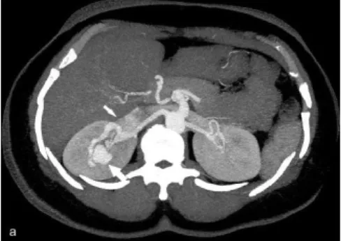
performed with a 16 channel MDCT scanner (Lightspeed 16, GE Medical Systems, Milwaukee, WI, USA). The scan parameters were 16x 1.25-mm detector configuration, 1,25 mm sec-tion thickness, 1,25 mm reconstructi-on interval, gantry rotatireconstructi-on time 0.5 s, pitch 0.938, 400 mAs, 120 Kv. The re-gion of interest for scanning was ad-justed from suprarenal abdominal aor-ta to the iliac artery bifurcation. After insertion of 18 –gauge catheter in to an antecubital vein, 120 mL of iover-sol 300mg I/mL (Optiray, Mallinc-kordt, St. Louis, MO, USA) was injec-ted with an automatic injector at a rate of 4 mL/s. All CT data were transfer-red to a Workstation (Advantage Win-dows 4.2, GE Medical Systems) ) for three-dimensional (3D) reconstructi-ons (multiplanar reformat, maximum intensity projection and volume ren-dering). MDCT angiography was reve-aled early opacification of the right re-nal vein and inferior vena cava. Also en-largement of the interlobar renal artery branch was noted. In the upper pole of the right kidney, a cirsoid type RAVM approximately 3 cm in diameter was demonstrated (Figure 3a, b) MDCT
renal angiography confirmed the diag-nosis and thus no additional imaging was obtained. Because the patient had no further symptoms and refused the treatment, we could not perform ar-terial embolization. In the follow-up 3 months after, the patient remained free of symptoms. We will continue follow-up her.
Discussion
RAVMs are a rare cause of haematuria (1). RAVMs consist of multiple tor-tuous communications between arte-ries and veins without interlaying ca-pilleries. These tortous, varix-like ves-sels are immediately beneath the urot-helium, leading to haematuria as the presenting finding in as many as 72% of cases (5). The reported prevalance of RAVMs is as low as 0.04 % (8), but the true prevalance might be higher because many RAVMs remain
clini-cally asypmtomatic. Other presentati-ons may be systolic or diastolic hyper-tension and may also present as high output cardiac failure (4). Our patient never had hypertension or cardiac fa-ilure symptoms. Macmillan and Ro-binette proposed a clinical classifica-tion of congenital RAVMs, including three subtypes based on location and size: an angiomatous type with a size smaller than 1 cm and a peripheral lo-cation; a cirsoid type with a size lar-ger than 1 cm and peripelvic location; and an idiopathic type with a hilar lo-cation and size larger than 1 cm. The first and second types seem to corres-pond to true AVMs differing in size and location, and the third type seems to be identical to arteriovenous fistu-lae (1). Naganuma et al (2) demons-trated that RAVM exhibit findings si-milar to postbiopsy arteriovenous fis-tulas. The reported findings of post-biopsy arteriovenous fistula are (i) an area of color mosaic appaerence with tissue vibration, ii) increased flow ve-locities and decreased resistive inde-xes in the supplying artery, iii) arteri-alization of the draining vein, and iiii) no abnormalities or small cystic lesions on gray-scale US. Regardless of whet-her the renal AVM was spontaneous or secondary, they found that gray-scale and color Doppler US showed similar findings. Also they concluded that US was not diagnostic and color Doppler US should be performed immediately in patients with hematuria. They were able to identify cirsoid AVMs in five of five patients with Doppler US befo-re the performance of catheter angiog-raphy. Importantly, no lesions were de-tected with gray-scale US alone, even with the knowledge of the location of the AVM. The lesions in this series were all identified on CT, but definiti-on regarding the communicating renal artery and draining vein was poor (5). Angiographically, a true RAVM has a characteristic cirsoid apperance, with tortuous small channels and multip-le fistulous connections. The main an-giographic feature is the simultaneous appearence of contrast in the main nal artery and vein. RAVMs usually
re-
Figure 1. US image of the right kidney shows anecoic cystic mass in the upper pole (arrow).
Figure 2. Longitudinal color Doppler image of the upper pole of the right kidney shows a blood flow- rich area (thin arrow) with posterior color spots (tissue vibration, thick arrow)
Figure 3.Contrast-enhanced MDCT image demonstrates RAVM (thick arrow). Also early opacification of the right renal vein (thin arrow) and inferior vena cava is seen. Axial thick-slab maximum-intensity projection (a) and volume-rendered reconstructed display seen from oblique posterior perspective (b).
Figure 3.Contrast-enhanced MDCT image demonstrates RAVM (thick arrow). Also early opacification of the right renal vein (thin arrow) and inferior vena cava is seen. Axial thick-slab maximum-intensity projection (a) and volume-rendered reconstructed display seen from oblique posterior perspective (b).
67
Nilgün Işıksalan Özbülbül, Muharrem Tola, Özlem Yener
Journal Of Ankara University Faculty of Medicine 2009, 62(2)
ceive blood supply from two or more lobar vascular tributaries (11). On CT, RAVM is imaged as a mass of
vas-cular density located in the renal si-nus and surrounding the pelvicalice-al system. In addition, the renpelvicalice-al vein and left gonadal vein were often dila-ted. However, CT presentation of the-se lesions depends on the level of cont-rast medium in the blood stream, the speed of infusion, the amount of cont-rast material used, and the time elap-sed until images are taken (11). Few studies regarding imaging of AVMs exist and most series are small (5). To the best of our knowledge, diagnosis of RAVMs with MDCTA has not been reported. MDCT offers many advan-tages for image quality in comparison with single slice CT. MDCT scanners allow for fast investigation with high
spatial resolution. Small slice thickness improves the detection of small struc-tures and allows better discrimination of solid and cystic structures as partial-volume effect diminish. Different pha-se of contrast-uptake can be differenti-ated (arterial, cortico-meduller, neph-rographic and excretory phase). For this reason, MDCT of the kidney has become very valuable tool in urology, but a careful protocol stategy is man-datory (9). MDCT represents an im-portant clinical tool that is replacing, in many institutions, catheter based angiography in the evaluation of renal vasculature (14). DSA has been con-sidered the gold standart for evaluati-on of renal arteries; nevertheless, this procedure may carry some complica-tions which should also be considered for patients with seconder hypertensi-on. A noninvazive imaging technique
is therefore desirable. Thus, MDCTA is currently the preferred modality. Management of congenital RAVMs is generally conservative. Most congeni-tal AVMs are small and asymptoma-tic, and some close spontanenously. Transcatheter arterial embolization is the treatment of choice if the RAVM is accompained by significant hematu-ria, severe hypertension, hemorrhage, or high-output cardiac failure. Large congenital AVMs may require surgical removal (7). Kubota et al (10) empha-sized the necessity of careful follow-up, because spontaneous regression of RAVM may ocur relatively rarely. In conclusion, RAVMs are a rare cause of
haematuria. We suggest that when sur-gical or interventional theraphy is not considered, renal MDCTA should be performed to diagnosis of the AVM.
REFERENCES
1. Defreyne L, Govaere F, Vanlangenhove P et al. Cirsoid renal arteriovenous malforma-tion treated by endovascular embolizamalforma-tion with n-butyl-2-cyanoacrylate. Eur Radiol 2000;10:772–
2. Naganuma H, Ishida H, Konno K et al. Renal arteriovenous malformation: sonographic findings. Abdom Imaging 2001;26:661–63 3. Minetti E, Montoli A. Bilateral renal arte-riovenous malformation. N Engl J Med 2004;351:e9
4. Tarif N, Mitwalli AH, Samayer SA et al. Con-genital renal arteriovenous malformation presenting as severe hypertension. Nephrol Dial Transplant 2002; 17:291–94
5. Brown D, Brandes SB. Radiofrequency ab-lation of a recanalized renal arterioveno-us malformation. J Vasc Interv Radiol 2005;16:403–6
6. Wakefield MR. Renal arteriovenous malfor-mation. emedicine 2007. Topic 2861 7. King BF, Hattery RR. Congenital cirsoid
re-nal arteriovenous malformation (AVM) in-volving lower pole of the kidney. Radiograp-hics 1990;10:1101–4
8. Allie DE, Hebert CJ, Walker CM. Multidetec-tor Computed Tomography Angiography. Endovascular Today 2004 March
9. Coppenrath EM, Mueller-Lisse UG. Mul-tidetector CT of the kidney. Eur Radiol 2006;16:2603–11
10. Kubota H, Sakagami H, Kubota Y et al. Spontaneous disappearance of a re-nal arteriovenous malformation. Int J Urol 2003;10:547–49
11. Honda H, Onitsuka H, Naitou K et al. Re-nal arteriovenous malformations: CT featu-res. J Comput Assist Tomo 1991;15:261–4 12. Mishal J, Leibovici O, Bregman L et al.
Huge renal arteriovenous malformation mi-micking a simple para-pelvic cyst. Urol Int 2001;66:49–50
13. Turkeri LN, Daudi I, Abraham JL et al. Cir-soid arteriovenous malformation of kidney presenting as a mass suggestive of malig-nancy. Int J Urol 1998;5:96–9
14. Fraioli M, Catalano C, Bertoletti L, Nardis P. Multidetector-Row CT of Renal Arteries. In: Catalano C, Passariello R, ed. Multidetector-Row CT Angiography. Springer-Verlag Ber-lin Heildeberg, 2005;177–86