1 Department of Physical Medicine and Rehabilitation, Yerkoy/Yozgat Public Hospital, Yozgat, Turkey 2 Department of Department of Radiology, Yerkoy/Yozgat Public Hospital, Yozgat, Turkey
3 Department of Department Of Orthopaedic Surgery And Traumatology, Yerkoy/Yozgat Public Hospital, Yozgat, Turkey 4 Department of Department of Internal Medicine, Yerkoy/Yozgat Public Hospital, Yozgat, Turkey
5 Department of Department of Otorhinolaryngology, Yerkoy/Yozgat Public Hospital, Yozgat, Turkey
Yazışma Adresi /Correspondence: Fulya Bakilan,
Yerkoy Devlet Hastanesi Fiziksel Tıp ve Rehabilitasyon Bölümü Email: [email protected] Geliş Tarihi / Received: 21.01.2015, Kabul Tarihi / Accepted: 26.02.2015
Copyright © Dicle Tıp Dergisi 2015, Her hakkı saklıdır / All rights reserved
Dicle Tıp Dergisi / 2015; 42 (1): 18-21
Dicle Medical Journal doi: 10.5798/diclemedj.0921.2015.01.0523
ORIGINAL ARTICLE / ÖZGÜN ARAŞTIRMA
Assessment of ultrasound imaging and physical examination findings in greater
trochanteric pain syndrome
Büyük trokanterik ağrı sendromunda ultrasonografi ve fizik muayene bulgularının
değerlendirilmesi
Fulya Bakılan1, Gökhan Yüce2, Ahmet Çağdaş Bicen3, Kadir Serkan Yalçın4, Gökçe Tanyeri5
ÖZET
Amaç: Bu çalışmanın amacı, büyük trokanter bölgesin-de ağrısı olan hastalardaki trokanterik bursit ve gluteus medius tendinopati prevelansını ultrasonografi kullana-rak belirlemek ve gluteus medius tendinopatisi tanısının konulmasında, dirençli kalça abduksiyonu ve iç rotasyo-nunda ağrı varlığının değerlendirilmesinin önemini belir-lemektir.
Yöntemler: Bu çalışma büyük trokanterde ağrısı olan 75 hasta üzerinde retrospektif analiz ile gerçekleştirilmiştir. Hastaların fizik muayene kayıtlarında dirençli kalça ab-duksiyon ve iç rotasyonda ağrı varlığı araştırılmıştır. Fizik muayene kayıtlarının analizinden sonra hastaların kas is-kelet sistemi ultrasonografi görüntüleme kayıtları incelen-miş olup, trokanterik bursit ve gluteus medius tendinopati varlığı değerlendirilmiştir.
Bulgular: Büyük trokanterde ağrısı olan 75 hastanın; %41,4’ünde trokanterik bursit, %20’sinde gluteus medius tendinopatisi saptanmıştır. Dirençli kalça abduksiyonu ve iç rotasyonunda ağrı varlığı, gluteus mediusta tendinopati saptanan ve saptanmayan hastalarda gluteus mediusta tendinopati lehine, istatistiksel olarak belirgin farklı bulun-muştur.
Sonuç: Hastalarda büyük trokanter ağrısının etyolojisinin aydınlatabilmesinde fizik muayene ve ultrasonografi ile görüntüleme önemlidir, özellikle dirençli kalça abduksiyo-nu ve iç rotasyoabduksiyo-nunda ağrı varlığının değerlendirilmesi, gluteus mediusta tendinopati varlığının saptanmasında gereklidir.
Anahtar kelimeler: Bursit, Büyük trokanterik ağrı sendro-mu, Gluteus medius tendinopatisi
ABSTRACT
Objective: The aim of this study was to investigate the prevalence of greater trochanteric bursitis and gluteus medius tendinopathy using ultrasound in patients with greater trochanteric pain and assess the value of the pain on resisted hip abduction and pain on resisted hip inter-nal rotation in predicting the presence of gluteus medius tendinopathy.
Methods: The study was a retrospective analysis of 75 patients with greater trochanteric pain. The physical ex-amination records were identified as pain on resisted hip abduction and hip internal rotation. After observing physi-cal examination records, presence of greater trochanteric bursitis or gluteus medius tendinopathy were assessed in documented ultrasound findings.
Results: Of the 75 patients with greater trochanteric pain, trochanteric bursitis was found in 41.4% of patients, glu-teus medius tendinopathy was found in 20% of patients. A significant difference was found in both presence of pain on resisted hip abduction and internal rotation between patients with gluteus medius tendinopathy and patients with other abnormalities in favor of gluteus medius ten-dinopathy.
Conclusion: In order to determine the etiology of greater trochanteric pain, physical examination and ultrasound imaging is important, especially examination of pain on resisted hip abduction and hip internal rotation is essen-tial to detect gluteus medius tendinopathy.
Key words: Bursitis, gluteus medius tendinopathy, great-er trochantgreat-eric pain syndrome
F. Bakılan et al. US imaging and physical examination in greater trochanteric pain syndrome 19
Dicle Tıp Derg / Dicle Med J www.diclemedj.org Cilt / Vol 42, No 1, 18-21
INTRODUCTION
Greater trochanteric pain syndrome is a common cause of hip pain due to greater trochanteric bursitis or other diagnoses, such as gluteus medius tendi-nopathy, mostly peaks between the fourth and sixth decades of life in women [1-3]. The underlying mechanisms are multifactorial as mechanical, de-generative causes. Because the gluteus medius and minimus muscles stabilize the hip and are involved in externally rotation of hip, it may be associated with degenerative processes like shoulder rotator cuff muscles [4,5].
The symptoms are pain over the lateral aspect of the hip with walking, and tenderness over the upper part of the femur [6]. Patients are sometimes misdiagnosed as many other pathologies of the nearby structures or as pain referred from the lum-bar spine. Accurate diagnosis is important in relief of symptoms because the treatment varies depend-ing on the cause. Ultrasound is a simple inexpensive and radiation free test that has been used to demon-strate tendinopathy and bursitis [7,8].
The purpose of this study was to investigate the prevalence of greater trochanteric bursitis and glu-teus medius tendinopathy using ultrasound in pa-tients with greater trochanteric pain and assess the value of pain on resisted hip abduction, and pain on resisted hip internal rotation in predicting the pres-ence of gluteus medius tendinopathy.
METHODS
The study was a retrospective analysis of all cases of patients with greater trochanteric pain seen at Department of Physical Medicine and Rehabilita-tion and Department of Orthopedics in Yerkoy State Hospital (Yozgat, Turkey) between March 2014 and January 2015 by searching the computerized database and patient files. The medical records of 75 patients were reviewed for age, gender, clinical findings, hip radiographs, ultrasound findings.
Patients (n=75) aged 25-85 years with a com-plaint of greater trochanteric pain or tenderness for at least 6 weeks and patients who have physical ex-amination records, hip radiographs and ultrasound findings reports, included to the study. Patients with pregnancy, rheumatic diseases, morbid obesity, cor-tisone injection in the affected hip in the previous 3 months, history of surgery, trauma, avascular
ne-crosis, arthroplasty and infection in the affected hip were excluded.
The physical examination records were identi-fied as pain on resisted hip abduction and hip inter-nal rotation as predictors of a gluteus medius ten-dinopathy. Recent hip radiographs were evaluated by one musculoskeletal radiologist to exclude other causes of hip pain such as fracture, avascular necro-sis and arthronecro-sis.
After detecting physical examination records and evaluation of x-ray, musculoskeletal diagnos-tic ultrasound finding reports were detected. Ultra-sound were performed by the same radiologist who works in the same hospital for 2 years with five years of musculoskeletal ultrasound experience using an Aloka Prosound Alpha 7 machine with a 12-MHz linear probe (Aloka, Tokyo, Japan). In ultrasound finding reports, synovial thickening, anechoic or hypoechoic fluid in the greater trochanteric bursa defined as greater trochanteric bursitis; a thickened heterogeneous tendon with loss of the fibrillar pat-tern with or without intratendinous calcifications, enthesophytes, or tears defined as gluteus medius tendinopathy.
Statistical Analysis
Results were expressed as mean plus/minus standard deviation or median (range) values for all continu-ous variables. Differences subjects were assessed using the Chi-square test. P<0.05 was considered statistically significant. All statistical analyses were performed using the SPSS program version 15.0 (SPSS Inc., Chicago, Ill., USA)
RESULTS
A total of 75 patients (15 male, 60 female) with a mean age of 57.08±12.91 years were assessed. Tro-chanteric bursitis was found in 41.4% of patients, as for gluteus medius tendinopathy (figure 1,2), it was found in 20% of patients; only tendinopathy was found 16% of patients, tendinopathy with trochan-teric bursitis was found 4% of patients (Table 1).
Pain on resisted hip abduction was found 45.1% of patients with trochanteric bursitis, 86.6% of pa-tients with gluteus medius tendinopathy, 20.6% of patients with no bursitis or tendinopathy. Pain on resisted hip internal rotation was found 35.4% of
F. Bakılan et al. US imaging and physical examination in greater trochanteric pain syndrome 20
Dicle Tıp Derg / Dicle Med J www.diclemedj.org Cilt / Vol 42, No 1, 18-21
patients with trochanteric bursitis, 80% of patients with gluteus medius tendinopathy, 13.7% of pa-tients with no bursitis or tendinopathy. A significant difference was found in both pain on resisted hip ab-duction (p<0.001) and pain on resisted hip internal rotation (p<0.001) between “patients with gluteus medius tendinopathy” and “patients with no bursi-tis and tendinopathy”. Also a significant difference were found between “patients with gluteus medius tendinopathy” and “patients with trochanteric bursi-tis” found in both physical examinations (p<0.05). A significant difference was not found in pain on resisted hip internal rotation between “patients with trochanteric bursitis” and “patients with no bursitis and tendinopathy” (p>0.05). However a significant difference was found in pain on resisted hip abduc-tion between “patients with trochanteric bursitis” and “patients with no bursitis or tendinopathy” (p<0.05) (Table 2).
Table 1. The demographic characteristics and ultrasound findings in patients with greater trochanteric pain
Patients (n=75)
Age (year) 57.08±12.91
Gender (female/male) 60/15
Only trochanteric bursitis (n) (%) 31 (41.4) Gluteus medius tendinopathy (n) (%)
Only tendinopathy 12 (16)
With trochanteric bursitis 3 (4) No bursitis or tendinopathy (n) (%) 29 (38.6)
Total (n) (%) 75 (100)
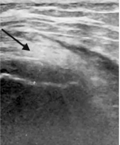
Figure 1. Gluteus medius tendinosis ultrasound images of long axis to lateral facet of greater trochanter show thick and hypoechoic gluteus medius tendon (arrow) with loss of normal fibrillar pattern and mucoid degeneration.
Table 2. Comparison of the presence of each physical finding between patients with trochanteric bursitis, gluteus me-dius tendinopathy and no bursitis or tendinopathy
Trochanteric bursitis
(n) (%) tendinopathy (n) (%)Gluteus medius tendinopathy (n) (%)No bursitis or
Pain on resisted hip abduction 14 (45.1%) * 13 (86.6%)** 6 (20.6%)***
Pain on resisted hip internal rotation 11 (35.4%) † 12 (80%) †† 4 (13.7%) †††
Total 31 (100%) 15 (100%) 29 (100%)
p<0.05 between * and **, p<0.05 between * and ***, p<0.001 between ** and ***, p<0.05 between † and ††, p>0.05 between † and †††, p<0.001 between †† and †††
DISCUSSION
Accurate diagnosis is important in greater trochan-teric pain. The pain may result from greater tro-chanteric bursitis, gluteus medius tendinopathy, il-iotibial band disorders [9] or may be referred pain
such as, knee osteoarthritis, and low back pain [10]. The treatment varies depending on the cause. The therapy modalities of greater trochanteric bursitis or gluteus medius tendinopathy include physiotherapy, nonsteroidal antiinflammatory medication, and lo-cal injection of corticosteroids [11].
Figure 2. Gluteus medius tendino-sis with bursal effusion ultrasonog-raphy images long axis to greater trochanter show tendinosis of the gluteus medius insertion (below ar-row) with loss of echogenicity due to edema and mucoid degenera-tion. There is also a minimal effu-sion in the trochanteric bursa (up-per arrow).
F. Bakılan et al. US imaging and physical examination in greater trochanteric pain syndrome 21
Dicle Tıp Derg / Dicle Med J www.diclemedj.org Cilt / Vol 42, No 1, 18-21
The most important step in the diagnosis is the anamnesis and physical examination. In our study pain on resisted hip abduction and hip internal ro-tation were investigated retrospectively in patients. Both tests were found positive in majority of pa-tients who have gluteus medius tendinopathy. Our results were similar to previous studies, in a study, the ratio of pain on resisted hip abduction was found 65% in patients who have gluteus medius tendi-nopathy in magnetic resonance imaging [12]. To determine the etiology of greater trochanteric pain, physical examination is important, especially ex-amination of pain on resisted hip abduction and hip internal rotation is essential to detect gluteus me-dius tendinopathy.
An imaging method is needed for differential diagnosis of greater trochanteric pain, ultrasound is established as an essential tool for the greater trochanteric pain syndrome. Ultrasound is signifi-cantly important for characterizing tendon abnor-malities, demonstrating fluid in the bursa at the greater trochanter [9,13-15]. In our study, we found trochanteric bursitis more than gluteus medius ten-dinopathy in contrast to other studies. 41.4% of the patients had trochanteric bursitis alone, 16% of the patients had gluteus medius tendinopathy alone and %4 of the patients had gluteal tendinopathy together with bursitis. The results of other studies differ from our study. In the study of Long et al., 877 patients were analyzed with greater trochanteric pain. 8.1% of the patients had trochanteric bursitis alone, 41.3% of the patients had gluteal tendinopathy alone and 8.6% of the patients had gluteal tendinopathy to-gether with bursitis [16]. Differences between the studies may be referred to number of patients. In the study of Bird et. al., gluteal tendinopathy together with bursitis was 10%, gluteal tendinopathy alone was 83.3% of the 24 patients according to magnetic resonance imaging (MRI) [12]. The difference from our study may be associated with MRI findings of gluteal tendinosis or bursitis are often present also in asymptomatic patients [17].
There are two limitations in our study. It is an retrospective study and it lacks control group. Con-sequently assessment of physical examination in as-ymptomatic patients was not performed.
In conclusion, to determine the accurate cause of greater trochanteric pain, both physical
examina-tion and ultrasound imaging are significantly impor-tant.
Conflicts of interest: There are no conflicts of
in-terest and funding.
REFERENCES
1. Walsh G, Archibald CG. MRI in greater trochanteric pain syndrome. Australas Radiol 2003;47:85-87.
2. Bard H, Vuillemin-Bodaghi V, Mutschler C. Tendinopathies du moyen et du petit glutéal: étude pilote de critères cli-niques. Rev Rhum Ed Fr 2006;73:552.
3. Tortolani PJ, Carbone JJ, Quartararo LG. Greater trochan-teric pain syndrome in patients referred to orthopedic spine specialists. Spine J 2002;2:251-254.
4. Gottschalk F, Kourosh S, Leveau B. The functional anatomy of tensor fasciae latae and gluteus medius and minimus. J Anat 1989;166:179-189.
5. Kagan A 2nd. Rotator cuff tears of the hip. Clin Orthop Relat Res 1999;368:135-140.
6. Collée G, Dukmans BAC, Vandenbroucke JP. Greater tro-chanteric pain syndrome (trotro-chanteric bursitis) in low back pain. Scand J Rheumatol 1991;20:262-266.
7. Adler RS, Sofka CM. Percutaneous ultrasound guided in-jections in the musculoskeletal system. Ultrasound Q 2003;19:3-12.
8. Weidner S, Kellner W, Kellner H. Interventional radiology and the musculoskeletal system. Best Pract Res Clin Rheu-matol 2004;18:945-956.
9. Connell DA, Bass C, Sykes CA, et al. Sonographic evalua-tion of gluteus medius and minimus tendinopathy. Eur Ra-diol 2003;13:1339-1347.
10. Segal NA, Felson DT, Torner JC, et al. Greater trochanteric pain syndrome: epidemiology and associated factors. Arch Phys Med Rehabil 2007;88:988-992.
11. Ege Rasmussen KJ, Fanø N. Trochanteric bursitis: treat-ment by corticosteroid injection. Scand J Rheumatol 1985;14:417-420.
12. Bird PA, Oakley SP, Shnier R, Kirkham BW. Prospective Evaluation of Magnetic Resonance Imaging and Physical Examination Findings in Patients With Greater Trochanter-ic Pain Syndrome. Arthritis & Rheum 2001;44:2138-2145. 13. Kong A, Van der Vliet A, Zadow S. MRI and US of gluteal
tendinopathy in greater trochanteric pain syndrome. Eur Radiol 2007;17:1772-1783.
14. Garcia FL, Picado CH, Nogueira-Barbosa MH. Sonograph-ic evaluation of the abductor mechanism after total hip ar-throplasty. J Ultrasound Med 2010;29:465-471.
15. Fearon AM, Scarvell JM, Cook JL, Smith PN. Does ultra-sound correlate with surgical or histologic findings in great-er trochantgreat-eric pain syndrome? A pilot study. Clin Orthop Relat Res 2010;468:1838-1844.
16. Long SS, Surrey DE, Nazarian LN. Sonography of Greater Trochanteric Pain Syndrome and the Rarity of Primary Bur-sitis. AJR 2013;201:1083-1086.
17. Blankenbaker DG, Ullrick SR, Davis KW, et al. Correlation of MRI findings with clinical findings of trochanteric pain syndrome. Skeletal Radiol 2008;37:903-909.