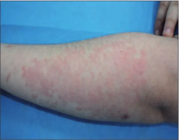
ighteen-year-old female patient with with red scaly plaques on her forearms, legs and scalp, was referred to our out-patient clinic (Fig-ure 1). The disease duration was over 10 years. The female patient was previously diagnosed as psoriasis. She was treated with various topi-cal anti-psoriatic agents unsuccessfully. The lesions were not completely cleared. The patient was evaluated and clinical differential diagnosis in-cluded a number of erythematosquamous skin diseases. Due to the shape and arrangement of lesions , the diagnosis of seborrheic dermatitis, pityr-iasis rosea, lichen planus, pityrpityr-iasis lichenoides et varioliformis acuta, pityriasis lichenoides chronica and parapsoriasis were excluded. And pre-liminary diagnosis of pityriasis rubra pilaris (PRP) and psoriasis were con-cluded. Skin punch biopsy was performed from her left forearm. Histopathological examination showed parakeratosis, loss of granular layer limited to small foci with diffuse hypergranulosis, and perivascular lym-phocyte, histiocytes and extravasated erythrocytes in upper dermis (Haematoxylin-Eosin, original magnification x100) (Figure 2). These histopathological
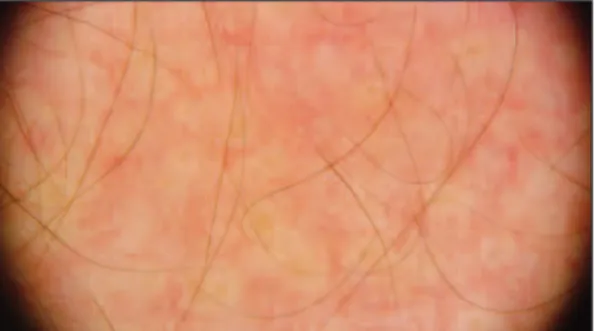
fea-tures were not fully con-sistent with psoriasis and did not exclude the diagnosis of PRP. Later, we performed dermo-scopic examination on erythematous plaques and it revealed round/ oval yellowish areas sur-rounded by vessels of mixed linear and dotted morphology (Figure 3). These findings were consistent with
dermo-Turkiye Klinikleri J Dermatol 2016;26(1)
69
Dermoscopy May be a Valuable Additional
Tool in Diagnosis of Pityriasis Rubra Pilaris:
Letter to the Editor
Ali BALEVİ,a
İlknur TÜRKMEN,b
Yeliz UÇAR TAVLI,a
Mustafa ÖZDEMİRa aDepartment of Dermatology and Venereology,
Medipol Mega University Hospital, bDepartment of Pathology, İstanbul Medipol University Faculty of Medicine, İstanbul Geliş Tarihi/Received: 09.02.2015 Kabul Tarihi/Accepted: 20.01.2016
This case report was presented as a poster at 23rdEuropean Academy of
Dermatology & Venereology Congress 08-12 October 2014,
Amsterdam, The Netherlands.
Yazışma Adresi/Correspondence: Ali BALEVİ
Medipol Mega University Hospital, Department of Dermatology and Venereology, İstanbul, TÜRKİYE/TURKEY [email protected] K
Keeyy WWoorrddss:: Pityriasis rubra pilaris; psoriasis; dermoscopy
A
Annaahh ttaarr KKee llii mmee lleerr:: Pitriyazis rubra pilaris; psöriyazis; dermoskopi
TTuurrkkiiyyee KKlliinniikklleerrii JJ DDeerrmmaattooll 22001166;;2266((11))::6699--7700
doi: 10.5336/dermato.2015-44134 Cop yright © 2016 by Tür ki ye Kli nik le ri
EDİTÖRE MEKTUP
FIGURE 1: Red scaly plaque on her left forearm.
(See color figure at http://www.turkiyeklinikleri.com/journal/dermatoloji-dergisi/1300-0330/)
scopic features of PRP. And the patient was diag-nosed as PRP type 4. Papulo-squamous skin dis-eases are variable, however they are very close in their clinical features. Clinical evaluation of skin lesions is based on common sense and experience of the dermatologist to differentiate features of each disease.1 However, unusual presentations at
times do exist and may cause difficulties in the dif-ferentiation among these entities. In those cases, histopathology contributes significantly to the
ac-curate diagnosis.2 However, in rare instances,
histopathology may be insufficient in the diagno-sis of papulo-squamous diseases. The dermoscope is a low-cost, noninvasive device that is readily available in daily clinical practice. Although the diagnosis of psoriasis is usually straightforward, at times it may be challenging to differentiate it from PRP. Recently, dermoscopy has been reported to be helpful in the distinction between these 2 enti-ties. In the literature, it was stated that dermo-scopic pattern of psoriasis consisted of uniformly distributed, dotted vessels on a light red back-ground and red globules, twisted hairpin and glomerular vessels could also be seen under der-moscopy.3,4However, the dermoscopic pattern of
PRP consisted of round/oval yellowish areas sur-rounded by vessels of mixed morphology.5 The
dermoscopic findings of erythematous plaques of the patient presented herein is consisted with the PRP findings of the latter study. As a result of clin-ical and dermoscopic features, the patient was di-agnosed as PRP type 4 and 25 mg oral dose of acitretin was administered for the treatment.
In conclusion, still retaining the clinico-pathological correlation as the ‘gold standard’ in doubtful cases, dermatoscopy may be used to dif-ferentiate PRP from psoriasis. And additionally, dermoscopy might permit the differential diagno-sis between both entities and avoid unnecessary biopsies in some cases. However, further larger studies are required to confirm the diagnostic value of dermoscopy in terms of differentiating PRP from psoriasis.
Ali BALEVİ et al. DERMOSCOPY MAY BE A VALUABLE ADDITIONAL TOOL IN DIAGNOSIS OF PITYRIASIS RUBRA PILARIS...
Turkiye Klinikleri J Dermatol 2016;26(1)
70
FIGURE 2: Parakeratosis, loss of granular layer is limited to small foci with
diffuse hypergranulosis, in upper dermis perivascular lymphocyte, histiocytes and extravasated erythrocytes (HE, original magnification x100). (See color figure at http://www.turkiyeklinikleri.com/journal/dermatoloji-dergisi/1300-0330/)
FIGURE 3: Dermoscopic findings revealed round/oval yellowish areas
sur-rounded by vessels of mixed linear and dotted morphology.
(See color figure at http://www.turkiyeklinikleri.com/journal/dermatoloji-dergisi/1300-0330/)
1. Mashaly HM, Masood A, Mohamed A. Classi-fication of papulo-squamous skin diseases using image analysis. Skin Res Technol 2012;18(1):36-44.
2. Lallas A, Kyrgidis A, Tzellos TG, Apalla Z, Karakyriou A, Karatolias A, et al. Accuracy of dermoscopic criteria for the diagnosis of
pso-riasis, dermatitis, lichen planus and pityriasis rosea. Br J Dermatol 2012;166(6):1198-205. 3. Lallas A, Apalla Z, Karteridou A, Lefaki I.
Der-moscopy for discriminating between and pso-riasis. J Dermatol Case Rep 2013;7(1):20-2. 4. Kim G, Jung HJ, Ko HC, Kim MB, Lee WJ,
Lee SJ, et al. Dermoscopy can be useful in
differentiating scalp psoriasis from sebor-rhoeic dermatitis. Br J Dermatol 2011; 164(3):652-6.
5. Lallas A, Apalla Z, Tzellos T, Lefaki I. Photo-letter to the editor: dermoscopy in clinically atypical psoriasis. J Dermatol Case Rep 2012;6(2):61-2.