© TÜBİTAK
doi:10.3906/kim-1906-26 h t t p : / / j o u r n a l s . t u b i t a k . g o v . t r / c h e m /
Research Article
Synthesis of Hofmann-type Zn(H
2O)
2Ni(CN)
4.nG (G = water and 1,4-dioxane)
clathrates and the determination of their structural properties by various
spectroscopic methods
Zeki KARTAL1,∗,, Onur ŞAHİN2,, Abdülkerim YAVUZ3,
1Department of Physics, Faculty of Arts and Sciences, Kütahya Dumlupınar University, Kütahya, Turkey 2Scientific and Technological Research Application and Research Center, Sinop University, Sinop, Turkey
3Institute of Science and Technology, Kütahya Dumlupınar University, Kütahya, Turkey
Received: 18.06.2019 • Accepted/Published Online: 17.10.2019 • Final Version: 09.12.2019
Abstract: Two new 2-dimensional cyanide-bridged coordination polymers [Zn(H2O)2Ni(CN)4 6(H2O) and Zn(H2O)2
Ni(CN)4 3(C4H8O2) ], which were similar to Hofmann-type clathrates, were synthesized based on [Ni(CN)4]2− and
Zn2+ as building blocks. These substances were synthesized as compounds in crystalline form. Thes tructures of the
crystalline compounds were characterized via their spectral analyses. General information about the structures of the newly obtained Hofmann-type clathrates was obtained from their vibration spectra by considering significant changes in the vibration peaks of the cyanide group, water ligand molecule, and guest molecules (water and 1,4-dioxane). The thermal behavior of the Hofmann-type clathrates was investigated in the range of 25–500 °C. In addition, experimental data on the magnetic properties of the Hofmann-type clathrates were obtained using the Gouy method under normal conditions. Information on the properties of the structures of the Hofmann clathrates was obtained by applying the single crystal diffraction technique. The asymmetric unit of the first Hofmann-type clathrate contained 1 Zn(II) ion, 1 Ni(II) ion, 1 cyanide ligand, 1 water ligand molecule, and 2 guest water molecules. The asymmetric unit of the second Hofmann-type clathrate contained 1 Zn(II) ion, 1 Ni(II) ion, 2 cyanide ligands, 1 water ligand molecule, and 2 half guest 1,4-dioxane molecules.
Key words: Hofmann-type clathrates, vibrational spectra, single crystal X-ray diffraction analysis, host structure, guest
molecules.
1. Introduction
There are many important chemical groups in organic chemistry. They play a major role in many important events in our universe and in the formation of the environment we live in. The cyanide group is one of these important chemical groups and is represented by the formula C≡N. The cyanide group forms a wide variety of compounds with metal atoms and other molecules. The Ni(CN)4 group is an important group in terms of both
chemical and physical properties formed by binding of the Ni atom to the carbon atom of the cyanide structure. Some of these compounds are 1-dimensional (1D), some are 2D, and some are 3D. Based on the potassium-Ni cyanide compound, which is one of the cyanide compounds, many new compounds have been synthesized and are still being synthesized. Hofmann-type compounds are among the most important compounds synthesized based on the potassium-Ni cyanide compound.
∗Correspondence: [email protected]
Since the discovery of an interesting compound by Hofmann in 1897, a number of host structures called Hofmann-type compounds have been synthesized and these compounds are given by the general formula M(II)LM'(II)(CN)4. In this formula, M and M'represent 1 transition metal atom, while L represents either 2
ligand molecules capable of a single chemical bond or a single ligand molecule capable of 2 chemical bonds [1,2]. If some suitable molecules, as guests, enter the cavities of the Hofmann-type compounds, these new structures are called Hofmann-type clathrates and are given by the formula M(II)LM'(II)(CN)4•nG. In this formula, G
shows the guest molecule in the Hofmann-type clathrates, whereas n shows the number of the guest molecules [3,4].
Different ligand molecules containing nonmetal atoms, such as N atoms, sulfur (S) atoms, and O atoms, are used to obtain Hofmann-type complexes and clathrates. These nonmetal atoms in the ligand molecules are electron-giving atoms in the formation of Hofmann-type complexes and clathrates. When the transition metal atoms M'(II), such as nickel (Ni), palladium, and platinum (Pt), are used to obtain Hofmann compounds, the compound formed is called a Hofmann-type complex. If the transition metal atoms zinc , cadmium, and mercury are used to obtain the Hofmann compounds, the resulting compounds are called Hofmann-Td-type
complexes and clathrates [1–4].
Broad and descriptive information about Hofmann-type compounds and clathrates can be found in various studies [1–7].
The various structural properties of Hofmann-type complexes and clathrates can be explained by various spectroscopic methods, such as vibrational (infrared (IR) and Raman), nuclear magnetic resonance, electron spin resonance, thermal analysis, single-crystal X-ray diffraction (SC-XRD) techniques, and magnetic moment spectroscopy. The formation of Hofmann-type complexes and clathrates and/or interactions of the guest molecule with the host structure can be easily understood by means of vibrational spectroscopy [5–7].
Hofmann-type complexes are compounds with 2D polymeric layers consisting of M'(CN)−24 ions bonding to one another with M(L)+2 cations. These multilayer transition metal complexes are chemically and
biolog-ically very important macromolecules consisting of metal-metal or metal-ligand-metal bridges with 1, 2, or 3 dimensions [8,9].
The aim of this study was to obtain new Hofmann-type clathrates in crystal structures using the water molecule as a ligand molecule and the water molecule and the 1,4-dioxane molecule as guest molecules. This was done because the water molecule is a substance that is both very abundant and very cheap compared to other ligand molecules.
Water is known as a universal solvent due to its ability to dissolve many substances in our environment, as well as being a good ligand molecule [10]. The 1,4-dioxane material is similar to water in terms of its chemical functions, and it also acts as a ligand molecule in some chemical reactions, while in others it acts as a guest molecule and is also a good solvent liquid. Because 1,4-dioxane is present in greater amounts than other isomers (for example, 1,2-dioxane and 1,3-dioxane), it is usually only referred to as dioxane (D) [11].
The D molecule is structurally centrosymmetric, meaning that it is in the form of a typical chair conformation shown by cyclohexane and its derivatives. However, the D molecule is structurally highly flexible and can easily change to boat conformation due to the changes in its environmental conditions [12].
Solutions of Zn(CH3COO)2 and K2Ni(CN)4•H2O in distilled water and D (C4H8O2) compound were
2. Experimental 2.1. Materials
All chemicals used to obtain the crystals of the Hofmann-type clathrates, namely potassium tetracyanonickelate hydrate {K2[Ni(CN)4]•H2O, Fluka, 96%}, [1,4-dioxane, (C4H8O2) , Fluka, 98%], Zn(II) acetate anhydrous
[Zn(CH3COO)2, Alfa Aesar, 99.9%], and ammonia solution (NH3, Merck, 25%), were used without any further
modifications.
2.2. Syntheses of Hofmann-type clathrates Zn(H2O)2Ni(CN)4•nG(G =H2O and C4H8O2) For synthesis of the compounds obtained in this study, the synthesis method of Kartal et al. [7] was used. The colorless, transparent crystal for the Hofmann-type clathrate Zn(H2O)2Ni(CN)4•n(H2O) (hereafter referred
to as 1) formed over a period of 6 weeks.
The other crystal was obtained in a similar manner as crystal 1. In contrast to crystal 1, while the other crystal was obtained in the third step, about 5 mmol of guest D molecules was added to the mixture medium. The colorless, transparent crystal for the Hofmann-type clathrate Zn(H2O)2Ni(CN)4•n(C4H8O2) (hereafter
referred to as 2) formed over a period of 7 weeks.
2.3. Instrumentation
The FT-IR spectra of crystals 1 and 2 were obtained with a Bruker Optics Vertex 70 FT-IR spectrometer (Billerica, MA, United States) at a resolution of 2 cm−1 and room temperature, at a wavenumber range of 3500–400 cm−1. The FT-Raman spectra of crystals 1 and 2 were obtained with a Bruker Senterra dispersive Raman microscope (Bruker Optics), on the 532-nm line of a 3B diode laser and at room temperature, at a wavenumber range of 3500–150 cm−1. The data of the crystal structures of crystals 1 and 2 were collected with a D8-QUEST diffraction meter (Bruker Optics) equipped with a graphite-monochromatic Mo-Kα ( λ =
0.71073 Å) radiation. The thermal gravimetric analysis (TGA) and differential thermal analysis (DTG) curves of crystals 1 and 2 were recorded in a static air atmosphere at a heating rate of 10 °C/min at temperatures of 25–500 °C, using Pt crucibles with a SII EXSTAR 6000 TG/DTA 6300 thermal analyzer (LabMakelaar Benelux B.V., Zevenhuizen, the Netherlands). Magnetic properties of crystals 1 and 2 were recorded with a Sherwood Scientific Magway MSB MK1 model magnetic balance (Sherwood Scientific Ltd., Cambridge, UK).
The amounts of Ni and Zn in the structures of crystals 1 and 2 were determined using a PerkinElmer optima 4300 DV ICP OES (PerkinElmer Inc., Waltham, MA, USA), and the amounts of C, H, and N were determined using a CHNS-932 element analyzer (LECO Corp., St. Joseph, MI, USA). According to the results of the elemental analysis, the number of guest molecules in the clathrates was n = 6 for crystal 1 and n = 3 for crystal 2. The results of all of the analyses for crystal 1 were as follows: [found/(calculated)%] C, 12.62/(12.91)%; H, 3.98/(4.33)%; N, 15.65/(15.05)%; Ni, 15.21/(15.77)%, and Zn, 17.24/(17.56)%. The results of all of the analyses for crystal 2 were as follows: [found/(calculated)%] C, 36.09/(36.36)%; H, 4.93/(5.34)%; N, 10.93/(10.60)%; Ni, 11.29/(11.11)%, and Zn, 12.64/(12.37)%.
2.4. SC-XRD analyses of crystals 1 and 2
Suitable crystals of 1 and 2 were selected for data collection, which was performed on a D8-QUEST diffractome-ter equipped with a graphite-monochromatic Mo-Kα radiation at 296 K. The structure was solved by direct
methods using SHELXS-2013 [13] and refined by the full-matrix least-squares method on F2 using
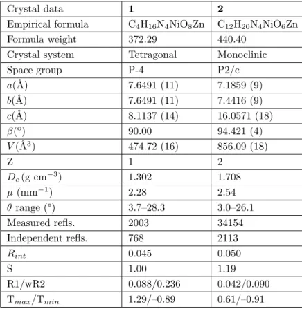
SHELXL-2013 [14]. All nonhydrogen atoms were refined with anisotropic parameters. The H atoms of the C atoms were located from different maps and then treated as riding atoms with C-H distances of 0.96 Å. The other H atoms were located on a difference map refined freely. The following procedures were implemented in the analysis: data collection: Bruker APEX2 [15], program used for molecular graphics: MERCURY program [16], and soft-ware used to prepare material for publication: WinGX [17]. The experimental conditions for the solution of the structures of crystals 1 and 2 and the data of their crystal structures are shown in Table 1.
Table 1. Crystal data and structure refinement parameters for crystals 1 and 2.
Crystal data 1 2
Empirical formula C4H16N4NiO8Zn C12H20N4NiO6Zn
Formula weight 372.29 440.40
Crystal system Tetragonal Monoclinic
Space group P-4 P2/c a(Å) 7.6491 (11) 7.1859 (9) b(Å) 7.6491 (11) 7.4416 (9) c(Å) 8.1137 (14) 16.0571 (18) β(º) 90.00 94.421 (4) V (Å3) 474.72 (16) 856.09 (18) Z 1 2 Dc(g cm−3) 1.302 1.708 µ (mm−1) 2.28 2.54 θ range (°) 3.7–28.3 3.0–26.1 Measured refls. 2003 34154 Independent refls. 768 2113 Rint 0.045 0.050 S 1.00 1.19 R1/wR2 0.088/0.236 0.042/0.090 Tmax/Tmin 1.29/–0.89 0.61/–0.91
3. Results and discussion
3.1. Crystallographic analysis of crystals 1 and 2
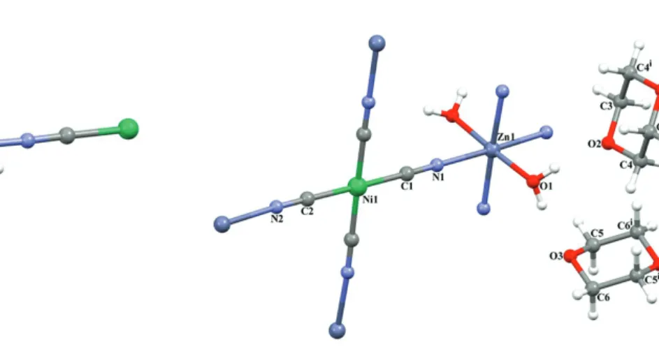
SC-XRD studies of crystals 1 and 2 showed that the structures of these crystals had a 2D polymeric coordination compound structure. The asymmetric unit of heterometallic crystal 1 consisted of 1 Zn(II) ion, 1 Ni(II) ion, 1 cyanide ligand, 1 coordinated water molecule, and 2 noncoordinated water molecules, as shown Figure 1. The asymmetric unit of heterometallic crystal 2 consisted of 1 Zn(II) ion, 1 Ni(II) ion, 2 cyanide ligands, 1 coordinated water molecule, and 2 half noncoordinated D molecules, as shown Figure 2. In crystals 1 and 2, each Zn(II) ion was located on the inversion center and coordinated by 4 N atoms (Zn1-N1 = 2.375(17) Å in crystal 1 and Zn1-N1 = 2.131(3) Å in crystal 2) from cyanide ligands, and 2 O atoms (Zn1-O1 = 2.33(2) Å in crystal 1 and Zn1-O1 = 2.117(3) Å in crystal 2) from water molecules, thus showing a distorted octahedral
coordination geometry. Each Ni(II) ion was coordinated by 4 carbon atoms (Ni1-C1 = 1.85(2) Å in crystal 1 and Ni1-C1 = 1.881(3) Å and Ni1-C2 = 1.877(3) Å in crystal 2) from cyanide ligands, thus showing a square planar coordination geometry. The Zn(II) and Ni(II) ions were bridged by cyanide ligands to generate grid-like 2D sheets, with a grid dimension of 5.409 Å in crystal 1 and 5.163 ×5.182 Å in crystal 2, as shown Figures 3 and 4 (defined by Zn•••Ni distance). These values are clearly seen in Tables 2 and 3. The most striking structural feature of crystals 1 and 2 was that they possessed a 3D network, as shown in Figures 5 and 6, which contained 1D channels along the a axis (7.649 ×8.114 Å2 in crystal 1 and 7.186 ×8.033 Å2 in crystal 2)
(defined by Zn•••Zn distance), which were filled with guest water molecules in crystal 1 and D molecules in crystal 2. The unit cells of crystals 1 and 2 are shown in Figures 7a and 7b, respectively. In order to provide a more comfortable view, the hydrogen atoms have been deleted in Figure 7b.
Figure 1. Molecular structure of crystal 1 showing the
atom numbering scheme.
Figure 2. Molecular structure of crystal 2 showing the
atom numbering scheme [(i) –x + 1, –y + 2, –z + 1].
Table 2. Selected bond distances for crystals 1 and 2 (Å, °). Crystal 1 Zn1-N1 2.375(17) Zn1-O1 2.33(2) Ni1-C1 1.85(2) Crystal 2 Ni1-C2 1.877(3) Ni1-C1 1.881(3) Zn1-O1 2.117(3) Zn1-N1 2.131(3) Zn1-N2vi 2.165(3) O1-Zn1-O1v 177.96(16) O1v-Zn1-N1 89.12(11) O1-Zn1-N1 92.35(11) N1-Zn1-N1v 88.33(15)
O1-Zn1-N2vii 88.27(12) O1-Zn1-N2vi 90.27(11)
N1-Zn1-N2vi 177.39(12) N1-Zn1-N2vii 91.71(10)
Symmetry codes: (v) –x+1, y,−z+1/2; (vi) x+1, y+1, z; and (vii) –x, y+1,−z+1/2 for crystal 2.
Table 3. Hydrogen-bond parameters for crystal 2 (Å, °).
D-H•••A D-H H•••A D•••A D-H•••A
C4—H4B•••O1viii 0.97 2.44 3.328 (5) 151
O1—H1A•••O3 0.82 (6) 1.95 (6) 2.765 (4) 172 O1—H1B•••O2 0.82 (6) 1.91 (6) 2.728 (4) 173
Symmetry code: (viii) –x+1,−y+1, −z+1 for crystal 2.
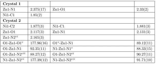
Figure 5. The 3D open framework filled by H2O
molecules in crystal 1.
Figure 6. The 3D open framework filled by the guest D
molecules in crystal 2.
3.2. Spectral characterization of crystals 1 and 2
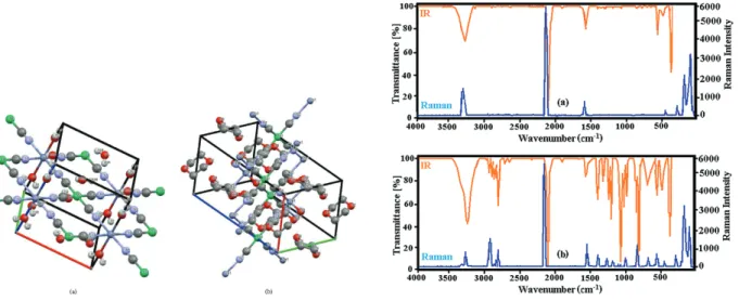
The vibrational (IR and Raman) spectra of crystals 1 and 2 are given in Figures 8a and 8b, respectively. The presence of characteristic vibration peaks of the water ligand molecule and guest water molecule with guest D
molecules in the vibrational spectra of crystals 1 and 2 indicated that these involved molecules played a major role in the formation of crystals 1 and 2.
Figure 7. Unit cells of crystal 1 (a) and 2 (b). Figure 8. Vibrational spectra of crystal 1 (a) and 2 (b).
The data of the vibration spectra of crystals 1 and 2 were interpreted separately for the vibrations of the H2O ligand molecule, [Ni(CN)4]2− ion, and guest H2O with D molecules. According to this interpretation, the
effects of the formation of crystal structures on the spectral data of all of the molecules were clearly revealed.
3.2.1. Vibrations of the H2O ligand and guest molecules
The water molecule is not a linear molecule because it has an angle of about 104.52°between the hydrogen bonds forming the water molecule. Furthermore, since the O atom that forms the water molecule has higher electronegativity than the hydrogen atoms in the structure of the water, the O atom carries some negative charge, while the hydrogen atoms have some positive charges. As a result, the water molecule is a polar molecule with an electrical dipole moment [18–20].
The water molecule has 3 vibration modes, namely asymmetric stretching, symmetric stretching, and the bending vibrations of the O-H bond. These vibration modes are theoretically calculated in vibrational spectroscopy at wavenumbers of 3625, 3520, and 1641 cm−1, respectively [18,20]. These vibration modes were obtained experimentally in IR and Raman spectroscopies, respectively, as 2 vibration peaks at 3431 and 3448 cm−1 with wavenumbers of 1641 and 1648 cm−1 in our laboratory studies. Due to the overlapping of the asymmetric and symmetrical stretching vibrations of the water molecule, a very broad peak with a maximum value at wavenumbers of 3431 and 3448 cm−1 was generated in the IR and Raman spectroscopies, respectively. Similar situations have been observed in the work of some previous researchers [18,19].
Many scientific studies can be found in the literature about the various properties of the water molecules and different studies in which water molecules were used as ligand molecules [21–25].
The broad peaks seen at 3327 and 3336 cm−1 for crystal 1 and 3304 and 3312 cm−1 for crystal 2, respectively, in the IR and Raman spectra of the crystals correspond to the asymmetric and symmetric stretching vibration peaks overlapping with the water ligand molecule. These peaks were shifted to low wavenumbers of 104 and 112 cm−1 for crystal 1 and 127 and 136 cm−1 for crystal 2, according to the liquid water molecule in the IR and Raman spectra, respectively. The reason for these shifts was the binding of the water ligand
molecule from the O atom to the Zn transition metal atom. Another shift to a high wavenumber also occurred in the H-O-H bending vibrations of the water ligand molecule. The reason for these shifts was that the water ligand molecule bound to the Zn transition metal atom. The values of shifting to high frequencies in the IR and Raman spectra of the crystals were 18 and 21 cm−1 for crystal 1 and 21 and 27 cm−1 for crystal 2, respectively. Shifts to these high and low wavenumbers were also observed in studies conducted by other researchers with different ligand molecules [5–7].
There were also some peaks that could not be identified in the vibration spectra of crystals 1 and 2. It was thought that these peaks were composed of their combinations or overtones with the other basic peaks of the water molecule at smaller wavenumbers. Similar situations have also been observed in other studies [19,24,25]. In addition, because weak interactions were formed between all of the guest water molecules in crystal 1, the free OH stretching vibration peak, which should be observed at about 3600 cm−1, was not observed in the IR and Raman spectra of crystal 1.
3.2.2. [Ni(CN)4]2−group vibrations
The Ni[(CN)4]2−ion has 21 basic vibration modes and a D4h symmetry group. The IR active vibrational
modes of Ni[(CN)4]2− ions are ν (C≡N), ν (Ni–CN), π (Ni–CN), and δ (Ni–CN). It was clear that the resulting
Hofmann-type clathrates had a square planar structure because the 4 vibration bands of the Ni[(CN)4]2− ions
that defined this structure were found in the IR spectra of crystals 1 and 2. The vibration frequencies of the Ni(CN)4 groups in the structures of crystals 1 and 2 were interpreted by making use of the studies conducted
by McCullough [26].
The characteristic vibration bands belonging to the Ni(CN)4 group in K2[Ni(CN)4]•H2O compound
with 1 and 2 crystals were obtained. These vibration bands and the frequency shift values that occur due to the formation of Hofmann-type compounds are given in Table 4. The frequency shifts in some vibration modes of a molecule may be to a low frequency region or high frequency region due to changes in environmental conditions or symmetry conditions during the formation of the compounds. In Table 4, some of these frequency shifts are shown to have a positive sign for those that shifted to the high frequency region. In addition, some other frequency shifts shifted to the low frequency region and they are shown with negative signs.
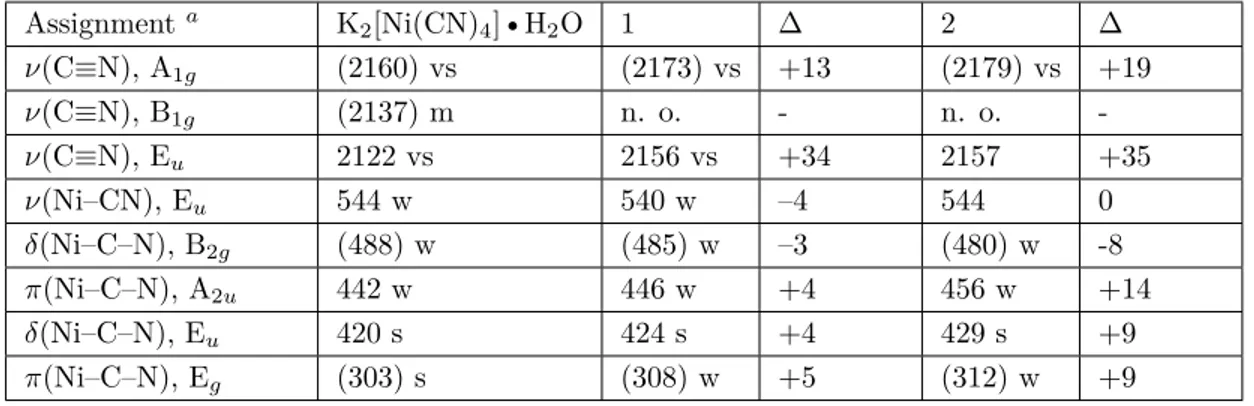
Table 4. Vibrational wavenumbers (cm−1) Ni(CN)4 group in crystals 1 and 2. Bands in the Raman spectra appear
within parentheses. Assignmenta K 2[Ni(CN)4]•H2O 1 ∆ 2 ∆ ν(C≡N), A1g (2160) vs (2173) vs +13 (2179) vs +19 ν(C≡N), B1g (2137) m n. o. - n. o. -ν(C≡N), Eu 2122 vs 2156 vs +34 2157 +35 ν(Ni–CN), Eu 544 w 540 w –4 544 0 δ(Ni–C–N), B2g (488) w (485) w –3 (480) w -8 π(Ni–C–N), A2u 442 w 446 w +4 456 w +14 δ(Ni–C–N), Eu 420 s 424 s +4 429 s +9 π(Ni–C–N), Eg (303) s (308) w +5 (312) w +9
ν : Stretching, δ: in plane bending, π: out-of-plane bending, vs: very strong, s: strong, m: medium, w: weak, vw: very weak, n. o.: not observed.aTaken from [26].
electronegativity of the transition metal atom in that compound, its environment, and the number of bonds it formed with the oxidation state. The frequency values of the C≡N vibrations in a chemical compound depend on the type of metal, electronegativity, chemical environment, oxidation state, and number of bonds it makes [5–7,24,25].
These shifts in the stretching vibration of the cyan group showed that the N atom of the cyan group was bound to the Zn transition metal atom. These shifts in the stretching vibration of the cyan group were due to the coupling effect of C≡N and metal-N bond stretching vibrations [27,28].
3.2.3. Guest D molecule vibrations
The D molecule has a centrosymmetric chair conformation, which is typical, such as relatives of cyclohexane [29,30]. The D molecule is conformationally flexible, and the boat conformation is easily adopted, as required for chelating to metal cations. However, as shown in Figures 4 and 6, the guest D molecule in crystal 2 has a chair conformation.
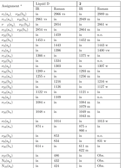
The frequencies of the vibrational bands arising from the enclathrated D observed in the spectrum of crystal 2 are given in Table 5. The wavenumbers of the D in the liquid and gas phases found by Shimanouchi are also given in Table 5 for comparison [30]. The vibrational studies showed that the D molecule had a chair conformation with a center of symmetry of C2hin the gas and liquid phases [29,30]. In our clathrate (in crystal 2), several vibrational bands of the guest D molecule [for example: ν17(au) , ν33(bu) , ν18(au) , and ν35(bu) ]
were split into doublets. When the vibrations of the free liquid phase D were compared with the vibrations of the guest D molecule in crystal 2, they showed a large variation relative to each other. This was probably due to the interactions between the vibrations of the guest D molecule and the vibrations of the H2O ligand molecule,
and the hydrogen bond formed between the guest D molecule and the ligand water molecule (see Table 3). These results indicated that the interactions between the host structure Zn(H2O)2Ni(CN)4 and the guest (D)
molecule, which formed crystal 2, were strong (the crystal field effects). In Figures 2 and 6, the presence of hydrogen bonds between the ligand water molecule and the guest molecule D is clearly apparent in the packed structure of crystal 2.
Similar observations have also been seen for Hofmann-type D clathrates [31–33] and Hofmann-Td-type
D clathrates [34].
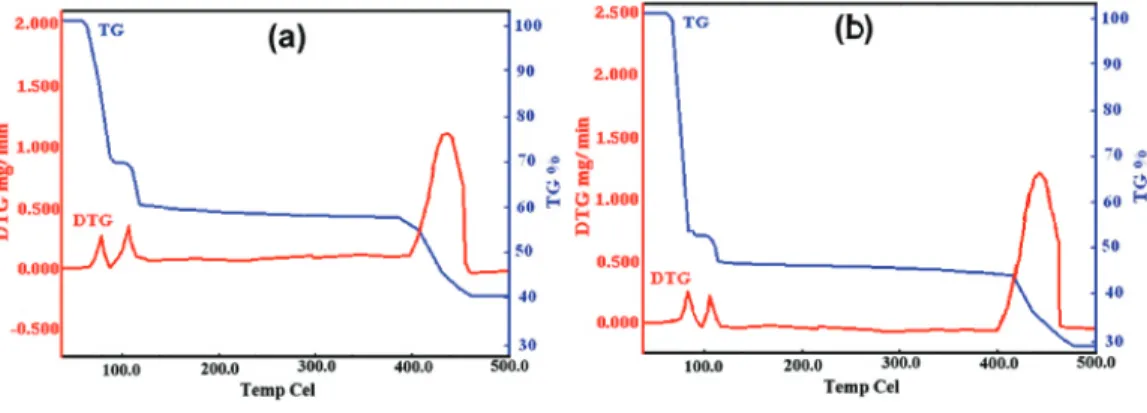
3.3. Thermal behavior of crystals 1 and 2
The TGA and DTG graphics of crystals 1 and 2 are shown in Figures 9a and 9b. When heated, crystals 1 and 2 gradually lost their guest molecules. In the structure of crystal 1, water molecules were present as both ligand and guest molecules. In the first step of the thermal analysis, the 6 guest water molecules, which had not bonded to the crystalline structure, were separated from the crystal structure due to the increased temperature of the environment. This first step of the thermal analysis took place for crystal 1 in a low temperature range of 74–92 °C, with a maximum peak of temperature of about 84 °C [found/(calc.)% = 28.85/(29.03)%].
In the second step of the thermal analysis, the 2 ligand water molecules, which had bonded to the crys-talline structure, were separated from the crystal structure due to the increased temperature of the environment. This second step of the thermal analysis took place for crystal 1 in a temperature range of 98–116 °C, with a maximum peak of temperature of about 108 °C [found/(calc.)% = 8.87/(9.68)%].
In the third stage of the thermal analysis, the Ni(CN)4 bridges forming the structure of crystal 1 were
broken down and the Zn and Ni oxides remained in a temperature range of 398–469 °C, with a maximum peak of temperature of about 448 °C [found/(calc.)% = 40.87/(41.92)%].
Table 5. Vibrational wavenumber (cm−1) of the D in crystal 2. Assignmenta Liquid D 2 IR Raman IR Raman ν1(ag), ν20(bg) ia 2966 vs ia 2989 m ν11(au), ν28(bu) 2961 vs ia 2949 m ia ν 2(ag), ν21(bg) ia 2854 ia 2861 w ν12(au), ν29(bu) 2854 vs ia 2864 m ia ν22(bg) ia 1459 ia n.o. ν30(bu) 1453 s ia 1452 m ia ν3(ag) ia 1443 ia 1443 w ν4(ag) ia 1396 ia 1400 vw ν14(au) 1366 s ia 1375 w ia ν23(bg) ia 1334 ia n.o. ν5(ag) ia 1303 ia 1307 w ν32(bu) 1289 s ia 1293 m ia ν15(au) 1255 s ia 1256 m ia ν24(bg) ia 1216 ia 1216 w ν25(bg) ia 1126 ia 1127 w ν16(au) 1122 vs ia 1121 s ia ν6(ag) ia 1109 ia n.o. ν17(au) 1084 s ia 1084 m 1079 m ia ν33(bu) 1048 s ia 1049 m 1043 m ia ν7(ag) ia 1014 ia 1013 w ν18(au) 874 s ia 875 s 866 s ia ν26(bg) ia 853 ia n.o. ν8(ag) ia 834 ia 831 w ν35(bu) 614 s ia 611 m 622 m ia ν27(bg) ia 486 ia Obs. N9(ag) ia 432 ia Obs. N10(ag) ia 424 ia Obs.
S, Strong; vs, very strong; m, medium; w, weak; vw, very weak; ia, inactive; n.o., not observed; Obs. = obscured.aTaken from [30].
Similar thermal degradation steps were observed for crystal 2. In the first stage of the thermal analysis of crystal 2, the 3 guest D molecules, each of which formed a hydrogen bond with the ligand water molecule, were separated from the crystal structure due to an increase in the temperature of the environment. This first step of the thermal analysis for crystal 2 was in the range of 86–98 °C, with a maximum temperature peak of about 94 °C [found/(calc.)% = 48.87/(50.00)%].
Figure 9. Thermal graphics of crystals 1 (a) and 2 (b).
In the second step of the thermal analysis, the 2 ligand water molecules, which had bonded to the crys-talline structure, were separated from the crystal structure due to the increased temperature of the environment. This second step of the thermal analysis took place for crystal 2 at a temperature range of 100–119 °C, with a maximum peak of temperature of about 110 °C [found/(calc.)% = 6.23/(6.82)%].
In the final stage of the thermal analysis, the Ni(CN)4 bridges forming the structure of crystal 2 were
broken down and the Zn and Ni oxides remained in a temperature range of 402–474 °C, with a maximum peak of temperature of about 441 °C [found/(calc.)% = 28.36/(29.53)%].
These thermal decomposition results showed that crystals 1 and 2 were 2 new examples of Hofmann-type clathrates. Similar decomposition stages were observed for other Hofmann-type clathrates [5,33,35–39].
3.4. Magnetic moments of crystals 1 and 2
Since crystals 1 and 2 both had a square planar coordination structure, and their structures had a square planar Ni2+ (d8) ion and an octahedral Zn2+ (d10) ion, the total number of unpaired electrons for both of
these crystals was zero. For crystals 1 and 2, the theoretically calculated values of the magnetic moments were 0.000 BM, and the experimental magnetic moment values at room temperature under normal conditions were measured as 0.002 and 0.005 BM, which were very close to the theoretical value, respectively.
4. Conclusions
In this study, 2 new Hofmann-type Zn(H2O)2Ni(CN)4•6(H2O) and Zn(H2O)2Ni(CN)4•3(C4H8O2) clathrates
were successfully synthesized in crystal form. Since the reaction medium contained a sufficient number of water molecules as both the ligand and the guest molecule, a Hofmann-type clathrate with the formula Zn(H2O)2Ni(CN)4•2(H2O) was formed in the form of a powder in an aqueous medium. In these
Hofmann-type clathrates, the H2O ligand molecule acted only as a monodentate ligand molecule by binding to the Zn
transition metal atoms from the O atom. At the same time, the guest H2O molecules in crystal 1 formed O-O
and O-hydrogen bonds among themselves. These bonds played a very important role in the stability of the crystalline structure.
While the molecules and the groups that made up the structure of both crystal 1 and crystal 2 were the same, the volumes of the crystals were determined by the type and number of molecules entering them. For example, the volume of crystal 2 was greater than the volume of crystal 1, because the D molecule was considerably larger than the water molecule.
Furthermore, due to weak interaction forces between the ligand water molecules constituting the host structure, it was thought that the volume of the host structure could easily change depending on the structures and volumes of the guest molecules entering the volume of the host structure. Therefore, because of the easily flexible volumes of the host structures formed by the ligand water molecule, it can be considered that they were more suitable for the storage of certain gases than host structures formed by larger ligand molecules.
In addition, in crystals 1 and 2, the Ni(II) ions were surrounded by 4 carbon atoms of 4 cyanide groups in a square planar arrangement, while the Zn(II) ions were surrounded by 4 N atoms of 4 cyanide groups and 2 O atoms of 2 water molecules in octahedral arrangement.
All of the properties and spectral data of the crystalline compounds obtained in this study showed that they were in the structure of polymeric clathrates with 2 transition metal atoms of Hofmann-type clathrates. In some future studies, the crystalline structures of Hofmann-type compounds or clathrates could be also obtained with other transition metal atoms or different guest molecules using the same water ligand molecule. Thus, the effects of different transition metal atoms and different guest molecules on the structural properties of the new Hofmann-type clathrates can be investigated.
Supplementary material
Crystallographic data for the structural analysis have been deposited at the Cambridge Crystallographic Data Centre, CCDC No. 1851096 for 1 and 1854594 for 2. Copies of this information may be obtained free of charge from the Director, CCDC, 12 Union Road, Cambridge CB2 1EZ, UK (fax: +44-1223-336033; e-mail: [email protected] or www: http://www.ccdc.cam.ac.uk).
Acknowledgments
The authors wish to particularly thank Kütahya Dumlupınar University for their technical support (Department of Physics and Chemistry) and financial support under project number 2017/25. The authors acknowledge the Scientific and Technological Research Application and Research Center, Sinop University, Turkey, for the use of their Bruker D8 QUEST diffractometer.
References
1. Iwamoto T. Recent developments in the chemistry of Hofmann-type and the analogous clathrates. Journal of Molecular Structure 1981; 75: 51-65. doi: 10.1016/0022-2860(81)85150-2
2. Iwamoto, T. The Hofmann-type and related inclusion compounds. In: Atwood JL, Davies JED, MacNicol DD (editors). Inclusion Compounds. London, UK: Academic Press, 1984, pp. 29-57.
3. Iwamoto T. Inclusion compounds of multi-dimensional cyanometal complex host. In: Atwood JL, Davies JED, MacNicol DD (editors). Inclusion Compounds. Oxford, UK: Oxford University Press, 1991, pp. 177-212.
4. Iwamoto T. Supramolecular chemistry in cyanometallate systems. In: Atwood, JL, Davies JED, MacNicol DD, Vogtle F (editors). Comprehensive Supramolecular Chemistry. Oxford, UK: Pergamon Press, 1996, pp. 643-690. 5. Kartal Z. Vibrational spectroscopic investigation on some M(Benzonitrile)2Ni(CN)4 complexes (M = Ni, Zn, Cd,
and Hg). Brazilian Journal of Physics 2012; 42: 6-13. doi: 10.1007/s13538-011-0054-x
6. Kartal Z, Yavuz A. The synthesis and the spectroscopic, thermal, and structural properties of the
M2[(fumarate)Ni(CN)4].2(1,4-dioxane) clathrate (M = Co, Ni, Cd and Hg). Journal of Molecular Structure 2018;
7. Kartal Z, Şahin O, Yavuz A. The synthesis of two new Hofmann-type M(3-aminopyridine)2Ni(CN)4 [M = Zn(II) and Cd(II)] complexes and the characterization of their crystal structure by various spectroscopic methods. Journal of Molecular Structure 2018; 1171: 578-586. doi: 10.1016/j.molstruc.2018.06.042
8. Endres H, Keller HJ, Lehmann R, Poveda A, Rupp HH et al. Linear chain bis( α , β -dionedioximato)metal com-pounds of the nickel triad: solid state design by molecular engineering. Zeitschrift für Naturforschung B 1977; 32 (B): 516-527. doi: 10.1515/znb-1977-0508
9. Farrugia L, Evans C. Metal-metal bonding in bridged ligand systems: experimental and theoretical charge densities in Co-3(mu(3)-CX)(CO)(9) (X = H, Cl). Comptes Rendus Chimie 2005; 8: 1566-1583.
doi: 10.1016/j.crci.2004.11.040
10. Graham Solomons TW. Organic Chemistry. New York, NY, USA: Wiley, 1996. 11. Budavari S. The Merck Index. Rahway, NJ, USA: Merck and Co., 1989.
12. Fowles GW, Rice DA, Walton RA. The Raman spectra of metal halide complexes containing 1,4-dioxane. Spec-trochimica Acta Part A: Molecular Spectroscopy 1970; 26A: 143-151. doi: 10.1016/0584-8539(70)80257-4
13. Sheldrick GM. A short history of SHELX. Acta Crystallographica 2008; A64: 112-122. doi: 10.1107/S0108767307043930
14. Sheldrick GM. Crystal structure refinement with SHELXL. Acta Crystallographica 2015; C71: 3-8. doi: 10.1107/S2053229614024218
15. Bruker AXS Inc. Bruker APEX2 (Version 2014.11.0). Madison, WI, USA: Bruker AXS Inc., 2014.
16. Macrae CF, Bruno IJ, Chisholm JA, Edgington PR, Patrick MC et al. Mercury CSD 2.0 – New features for the visualization and investigation of crystal structures. Journal of Applied Crystallography 2008; 41: 466-470. doi: 10.1107/S0021889807067908
17. Farrugia LJ. WinGX and ORTEP for Windows: an update. Journal of Applied Crystallography 2012; 45: 849-854. doi: 10.1107/S0021889812029111
18. Praprotnik M, Janežič D, Mavri J. Temperature dependence of water vibrational spectrum: a molecular dynamics simulation study. Journal of Physical Chemistry A 2004; 108: 11056-11062. doi: doi.org/10.1021/jp046158d 19. Carey DM, Korenowski GM. Measurement of the Raman spectrum of liquid water. Journal of Chemical Physics
1998; 108 (7): 2669-2675. doi: 10.1063/1.475659
20. Zhang C, Khaliullin, RZ, Bovi D, Guidoni L, Ku¨hne TD. Vibrational signature of water molecules in asym-metric hydrogen bonding environments. Journal of Physical Chemistry Letters 2013; 4 (19): 3245-3250. doi: /10.1021/jz401321x
21. Yuge H, Kim CH, Iwamoto T, Kitazawa T. Hofmann-H2O-type and Hofmann-H2O-Td-type host structures
ac-comodating 1,4-dioxane: crystal structures of bis (morpholine- N ) cadmium(II) tetracyanonickelate(II), trans-diaquacadmium(II) tetracyanonickelate(II)-(1,4-dioxane)(1/2) and trans-trans-diaquacadmium(II) tetracyanocadmate(II) (1,4-dioxane)(1/2). Inorganica Chimica Acta 1997; 257: 217-224. doi: 10.1016/S0020-1693(96)05484-9
22. Lu R, Chen Y, Zhou H, Yuan A. M(H2O)2[Ni(CN)4]• x H2O. Acta Chimica Sinica 2010; 68 12: 1199-1204.
23. Rodríguez-Hernández J, Lemus-Santana AA, Vargas CN, Regue E. Three structural modifications in the series of layered solids T(H2O)2[Ni(CN)4].xH2O with T = Mn, Co, Ni: their nature and crystal structures. Comptes
Rendus Chimie 2012; 15: 350-355. doi: 10.1016/j.crci.2011.11.004
24. Alowasheeir A, Tominaka S, Ide Y, Yamauchi Y, Matsushita Y. Two-dimensional cyano-bridged coordination polymer of Mn(H2O)2[Ni(CN)4]: structural analysis and proton conductivity measurements upon dehydration
25. Azhar A, Young C, Kaneti YV, Yamauchi Y, Badjah AY et al. Cyano-bridged Cu-Ni coordination polymer nanoflakes and their thermal conversion to mixed Cu-Ni oxides. Nanomaterials 2018; 8: 968. doi: 10.3390/nano8120968 26. McCullough RL, Jones LH, Crosby GA. An analysis of the vibrational spectrum of the tetracyanonickelate(II) ion
in a crystal lattice. Spectrochimica Acta 1960; 16 (8): 929-944. doi: 10.1016/0371-1951(60)80057-4
27. Smékal Z, Císařová I, Mroziński J. Cyano-bridged bimetallic complexes of copper(II) with tetracyanonickelate(II). Crystal structure of [Cu(dpt)Ni(CN)4]. Polyhedron 2001; 20: 3301-3306. doi: 10.1016/S0277-5387(01)00942-1
28. Kürkçüoğlu GS, Karaağaç D, Yeşilel OZ, Taş M. Synthesis, spectroscopic and structural properties of heteropolynu-clear cyano-bridged complexes. Journal of Inorganic and Organometallic Polymers and Materials 2012; 22: 324-331. doi: 10.1007/s10904-011-9612-5
29. Ellestas OH, Klæboe P. The vibrational spectra of 1,4-dioxan-d0 and 1,4-dioxan-d8. Spectrochimica Acta Part A
Molecular Spectroscopy 1971; 27 (7): 1025-1048. doi: 10.1016/0584-8539(71)80186-1
30. Shimanouchi T. Tables of molecular vibrational frequencies. Consolidated volume II. Journal of Physical and Chemical Reference Data 1977; 6 (3): 993-1102. doi: 10.1063/1.555560
31. Dempster B, Uslu H. Infrared spectra and stability of Hofmann-type dioxane clathrates. Spectrochimica Acta Part A Molecular and Biomolecular Spectroscopy 1978; 34 (1): 71-75. doi: 10.1016/0584-8539(78)80188-3
32. Kartal Z. IR spectroscopic study of M(benzoic acid)2Ni(CN)4•(1,4-dioxane) clathrate (M = Ni, Cd and Co).
Zeitschrift für Naturforschung A 2005; 60 (A): 469-472. doi: 10.1515/zna-2005-0613
33. Kartal Z, Türk T. FT-IR spectroscopic and thermal study of M(1,6-hexanedithiol)Ni(CN)4.2(1,4-dioxane) clathrate
(M = Mn, Co, Ni and Cd). Journal of Molecular Structure 2012; 1014: 74-80. doi: 10.1016/j.molstruc.2012.01.031 34. Zengin T, KantarcıZ, Kasap E. An infrared and Raman spectroscopic study on the Hofmann-Td-type 1,4-dioxane
clathrates: M(NH3)2M′(CN)4•2C4H8O2(M = Mn or Cd, M' = Hg; M = Cd, M' = Cd). Journal of Molecular
Structure 1999; 482-483: 81-85. doi: 10.1016/S0022-2860(98)00839-4
35. Kartal Z, Sayın E. FTIR spectroscopic and thermal study of M(Cyclohexanethiol)2Ni(CN)4.(1,4-dioxane) clathrate
(M = Mn, Co, Ni and Cd). Journal of Molecular Structure 2011; 994: 170-178. doi: 10.1016/j.molstruc.2011.03.014 36. Şenyel M, Parlak C, Alver Ö. FT-IR spectroscopic investigation of some Hofmann type complexes:
M(1-phenylpiperazine)2Ni(CN)4 (M = Ni, Co, Cd, Pd or Mn). Spectrochimica Acta Part A Molecular and
Biomolecular Spectroscopy 2008; 70 367-375. doi: 10.1016/j.saa.2007.10.024
37. Yılmaz VT, Karadağ A. Thermal decomposition of Hofmann-type complexes of di- and triethanolamine. Ther-mochimica Acta 2000; 348 (1): 121-127. doi: 10.1016/S0040-6031(00)00348-8
38. Nishikiori S, Takahashi A, Ratcliffe CI, Ripmeester JA. X-ray and 2H NMR studies of structure and dynamics
in the Hofmann-type and the Hofmann-(en)-type pyrrole clathrates. Journal of Supramolecular Chemistry 2 2002; 483-496. doi: 10.1016/S1472-7862(03)00067-4
39. Parlak C, Alver Ö, Şenyel M. Vibrational spectroscopic study on some Hofmann type clathrates:
M(1-Phenylpiperazine)2Ni(CN)4.2G (M = Ni, Co and Cd; G = aniline). Journal of Molecular Structure 2009;